Method for preparing heart valve endothelial cells by inducing pluripotent stem cell differentiation, and application of heart valve endothelial cells

A technology for pluripotent stem cells and heart valve, applied in the field of stem cell differentiation, can solve the problems of cumbersome process, difficult to guarantee, complicated operation, etc., and achieve the effect of simple differentiation operation, short time-consuming and high differentiation efficiency

Active Publication Date: 2021-02-12
INST OF AQUATIC LIFE ACAD SINICA +1
View PDF4 Cites 5 Cited by
  • Summary
  • Abstract
  • Description
  • Claims
  • Application Information

AI Technical Summary

Problems solved by technology

[0008] (1) The biological valve is easy to decay and has a short lifespan;
[0009] (2) Calcification is prone to occur, so biological valves often face reoperation due to damage (Fu Jie et al., 2011)
[0011] There are many reasons for the lack of seed cells. The first is that heart valve cells are difficult to separate and cultured in vitro for a long time, so the number of cells is extremely limited, and it is difficult to meet the number requirements for seed cell planting.
The second is that it is difficult to obtain analogs of heart valve endothelial cells because of the lack of previous research on human heart valves.
[0014] (1) The differentiation efficiency is low, and the number of real endothelial cells obtained is very small;
[0015] (2) Most of the previous studies were aimed at obtaining vascular endothelial cells, which are difficult to be used for planting tissue-engineered heart valves due to tissue differences;
Simple differentiation to obtain endothelial cells cannot meet the requirements of heart valve seed cells;
[0017] (4) The real valve mesenchymal cells are derived from valve endothelial cells undergoing endothelial-mesenchymal transition, and these endothelial cells have not undergone endothelial-mesenchymal transition tests, so it is difficult to guarantee that they have this function;
[0018] (5) Valve mesenchymal cells will secrete extracellular matrix. Part of the endothelial cells planted on tissue engineered heart valves should maintain their endothelial properties, and the other part should be transformed into mesenchymal cells and secrete extracellular matrix to strengthen and stabilize The role of tissue engineered flaps, the current differentiated endothelial cells have not been able to do this
[0020] (1) The process of this method is very cumbersome, and the cells obtained in each step need to be sorted by flow cytometry. After the mesoderm cells are obtained, the MESP1-positive cells need to be sorted, and the induced cells need to be sorted by CD31 magnetic beads ;
[0021] (2) The cells after mesoderm sorting need to be plated on mouse embryonic fibroblasts in a fibronectin-coated plate, and then induced, the operation is very complicated and time-consuming;
It shows that the differentiation efficiency is not high, and it will increase the workload at the same time
A large amount of cells are also lost during flow sorting

Method used


Image

  • Method for preparing heart valve endothelial cells by inducing pluripotent stem cell differentiation, and application of heart valve endothelial cells
  • Method for preparing heart valve endothelial cells by inducing pluripotent stem cell differentiation, and application of heart valve endothelial cells
  • Method for preparing heart valve endothelial cells by inducing pluripotent stem cell differentiation, and application of heart valve endothelial cells

Examples


Embodiment 1

[0101] The method for inducing the differentiation of human embryonic stem cell line H9 to prepare heart valve endothelial cells comprises the following steps:

[0102] 1) The human embryonic stem cell line H9 was tiled and planted in a matrigel-coated culture dish to make it adhere to the wall; wherein, the tile density of pluripotent stem cells was 30,000 / cm 2 ; Human embryonic stem cell line H9 comes from Wicell Laboratory in the United States;

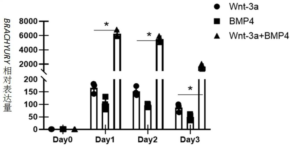
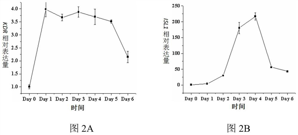
[0103] 2) The pluripotent stem cells were placed in the differentiation medium Essential 6 (Gibco Company) containing Wnt3a and bone morphogenetic protein 4 (BMP4) for 1 day, and then placed in the differentiation medium containing epidermal growth factor (bFGF) and bone morphogenetic protein 4(BMP4) Differentiation medium Essential 6 was cultured for 2 days to obtain cardiac mesoderm cells (LPM); wherein, in the differentiation medium Essential 6 containing Wnt3a and BMP4, the final concentration of Wnt3a was 25ng / mL and the conce...

Embodiment 2

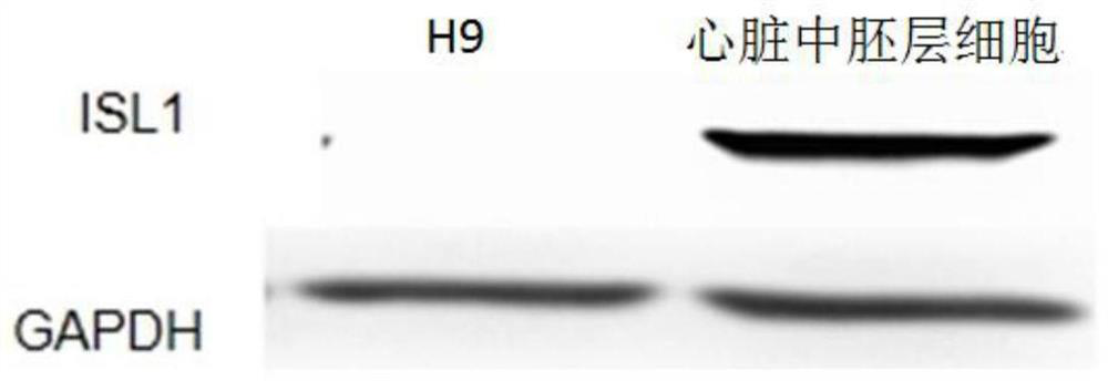
[0149] Morphological and molecular verification of H9-derived heart valve endothelial cells in the above-mentioned Example 1

[0150] 1. Cells and culture conditions

[0151] Heart valve endothelial cells (VEL) derived from H9 obtained in Example 1;

[0152] Umbilical vein endothelial cells (HUVEC) were purchased from the American Type Culture Collection (Rockville, MD, USA);

[0153] Human aortic endothelial cell line (HAEC) was purchased from Shanghai Cell Bank of Chinese Academy of Sciences;

[0154] Isolated human heart valve endothelial cells (VEC), from Wuhan Union Hospital;

[0155] Isolation of human heart valve endothelial cells Culture conditions: 37°C, 5% CO 2 Cell incubator, EGM-2BulletKit culture, EGM-2BulletKit purchased from Lonza Company of the United States.

[0156] 2. Experimental method

[0157] 2.1 Adhesive isolated human heart valve endothelial cells and H9-derived heart valve endothelial cells were photographed and recorded using a microscope.

[0...

Embodiment 3

[0173] Verification of the function of the H9-derived heart valve endothelial cells in the above example 1

[0174] 1. Materials

[0175] Matrigel and all culture dishes were purchased from Corning Company of the United States; Alexa Fluor 594AcLDL fluorescent dye of acetylated low-density lipoprotein was purchased from Invitrogen Company of the United States; nuclear dye DAPI was purchased from Sigma Company of the United States; normal saline DPBS was purchased from BI Company; EBM-2BulletKit was purchased from American Lonza Company; trypsin was purchased from BI Company.

[0176] 2. Cells

[0177] Human embryonic stem cell line H9, from Wicell Laboratory, USA; heart valve endothelial cells prepared in Example 1.

[0178] 3. Experimental method

[0179] 3.1 In vitro low-density lipoprotein uptake test method:

[0180] H9-derived heart valve endothelial cells were cultured to 30-40% confluency with H9 (as a negative control). After cells were serum starved for 12 hours,...


PUM


Abstract

The invention discloses a method for preparing heart valve endothelial cells by inducing differentiation of pluripotent stem cells, and application of the heart valve endothelial cells. The method comprises the following steps: planting pluripotent stem cells in a culture dish coated with matrix glue in a tiled manner, and culturing the pluripotent stem cells by using Essential 8 to enable the pluripotent stem cells to be attached to the wall of the culture dish; and culturing the pluripotent stem cells in a differential medium Essential 6 containing Wnt3a and bone morphogenetic protein 4, culturing the pluripotent stem cells in a differential medium Essential 6 containing epidermal growth factors and bone morphogenetic protein 4, digesting mesoderm cells of the heart, and putting the cardiac mesoderm cells in the culture dish into a differential medium Essential 6 containing bone morphogenetic protein 4, transforming growth factors and endothelial growth factors for continuous cultureto obtain heart valve endothelial cells. The method disclosed by the invention is high in differentiation efficiency, and more than 80% of valvular endothelial cells can be obtained under the condition that flow sorting is not carried out, so that sufficient seed cell sources can be guaranteed.

Description

technical field [0001] The invention relates to the field of stem cell differentiation, in particular to a method for inducing differentiation of pluripotent stem cells to prepare heart valve endothelial cells and its application. Background technique [0002] During a heart beat, blood flows from the low-pressure atria to the high-pressure ventricles, and eventually to the major arteries throughout the body. The heart valves act as one-way valves to maintain the one-way flow of blood. When the heart contracts, the mitral valve, which connects the left atrium to the left ventricle, and the tricuspid valve, which connects the right atrium to the right ventricle, prevent backflow of blood from the ventricles to the atria; when the heart relaxes, the aortic valve connects the left ventricle to the aorta , The pulmonary valve, which connects the right ventricle and the pulmonary artery, prevents the reverse flow of blood from the artery to the ventricle. So as to ensure the no...

Claims


Application Information

Patent Timeline
Patent Type & Authority Applications(China)
IPC IPC(8): C12N5/077
CPCC12N5/0657C12N2506/45C12N2501/415C12N2501/155C12N2501/115
Inventor 孙玉华程麟茜董念国
Owner INST OF AQUATIC LIFE ACAD SINICA
Why Eureka
Try Eureka