
Tissue-engineered bone scaffold for treating infectious large bone defects as well as preparation method and application of tissue-engineered bone scaffold

A tissue-engineered bone and infectious technology, which is applied in the field of tissue-engineered bone scaffolds and its preparation, can solve the problems of weakened recruitment effect, prolonged bone healing time, and failure of angiogenesis to achieve the effect of promoting angiogenesis and preventing infection

Active Publication Date: 2021-03-12
THE FIRST AFFILIATED HOSPITAL OF ARMY MEDICAL UNIV
View PDF2 Cites 0 Cited by
  • Summary
  • Abstract
  • Description
  • Claims
  • Application Information

AI Technical Summary

Problems solved by technology

Previous studies have shown that when host macrophages are absent, angiogenesis in grafts cannot proceed, and studies have also shown that when host macrophage recruitment is blocked, the recruitment effect of host endothelial progenitor cells (EPC) is significant Weakened, and the osteogenic repair ability of TEB was significantly reduced
The release of inflammatory factors in the early fracture can accelerate the bone remodeling process, but the long-term inflammatory response will lead to a decrease in the vascularization level of the injured site and prolong the bone healing time

Method used


Image

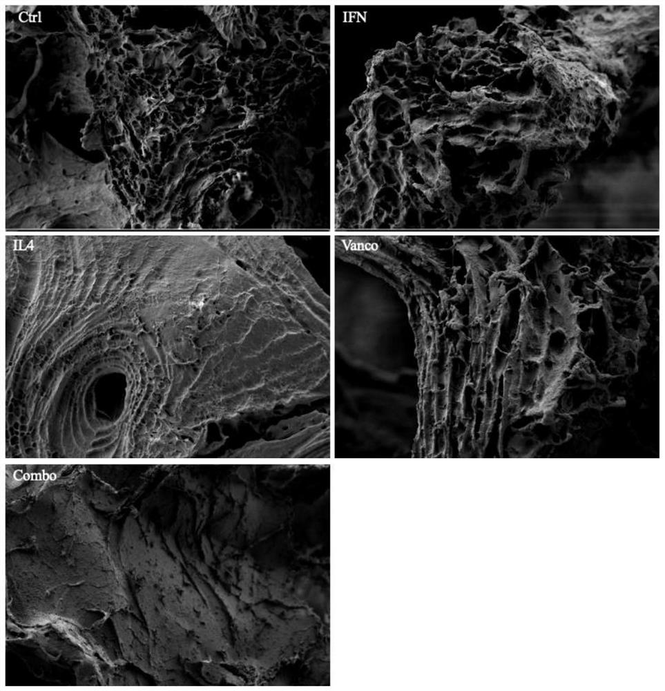
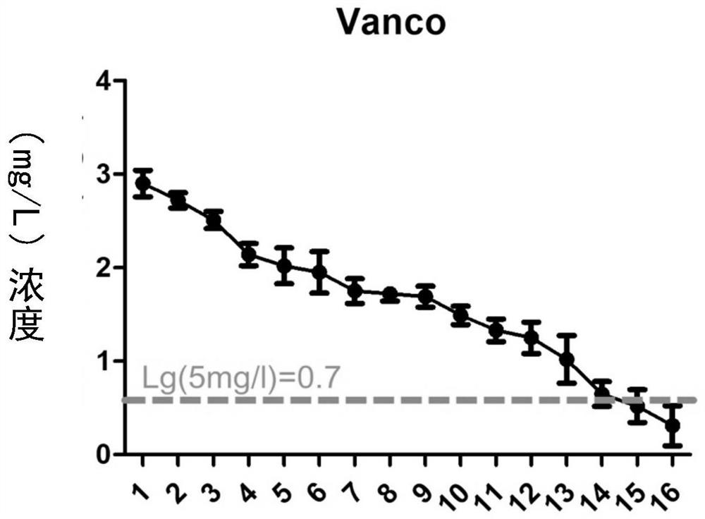
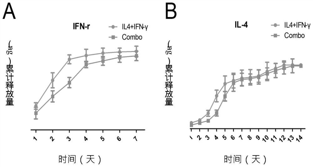
  • Tissue-engineered bone scaffold for treating infectious large bone defects as well as preparation method and application of tissue-engineered bone scaffold
  • Tissue-engineered bone scaffold for treating infectious large bone defects as well as preparation method and application of tissue-engineered bone scaffold
  • Tissue-engineered bone scaffold for treating infectious large bone defects as well as preparation method and application of tissue-engineered bone scaffold

Examples


Embodiment 1

[0036] Example 1 Decalcified bone matrix (decalcified bone matrix, DBM) material preparation and biotinylation

[0037] 1. Prepare the scaffold:

[0038] Relying on the bone bank of Southwest Hospital, the DBM scaffold was prepared from Yunnan Xiaoxiang pig tibia, proximal and distal cancellous bone of femur, and the size of the scaffold was about 3x3x3mm 3 . First decalcify the DBM, add 0.6M hydrochloric acid in a closed glass container, and put the DBM stent into it for soaking. Soak at room temperature for 24 hours, replace the hydrochloric acid once every 8 hours, when the cortical bone is translucent and flexible and elastic, the cancellous bone will automatically return to its original shape after being compressed in a spongy shape, take out the bone, rinse and soak repeatedly with sterile distilled water , until the pH value is 7, suck out the soaking solution, place the decalcified DBM scaffold material in a centrifuge tube, centrifuge at 300g for 5min, and irradiate...

Embodiment 2

[0045] Embodiment 2 Sustained-release kinetic detection

[0046]Put the second and third groups of composite materials in 10 ml of DMEM / F12 medium, place them in a 37°C incubator, change the liquid every day, and collect the medium, centrifuge at 10,000r / min for 10 minutes, and take the supernatant The vancomycin concentration was measured on a high-performance liquid chromatograph, and the release of IL-4 and IFN-γ was measured by ELISA.

Embodiment 3

[0047] Example 3 Effect of Composite Materials on Macrophage Polarization

[0048] Take the macrophages stimulated by M-CSF for 5 days, and use 1×10 6 The concentration per ml was inoculated to each group of materials by the double-sided static inoculation method, and the medium was added, and the medium was replaced after cultivating for 3 days. The medium was taken at 1, 4, and 7 days of culture, and the protein content was detected by ELISA. The materials of each group were collected at the same time point, and the mRNA was extracted after grinding with liquid nitrogen. After reverse transcription was performed according to the instructions of the Takara reverse transcription kit, Real-time PCR was performed. The primer sequences and the length of the amplified products are listed in Table 2. The amplification conditions were 95°C for 30s, [95°C for 5s (denaturation), 57°C for 20s (annealing), 72°C for 15s (extension)] x 40 cycles, 95°C for 30s, 57°C for 30s, 95°C for 30s...


PUM

PropertyMeasurementUnit
concentrationaaaaaaaaaa

Abstract

The invention discloses a tissue-engineered bone scaffold for treating infectious large-segment bone defects as well as a preparation method and an application of the tissue-engineered bone scaffold.The preparation method comprises the following steps: 1) soaking a decalcified bone matrix scaffold in a calcium chloride solution; 2) uniformly mixing the sodium alginate solution containing IFN-gamma with a proper amount of vancomycin solution to obtain a viscous solution; 3) slowly dripping a viscous solution into pores of the decalcified bone matrix in the step 1), and performing standing to form a composite scaffold; 4) cleaning the composite scaffold with deionized water, cryopreserving at -70 DEG C, and freeze-drying at low pressure until the weight does not change any more; and 5) immersing the material obtained in the step 4) into a fibrinogen-containing KH2PO4 solution, slowly stirring, immersing into a thrombin solution, slowly stirring for a period of time, taking out, and air-drying at room temperature to obtain the tissue engineering bone scaffold. The invention also discloses the tissue engineering bone scaffold prepared by the method and the application of the tissue engineering bone scaffold. The tissue engineering bone scaffold provided by the invention can sequentially release inflammatory factors, promote angiogenesis and prevent infection.

Description

Technical field: [0001] The invention relates to the technical field of tissue engineering, in particular to a bone scaffold for tissue engineering and its preparation method and application. Background technique: [0002] Infected large segmental bone defect is a major problem in the treatment of clinical bone tissue injuries. So far, there is no ideal treatment method. One of the important reasons is the lack of effective and reliable bone repair materials that take into account the functions of anti-infection and tissue reconstruction. The sequential release of inflammatory cytokines plays an important role in bone repair, anti-inflammation, and angiogenesis. Previous studies have shown that when host macrophages are absent, angiogenesis in grafts cannot proceed, and studies have also shown that when host macrophage recruitment is blocked, the recruitment effect of host endothelial progenitor cells (EPC) is significant weakened, and the osteogenic repair ability of TEB w...

Claims


Application Information

Patent Timeline
Patent Type & Authority Applications(China)
IPC IPC(8): A61L27/36A61L27/20A61L27/22A61L27/50A61L27/54
CPCA61L27/3608A61L27/365A61L27/3687A61L27/20A61L27/225A61L27/227A61L27/50A61L27/54A61L2430/02A61L2300/602C08L5/04C08L89/00
Inventor 邢军超贺思豪侯天勇窦策周江玲艾秋池许建中
Owner THE FIRST AFFILIATED HOSPITAL OF ARMY MEDICAL UNIV
Learn More