Isolated culture method of human umbilical cord mesenchymal stem cells

A quality stem cell, separation and culture technology, applied in the field of stem cells and regenerative medicine, can solve problems such as difficulty in meeting large-scale experimental research and clinical applications, prolonging the time of in vitro separation of cells, and affecting cell activity, so as to ensure cell proliferation, differentiation, shortening Enzyme action time, the effect of strong proliferation and differentiation ability

Inactive Publication Date: 2021-04-13
THE PEOPLES HOSPITAL WEIFANG CITY CN0
View PDF0 Cites 1 Cited by
  • Summary
  • Abstract
  • Description
  • Claims
  • Application Information

AI Technical Summary

Problems solved by technology

The traditional protease digestion method mainly uses collagenase, hyaluronidase and neutral protease to obtain cells, and the digestion time is 6 hours or more, which not only prolongs the separation time of cells in vitro, but also easily affects the cell viability (BeeravoluN , et al.Isolation and Characterization of Mesenchymal Stromal Cells from Human Umbilical Cord and Fetal Placenta.JVis Exp.2017;(122):55224; Zhang Z, et al. Human Umbilical Cord Mesenchymal StemCells Inhibit T Follicular Helper Cell Expansion Through the OS of Activation in Lupus-Prone B6. MRL-Faslpr Mice. Cell Transplant. 2017; 26(6):1031-1042.)
Although the tissue block attachment method has the advantages of simple process and low cost, its cell acquisition cycle is long and the cell acquisition rate is low, which is difficult to meet the needs of large-scale experimental research and clinical application, seriously affecting the progress of experiments

Method used


Image

  • Isolated culture method of human umbilical cord mesenchymal stem cells
  • Isolated culture method of human umbilical cord mesenchymal stem cells
  • Isolated culture method of human umbilical cord mesenchymal stem cells

Examples


Embodiment 1

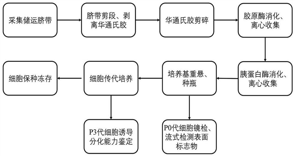
[0046] Such as figure 1 As shown, a method for extracting mesenchymal stem cells from umbilical cord Wharton's jelly tissue, the steps are as follows:

[0047] (1) Collection, storage and transportation of umbilical cord: Obtain the umbilical cord under aseptic conditions, after preliminary cleaning of the arterial and venous blood in the human umbilical cord, the two ends of the umbilical cord are ligated with rubber bands, soaked in sterile saline, stored and transported at low temperature (0-4°C) for 6 hours processed within;

[0048] (2) Separation of Wharton's jelly tissue: Rinse repeatedly with sterile normal saline to remove residual blood on the surface of the umbilical cord and in blood vessels, then use sterile scissors to divide the umbilical cord into small sections of about 3-4cm, and then place the small pieces of umbilical cord in normal saline On the soaked sterile gauze, use ophthalmic scissors to cut the adventitia of the umbilical cord longitudinally from t...

Embodiment 2

[0063] Example 2 Cell Culture Detection Test

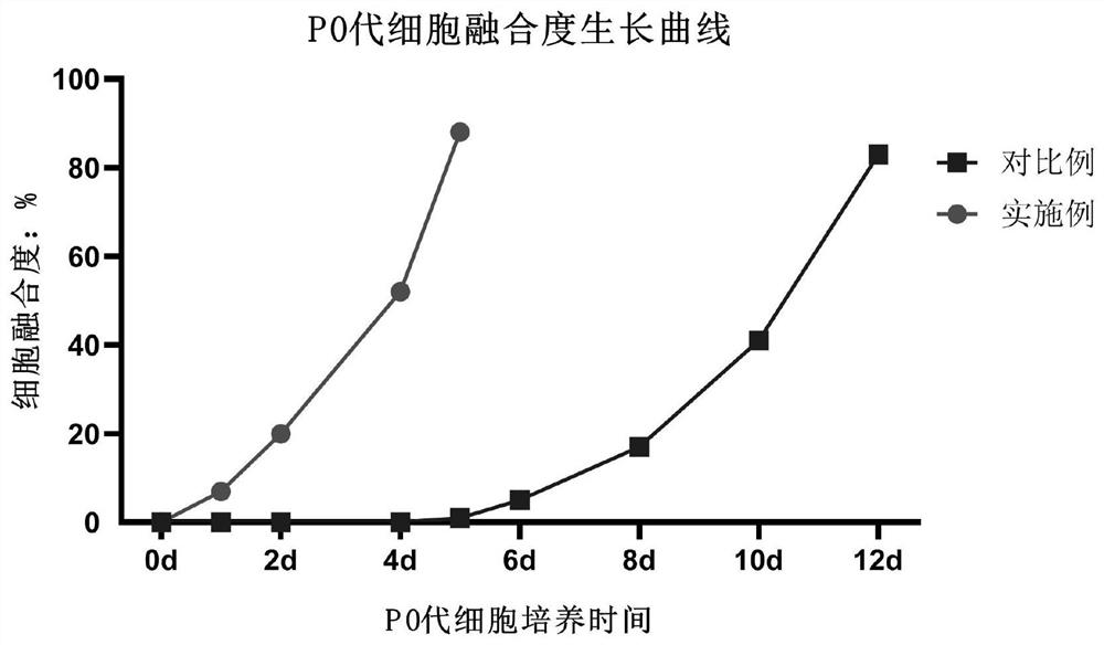
[0064] Observing the growth status of the P0 generation mesenchymal stem cells obtained in Example 1 and Comparative Example 1 and drawing the cell fusion degree curves in different time periods, the results are as follows image 3 Shown, visible embodiment 1 method cell fusion is faster.

[0065] Carry out cell count and draw growth curve according to embodiment 1P0-P5 generation mesenchymal stem cells, the result is as follows Figure 4 shown.

[0066] Choose embodiment 1 and comparative example 1 P0 generation after culture 3 days and the cell after P0 generation culture 6 days to observe under microscope, take pictures, as Figure 5 as shown, Figure 5 Among them, A is Example 1, and B is Comparative Example 1. It can be seen that about 20% of the cells in Example 1 have adhered to the wall by the second day of culture, while no cells have climbed out in Comparative Example 1.

[0067] Observe the pictures of Example 1 and...


PUM


Abstract

The invention discloses an isolated culture method of human umbilical cord mesenchymal stem cells. The isolated culture method comprises the following steps of: collecting storage and transportation umbilical cords, separating Wharton's jelly tissues, extracting and culturing cells, and carrying out cell passage and breed conservation cryopreservation. According to the isolated culture method disclosed by the invention, umbilical cord adventitia and blood vessels are removed to obtain pure Wharton's jelly tissues, so that the purity of the isolated mesenchymal stem cells is higher and the quantity of the isolated mesenchymal stem cells is larger; a two-step enzyme digestion method is utilized, so that the preparation period of primary cells is greatly shortened; and in addition, the cells are good in activity and strong in multiplication capacity, have good differentiation potential and high separation efficiency, and especially can meet the cell requirements of large-scale experimental research and clinical application.

Description

technical field [0001] The invention belongs to the field of stem cells and regenerative medicine, relates to the technology of separating and culturing stem cells, in particular to a method for separating, extracting and culturing mesenchymal stem cells from human umbilical cords. Background technique [0002] Mesenchymal stem cells are a special type of stem cells with strong self-renewal and multidirectional differentiation potential. Mesenchymal stem cells have received great attention in various fields of medicine because of their great potential in immune regulation and tissue repair. Mesenchymal stem cells can be obtained from a wide range of sources, such as bone marrow, synovium, fat, umbilical cord blood, umbilical cord and other tissues. In addition, after special induction, it can differentiate into various tissue cells such as bone cells, endothelial cells, chondrocytes, cardiomyocytes, and adipocytes. At present, the main research source of mesenchymal stem c...

Claims


Application Information

Patent Timeline
Patent Type & Authority Applications(China)
IPC IPC(8): C12N5/0775A01N1/02
CPCA01N1/0226A01N1/0284C12N5/0668C12N2509/00C12N2509/10
Inventor 徐栋花杨晶涵杨春娟褚锦锦张娇娇王菁华孙家梅张丽丽李海波
Owner THE PEOPLES HOSPITAL WEIFANG CITY CN0
Why Eureka
Try Eureka