Preparation and application of near-infrared viscosity fluorescent probe

A viscosity and probe technology, applied in fluorescence/phosphorescence, luminescent materials, material analysis through optical means, etc., can solve the problems of cell background fluorescence interference, etc., and achieve the effect of simple synthesis path and easy application

Active Publication Date: 2021-05-11
FUDAN UNIV
View PDF4 Cites 5 Cited by
  • Summary
  • Abstract
  • Description
  • Claims
  • Application Information

AI Technical Summary

Problems solved by technology

[0003] At present, the fluorescent probes for targeting mitochondria to detect viscosity are generally

Method used


Image

  • Preparation and application of near-infrared viscosity fluorescent probe
  • Preparation and application of near-infrared viscosity fluorescent probe
  • Preparation and application of near-infrared viscosity fluorescent probe

Examples


Embodiment 1

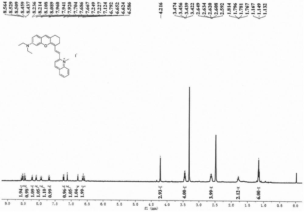
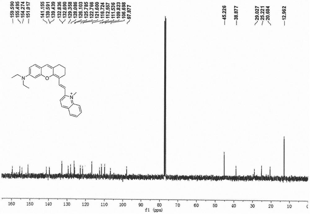
[0025] Embodiment 1: the synthesis of probe M550

[0026]

[0027] Synthesis of compound 1: DMF (2.6 mL, 33.9 mmol) and 10.0 mL CHCl were added to a 50 mL round bottom flask 3 , after stirring at 0°C for 15 min, slowly add PBr at this temperature 3 (2.7 mL, 28.2 mmol), and after stirring for another 45 min, cyclohexanone (2.0 mL, 19.2 mmol) was added. The reaction solution was naturally warmed to room temperature and reacted for 16 h. Stop the reaction, pour the resulting red reaction solution into ice, add solid NaHCO 3 Adjust pH=7. Separate the organic layer and wash the aqueous layer with 30.0 mL CH 2 Cl 2 Extract three times and combine the organic layers. Washed three times with saturated NaCl solution, anhydrous NaCl 2 SO 4 After drying, the solvent was distilled off under reduced pressure to obtain a crude product. The crude product was separated and purified by column chromatography (ethyl acetate / n-hexane=1 / 8, v / v) to obtain Compound 1 as a colorless oil. ...

Embodiment 2

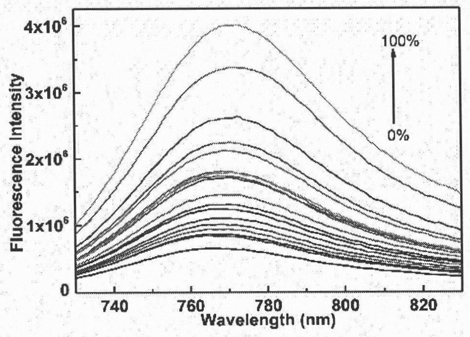
[0031] Example 2: Sensitivity of Probe M550 to Viscosity

[0032] The probe M550 in Example 1 was dissolved in DMSO to prepare a stock solution with a concentration of 2 mM. Take 10 μL of the above mother solution and add 2 mL of different volume ratios (ethanol:glycerol=90:10; 80:20; 70:30; 60:40; 50:50; 40:60; 30:70; 20:80; 10:90 ) in the ethanol / glycerin system, and then perform fluorescence detection, the results are shown in the attached image 3 : attached by image 3 It can be seen that with the increase of glycerol volume specific gravity, the fluorescence intensity of the probe increases gradually. This shows that the probe M550 can have a good response to the viscosity in the glycerol system.

Embodiment 3

[0033] Example 3: Mitochondrial colocalization of probe M550

[0034] The probe M550 in Example 1 was made into 1 mM mother solution with DMSO. Before the cell imaging experiment, first discard the supernatant in the culture dish, wash the cells carefully with PBS buffer three times, and then add the probe at a final concentration of 5 μM. The needle M550 was incubated at 37° C. for 30 min, then 1.0 μM Mito-Tracker Green FM was added, and the incubation was continued for 15 min, and then the cells were washed three times with fresh PBS (10 mM). By confocal laser scanning fluorescence microscopy, it was detected whether the probe M550 was localized in the mitochondria of cells. For probe M550, use 637nm laser as excitation light source, use red channel (730-800nm) to collect emission spectrum; for Mito-Tracker Green FM, use 488nm laser as excitation light source, use green channel (500-550nm) Collect emission spectra. Confocal fluorescence microscopy images such as Figure 4...


PUM


Abstract

The invention belongs to the field of small organic molecule fluorescent probes, and particularly relates to synthesis and biological application of a near-infrared excited and emitted mitochondria-targeted viscosity-sensitive fluorescent probe. The invention provides the mitochondria-targeted near-infrared excited and emitted viscosity fluorescent probe as shown in the following formula (1), and application of the fluorescent probe to detection of viscosity in a solution and detection of viscosity change of mitochondria in cells. The probe has near-infrared excitation and emission properties, can realize detection of viscosity in a solution and intracellular imaging, and has potential application value in the field of fluorescent biomarkers.

Description

technical field [0001] The invention belongs to the field of organic small molecule fluorescent probes, in particular to the synthesis of a near-infrared excited and emitted mitochondrial-targeted viscosity-sensitive fluorescent probe and its biological application. Background technique [0002] The prior art discloses that mitochondrial viscosity, as an important mitochondrial microenvironment, plays a very important role in the interaction between intracellular biomolecules, the transmission of chemical signals and the diffusion of metabolites in living cells. Abnormalities in mitochondrial viscosity are important contributors or indicators of many diseases and dysfunctions in cells such as cellular malignancies, atherosclerosis, hypertension, diabetes and Alzheimer's disease. Therefore, it is of great significance to accurately measure the change of local viscosity in the mitochondria of living cells. Viscosity-sensitive fluorescent probe molecules, due to their simple o...

Claims


Application Information

Patent Timeline
IPC IPC(8): C09K11/06C07D405/06G01N21/64
CPCC09K11/06C07D405/06G01N21/6428C09K2211/1029C09K2211/1088
Inventor 张炜佳黄晖骆沙曼唐美麟
Owner FUDAN UNIV
Why Eureka
Try Eureka