
Preparation method of micron-sized spherical particle section for electron microscope observation

A spherical particle, micron-scale technology, applied in the preparation of test samples, material analysis using wave/particle radiation, instruments, etc., can solve the problems of cumbersome processing steps, long polishing time, large consumption of raw materials, etc., and achieve polishing time Short, low cost, short time effect

Active Publication Date: 2021-05-28
CHONGQING UNIV
View PDF20 Cites 0 Cited by
  • Summary
  • Abstract
  • Description
  • Claims
  • Application Information

AI Technical Summary

Problems solved by technology

[0003] The existing ion beam polishing technology to prepare cross-section samples has the following shortcomings. First, the sample pretreatment steps are cumbersome, and carbon conductive glue is used as a dispersant, fixative, and filler, which consumes a lot of raw materials and high costs; second, ion polishing uses Cross-section polishing mode, long polishing time and low efficiency

Method used

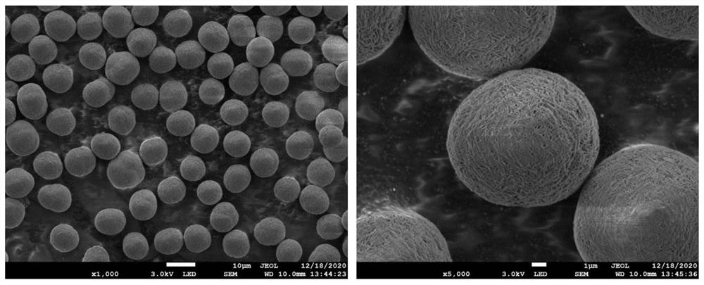

Image

  • Preparation method of micron-sized spherical particle section for electron microscope observation
  • Preparation method of micron-sized spherical particle section for electron microscope observation
  • Preparation method of micron-sized spherical particle section for electron microscope observation

Examples


Embodiment 1

[0034] To prepare a section of a micron-sized spherical particle for SEM observation, follow the steps below:

[0035] (1) Paste the double-sided conductive carbon tape on the aluminum metal substrate with a size of 8mm×8mm;

[0036] (2) Take a small amount of LiNiCoMnO with a toothpick 2 The battery positive electrode material powder is spread flat on the conductive tape, and the excess powder is blown away with a blower, and only the powder stuck on the conductive tape remains;

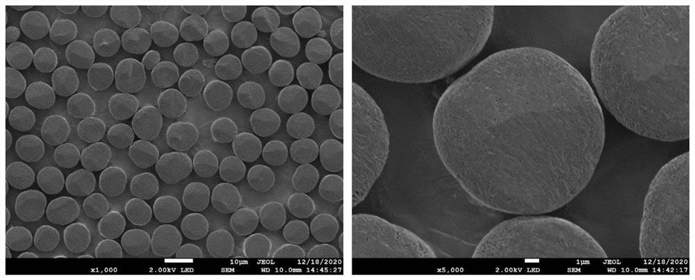
[0037] (3) Put the aluminum metal substrate with powder in the argon ion polishing machine for polishing, adopt the plane polishing mode, the ion gun angle is 2°, and the polishing voltage and time are 7kV 5min+4kV 10min+1kV 10min, and the satisfaction can be obtained. The cross-section observed by the scanning electron microscope, the scanning electron microscope picture of the spherical particle cross-section after ion polishing is as follows figure 2 shown.

Embodiment 2

[0039] To prepare a section of a micron-sized spherical particle for SEM observation, follow the steps below:

[0040] (1) Paste the double-sided conductive copper tape on the copper metal substrate with a size of 8mm×8mm;

[0041] (2) Take a small amount of LiNiCoMnO with a toothpick 2 The battery positive electrode material powder is spread flat on the conductive tape, and the excess powder is blown away with a hair dryer;

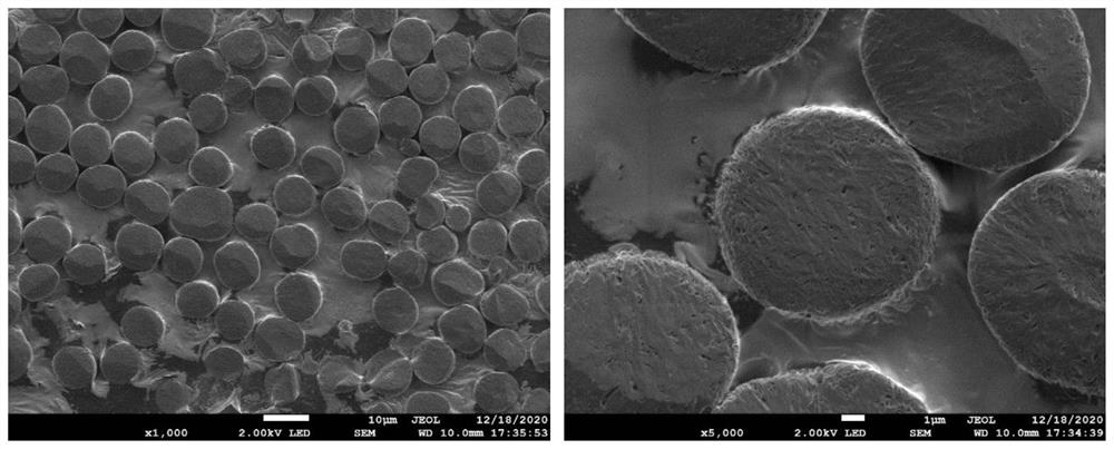
[0042] (3) Put the copper metal substrate with powder in the argon ion polishing machine for polishing, adopt the plane polishing mode, the ion gun angle is 0.5°, and the polishing voltage and time are 7kV 5min+4kV 10min+1kV 10min, and the satisfaction can be obtained. The cross-section observed by the scanning electron microscope, the scanning electron microscope picture of the spherical particle cross-section after ion polishing is as follows image 3 shown.

Embodiment 3

[0044] To prepare a section of a micron-sized spherical particle for SEM observation, follow the steps below:

[0045] (1) Paste the double-sided conductive copper tape on the aluminum metal substrate with a size of 8mm×8mm;

[0046] (2) Take a small amount of LiNiCoMnO with a toothpick 2 The battery positive electrode material powder is spread flat on the conductive tape, and the excess powder is blown away with a hair dryer;

[0047] (3) Put the copper metal substrate with powder in the argon ion polishing machine for polishing, adopt the plane polishing mode, the ion gun angle is 0°, and the polishing voltage and time are 7kV 5min+4kV 10min+1kV 10min, and the satisfactory The cross-section observed by the scanning electron microscope, the scanning electron microscope picture of the spherical particle cross-section after ion polishing is as follows Figure 4 shown.


PUM

PropertyMeasurementUnit
diameteraaaaaaaaaa
diameteraaaaaaaaaa

Abstract

The invention discloses a preparation method of a micron-sized spherical particle section for electron microscope observation. The preparation method comprises the following steps: (1), adhering a conductive adhesive tape to a conductive metal matrix; (2), taking micron-sized spherical particle powder to be flatly laid and adhered to the conductive adhesive tape, and blowing away redundant powder; and (3), placing the conductive metal matrix adhered with the powder in an ion beam polishing machine to be polished, using a plane polishing mode, the polishing angle being 0-2 degrees, the polishing voltage and time being 5-8 kV for 5-40 min, 3-5 kV for 5-20 min and 0.5-2 kV for 5-20 min; and obtaining the particle cross section meeting the scanning electron microscope observation requirement. According to the method, embedding treatment is not needed, and extra sample preparation consumables are not consumed; time is short, cost is low, and operability is high; the consumed time is only one tenth of that of a section polishing mode, and the polishing efficiency is high; the effective area of the obtained sample is large, and the selectable areas for scanning electron microscope observation are more.

Description

technical field [0001] The invention belongs to the technical field of scanning electron microscope sample preparation, and in particular relates to a method for preparing a cross-section of micron-sized spherical particles used for electron microscope observation. Background technique [0002] There has been a lack of high-quality preparation methods for cross-sectional samples of micron-sized spherical particles. Conventional methods include embedding mosaic grinding and polishing, which is cumbersome and time-consuming, and the polishing solution may contaminate, corrode or damage the surface of the sample. Improved methods include focused ion beam cutting, ultrathin sectioning, and ion beam polishing. Focused ion beam cutting has precise positioning, high cutting efficiency for individual particles, and can obtain a better observation surface. However, high-energy ions Cutting will inevitably introduce ion implantation contamination. In addition, the high cost of focus...

Claims


Application Information

Patent Timeline
Patent Type & Authority Applications(China)
IPC IPC(8): G01N23/2251G01N23/2202G01N1/32
CPCG01N23/2251G01N23/2202G01N1/32
Inventor 邓超文婷婷洪睿杨帅周仕远刘施峰
Owner CHONGQING UNIV
Learn More