Aptamer colloidal gold lateral chromatography test paper for detecting kanamycin
A kanamycin and lateral chromatography technology, which is used in food safety, nano-biosensing, analytical chemistry, medicine, and the environment. problem, to achieve the effect of high repeatability, high sensitivity detection, and improved hybridization efficiency
- Summary
- Abstract
- Description
- Claims
- Application Information
AI Technical Summary
Problems solved by technology
Method used
Image
Examples
Embodiment 1
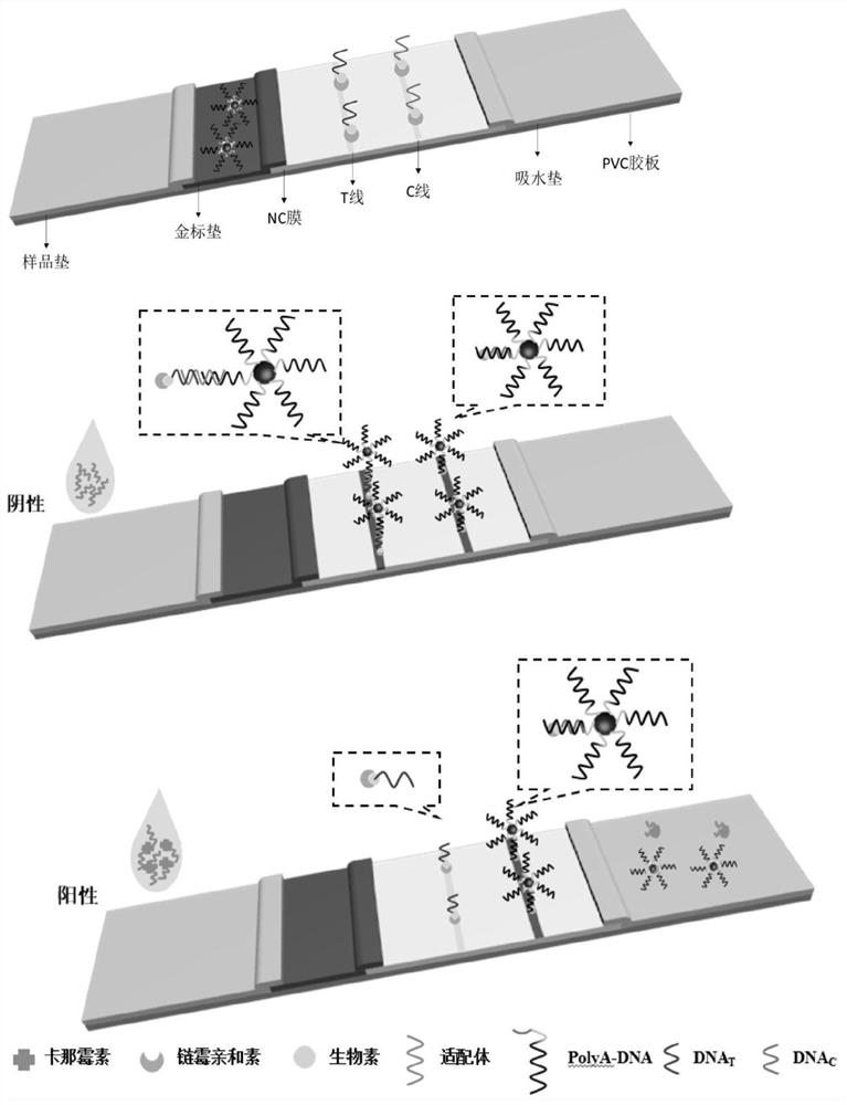
[0049] Example 1: A rapid detection test strip for kanamycin based on nucleic acid aptamer
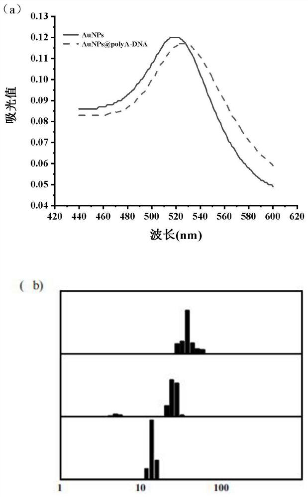
[0050] Preparation and functionalization of gold nanoparticles, construction of streptavidin-biotin-capApt and streptavidin-biotin-capDNA composite structures, assembly of nucleic acid aptamer test strips. The specific steps are:
[0051] 1. Preparation and functionalization of gold nanoparticles
[0052] (1) Preparation of gold nanoparticles (AuNPs)
[0053] The glassware used for the synthesis and storage of nanomaterials in the experiment was soaked in aqua regia (hydrochloric acid: nitric acid = 3:1) for 12 hours, washed with ultrapure water before use.
[0054] AuNPs were prepared by sodium citrate reduction method. details as follows:
[0055] 1) Dissolve 100mL of 0.01% HAuCl 4 Add it into a 250mL Erlenmeyer flask, heat and stir until the solution bumps, and keep for 1-2min.
[0056] 2) Quickly add 2 mL of 1% trisodium citrate solution to the Erlenmeyer flask, and continue ...
Embodiment 2
[0073] Embodiment two: with test strip to the mensuration of kanamycin standard solution
[0074] (1) Preparation of Kanamycin Standard Solution
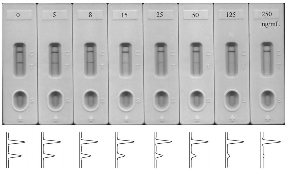
[0075] Kanamycin standard solution was diluted with Running buffer (4×SSC, pH7) to a final concentration of 5, 8, 5, 25, 50, 125 and 250 ng / mL. The nucleic acid aptamer (Aptamer) was diluted to 1 μM with ultrapure water.
[0076] (2) Establishment of standard curve for detection of kanamycin nucleic acid test strips:
[0077] Mix 99 μL of kanamycin standard solution with different concentrations and 1 μL of Aptamer solution and incubate for 20 min. After mixing and reacting, add the mixed solution to the sample pad for detection. After 3 min of reaction, measure (T / C) relative signal intensity and establish (T / C) C) Standard curve of the corresponding relationship between relative light signal intensity and different kanamycin concentrations.
[0078] The result is as image 3 As shown, when the concentration of kanamycin is 15n...
Embodiment 3
[0080] Example three: detection of kanamycin residues in milk samples
[0081] To test the recovery rate using a milk mock sample, the steps are:
[0082] (1) Pretreatment of AuNPs@polyA-DNA solution: Add 0.8-1.4 μL of the prepared and stored AuNPs@polyA-DNA onto the gold standard pad of the test strip, and store at 4°C.
[0083](2) Sample pretreatment: the milk sample was diluted 10 times and filtered through a 0.22 μm microporous membrane. Add different concentrations of kanamycin (50, 150, 250ng / mL) to the milk;
[0084] (3) Determination of the recovery rate of kanamycin in milk: 1 μL Aptamer (1 μM) was mixed with 99 μL milk solution containing different concentrations of kanamycin and incubated for 20 min, and then tested with test strips after the mixed reaction.
PUM
| Property | Measurement | Unit |
|---|---|---|
| particle diameter | aaaaa | aaaaa |
| particle diameter | aaaaa | aaaaa |
Abstract
Description
Claims
Application Information
 - R&D
- Intellectual Property
- Life Sciences
- Materials
- Tech Scout
- Unparalleled Data Quality
- Higher Quality Content
- 60% Fewer Hallucinations
Browse by: Latest US Patents, China's latest patents, Technical Efficacy Thesaurus, Application Domain, Technology Topic, Popular Technical Reports.
© 2025 PatSnap. All rights reserved.Legal|Privacy policy|Modern Slavery Act Transparency Statement|Sitemap|About US| Contact US: help@patsnap.com


