Method for inducing adipose tissue-derived stem cells to differentiate into testicular interstitial-like cells by single transcription factor

A technology of stromal stem cells and testicular interstitium, applied in the field of stem cell biology and regenerative medicine, to achieve the effect of good application prospects

Pending Publication Date: 2021-07-27
THE FIRST AFFILIATED HOSPITAL OF HENAN UNIV OF SCI & TECH
View PDF0 Cites 1 Cited by
  • Summary
  • Abstract
  • Description
  • Claims
  • Application Information

AI Technical Summary

Problems solved by technology

[0006] In order to solve the problem that the existing techniques for inducing differentiation of stem cells into Leydig-like cells cannot simultaneously take into account "safety, rapidity and high efficiency", the present invention provides a new method for inducing the differentiation of human-derived ADMSCs into Leydig-like cells i

Method used


Image

  • Method for inducing adipose tissue-derived stem cells to differentiate into testicular interstitial-like cells by single transcription factor
  • Method for inducing adipose tissue-derived stem cells to differentiate into testicular interstitial-like cells by single transcription factor
  • Method for inducing adipose tissue-derived stem cells to differentiate into testicular interstitial-like cells by single transcription factor

Examples


Embodiment 1

[0031] Example 1. Isolation and cultivation of human-derived ADMSCs.

[0032] Collect the resected tissue after circumcision in the operating room, remove the skin and retain the fat, cut it into pieces with sterile ophthalmic scissors, incubate with 0.2% collagenase I solution at 37°C for 1-2 hours, and culture with DMEM containing 10% fetal bovine serum Stop the digestion, filter aseptically, centrifuge at 250×g for 5 minutes, discard the supernatant, resuspend the bottom of the tube, centrifuge again, and resuspend the cells in DMEM medium containing 1% penicillin and streptomycin and 10% fetal bovine serum , inoculated into a culture dish, replaced with fresh culture medium after overnight culture, subcultured, and retained the third generation cells ( figure 1 ) were stored frozen at -80°C for later use.

Embodiment 2

[0033] Example 2. Optimal synthesis of modNR5A1 in vitro.

[0034] In the case of avoiding nuclease contamination, first use the pTEMPlz plasmid (purchased from Promega, USA) and 2×HiFi HotStart Ready Mix kit (purchased from Kapa biosystems, USA) to synthesize poly T-tailed DNA by Tail-PCR reaction. Linear DNA template (see Kondrat et al., Methods in molecular biology, 2017, 1521: 127-138). i.e. first pass AleI, AfeI Enzymes linearize the pTEMPlz plasmid, use a pair of primers (1 μM) of the NR5A1 gene and 2× KAPA HiFi HotStart ReadyMix (KAPA Biosystems, USA) to amplify its open reading frame (ORF) by PCR reaction, and use T4 DNA ligation The enzyme clones the amplified ORF into the pTEMPlz plasmid vector, and then transforms, screens positive clone colonies for resistance, and shakes the bacteria to amplify and extract the plasmid. The concentration is detected by the NanoDrop instrument and adjusted to 100-200 ng / μl, and placed in - Store at 80°C for later use.

[0035] Usi...

Embodiment 3

[0040] Example 3. Extracting urine-derived exosomes and loading them with modNR5A1 molecules.

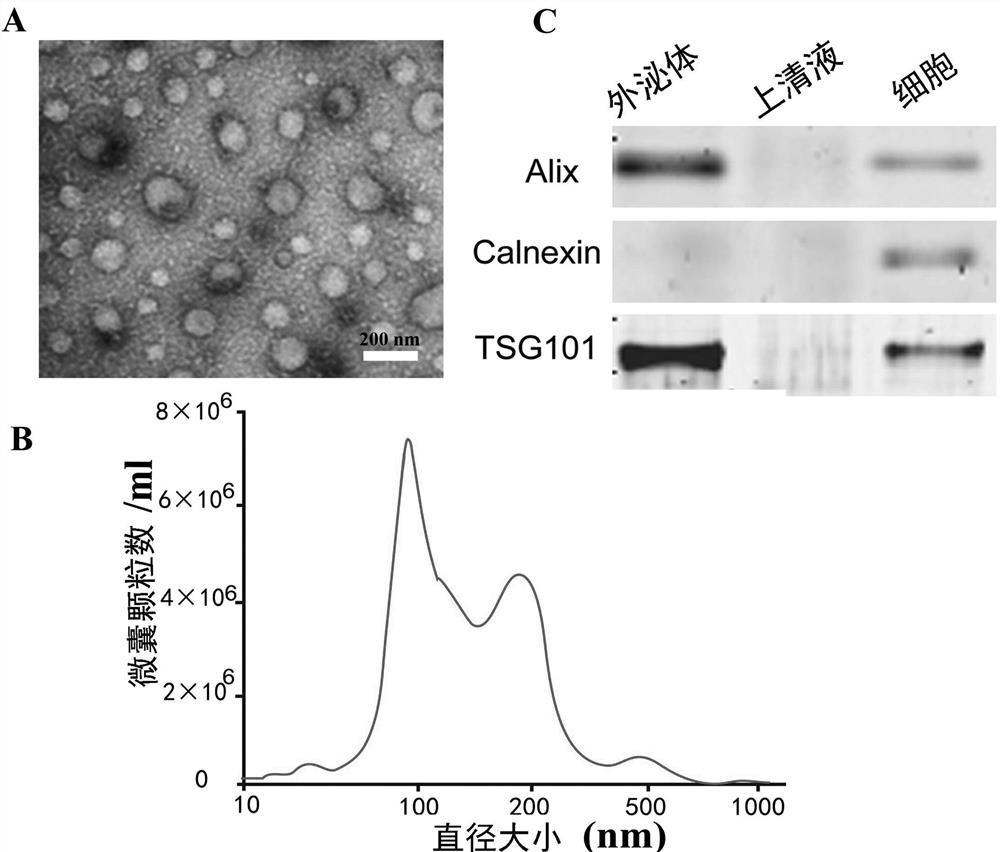
[0041] Collect autologous urine from circumcision patients, and separate exosomes in a 50ml sterile centrifuge tube at 4°C by ultracentrifugation. First, centrifuge at 2,000×g for 20 minutes to remove cells and debris, and then centrifuge at 10,000×g for 60 minutes to remove microcapsules. , and finally centrifuged at 100000×g for 2 hours; next, observe the shape and size of the extract by electron microscope ( figure 2 A), nanoparticle tracking analysis (Nanoparticle Tracking Analysis, NTA) to detect the molecular size of the extract ( figure 2 B), Western blotting method to detect the expression of exosomes markers ( figure 2 C), confirming that the extracts are exosomes.

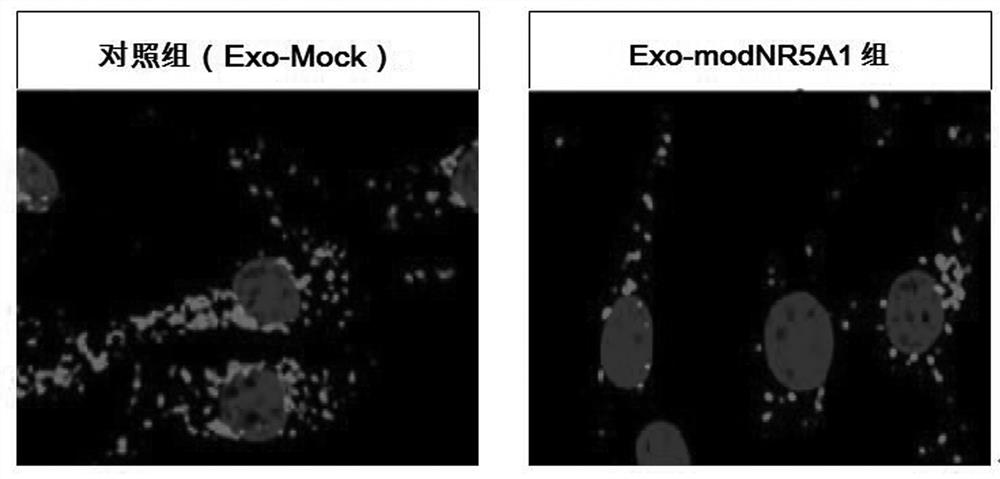
[0042] The extracted exosomes were placed on ice, mixed with the fluorescent probe PKH26 dye and incubated for 12 hours, so that the exosomes were labeled with PKH26. Then, through optimized sonication: 5...


PUM


Abstract

The invention relates to a method for inducing adipose tissue-derived stem cells to differentiate into testicular interstitial-like cells by a single transcription factor, and belongs to the fields of stem cell biology and regenerative medicine. According to the method, chemically modified mRNA of a transcription factor NR5A1 is synthesized in vitro, exosomes extracted from autologous urine of a patient are used as a delivery carrier to form an exosome-modNR5A1 compound, and then the exosome-modNR5A1 compound and ADMSCs derived from autologous tissue of the patient are co-cultured to induce the ADMSCs to be differentiated into Leydig-like cells with a testosterone secretion function. According to the method disclosed by the invention, the Leydig-like cells with the testosterone secretion function can be quickly and efficiently obtained, meanwhile, the cells can effectively react to the stimulation of luteinizing hormone, and the cells have a gene expression profile related to the androgen synthesis of mature Leydig cells. The Leydig cell obtained by the invention has a good application prospect in the aspect of treatment of male hypogonadism.

Description

technical field [0001] The invention belongs to the fields of stem cell biology and regenerative medicine, and relates to a method for directional differentiation of human adipose-derived mesenchymal stem cells into testis-like cells with testosterone secretion function. Background technique [0002] The number of Leydig cells accounts for 3-5% of the total testicular cells, but the testosterone secreted by them accounts for more than 90% of the testosterone in the body. Its structural damage or dysfunction is the main cause of insufficient male androgen secretion. Clinically, androgen replacement therapy is mainly used, but long-term androgen supplementation can lead to many complications, such as: abnormal spermatogenesis, induction of cardiovascular events, and increased risk of prostate cancer. In addition, it is difficult for simple drug therapy to simulate the physiological changes of normal androgen levels in the body. Studies have shown that by transplanting Leydig...

Claims


Application Information

Patent Timeline
IPC IPC(8): C12N5/071C12N5/0775C12N15/12A61K38/17A61K48/00A61P15/08
CPCA61K38/1709A61K48/0008A61K48/005A61P15/08C07K14/4702C12N5/0683C12N2501/60C12N2506/1384
Inventor 黄华张雯张剑李志军韩青江
Owner THE FIRST AFFILIATED HOSPITAL OF HENAN UNIV OF SCI & TECH
Why Eureka
Try Eureka