Biosensor based on silver@polydopamine nanosphere composite structure and preparation method
A technology of dopamine nanometer and biosensor, which is applied in material analysis using radiation diffraction, material analysis using radiation, Raman scattering, etc. It can solve problems such as low detection limit, low protein sensitivity, and poor biocompatibility. Achieve the effect of simple and green preparation process, enhanced Raman signal, and low cost
- Summary
- Abstract
- Description
- Claims
- Application Information
AI Technical Summary
Problems solved by technology
Method used
Image

Examples
preparation example Construction
[0043] A method for preparing a biosensor based on a silver@polydopamine nanosphere composite structure, comprising the following steps:
[0044] S1, synthesizing polydopamine;
[0045] S2, adding the purified polydopamine aqueous solution into a round-bottomed flask, slowly adding ice-cold silver nitrate aqueous solution dropwise under magnetic stirring, and reacting;
[0046] S3. Take the prepared silver@polydopamine aqueous solution into a centrifuge tube, add 4-MBA ethanol solution, vortex and store;
[0047] S4. Add 100 μL of 500 μM SH-PEG-COOH to step S3, and continue to store;
[0048]S5. Add EDC / NHS (EDC 40mg / ml; NHS 110mg / ml), and shake gently in the dark, then add cTn I-Antibody to react;
[0049] S6, adding BSA solution, reacting and sealing, that is, the prepared nanolabel;
[0050] S7. Add the nano-label and the serum to the centrifuge tube successively, vortex, place it at room temperature, and drop it onto the grooved glass slide for reaction.
[0051] Furth...
Embodiment 1
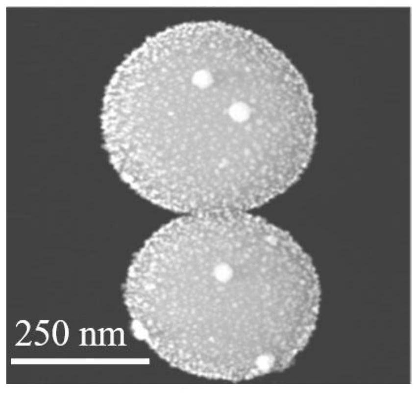
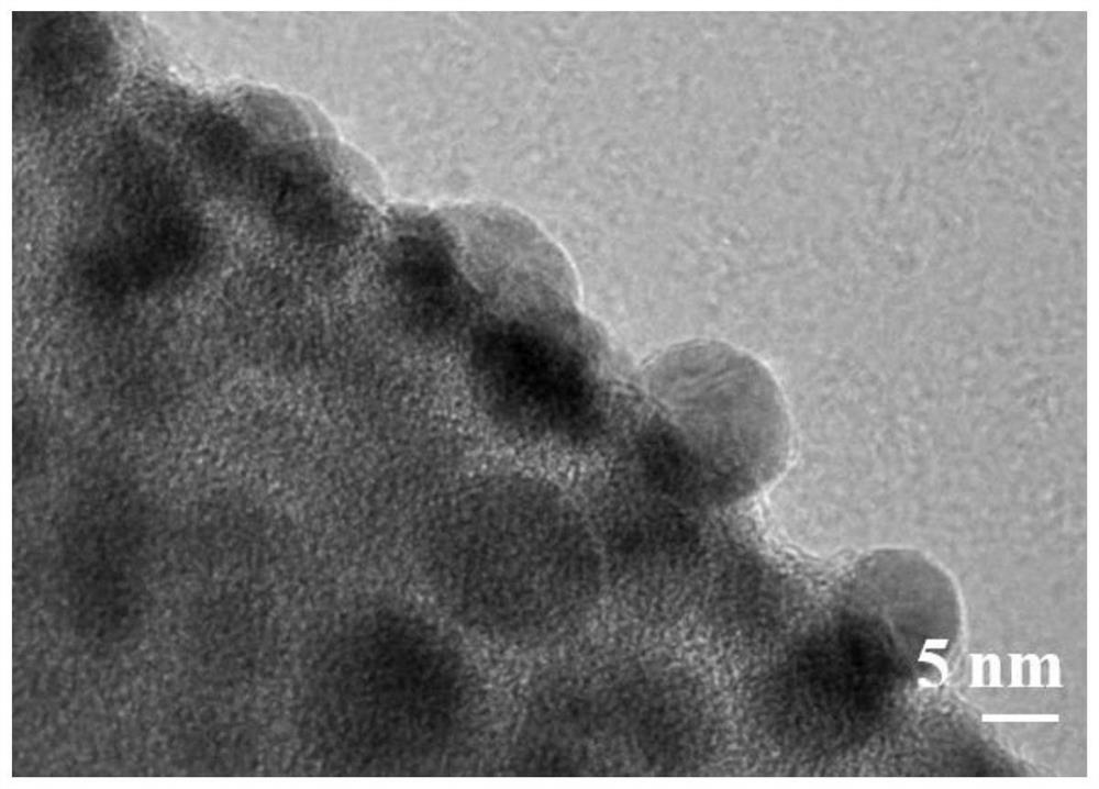
[0061] Preparation of three-dimensional silver@polydopamine nanocomposite material, the specific steps are as follows:
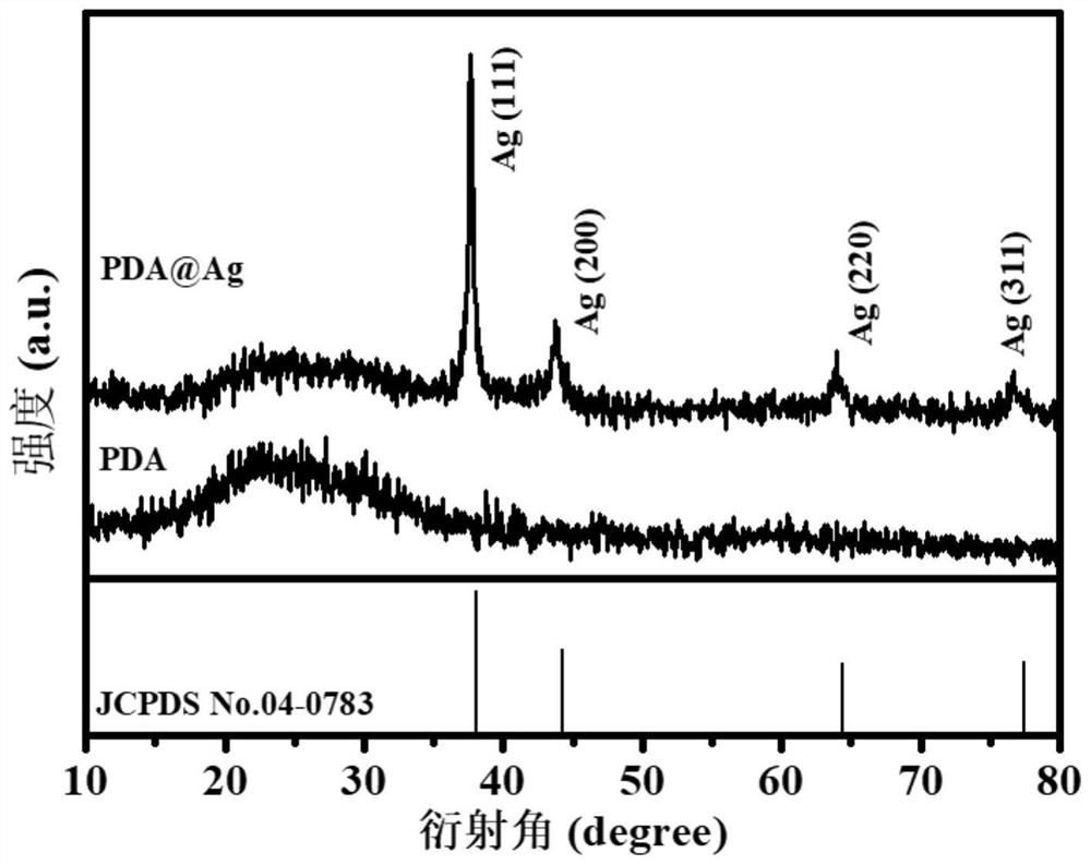
[0062] (1) Dissolve 0.5 g of dopamine hydrochloride in 10 mL of ultrapure water and set aside. Add 2mL of ammonia water, 40mL of absolute ethanol, and 90mL of ultrapure water into a round-bottomed flask, and stir with an open magnetic force for 30min at room temperature. Then quickly add the dopamine aqueous solution, open the reaction at room temperature for 24 to 48 hours, and centrifugally purify the polydopamine aqueous solution and ethanol at a ratio of 1:3, and finally disperse in the aqueous solution.
[0063] (2) Add 5 mL of the purified polydopamine aqueous solution into a round-bottomed flask, slowly add 8 mL of 10 mM ice-cold silver nitrate aqueous solution dropwise under magnetic stirring, and react for 1 h.
[0064] The specific steps for preparing a biosensor from the above-mentioned three-dimensional silver@polydopamine nanocomposite material...
Embodiment 2
[0074] The specific steps of preparing the three-dimensional silver@polydopamine nanocomposite material are the same as those in Example 1.
[0075] The specific steps for preparing a biosensor from the above-mentioned three-dimensional silver@polydopamine nanocomposite material are as follows:
[0076] (3) with embodiment 1.
[0077] (4) with embodiment 1.
[0078] (5) After 24 hours, at room temperature of 25°C, add EDC / NHS (EDC 40mg / ml; NHS 110mg / mL 5μL each), and after 15min in the dark, add 10μL 0.1mg / mL CK-MB- Antibody, react at 4°C for 24h.
[0079] (6) With embodiment 1.
PUM
| Property | Measurement | Unit |
|---|---|---|
| size | aaaaa | aaaaa |
Abstract
Description
Claims
Application Information
 - R&D
- Intellectual Property
- Life Sciences
- Materials
- Tech Scout
- Unparalleled Data Quality
- Higher Quality Content
- 60% Fewer Hallucinations
Browse by: Latest US Patents, China's latest patents, Technical Efficacy Thesaurus, Application Domain, Technology Topic, Popular Technical Reports.
© 2025 PatSnap. All rights reserved.Legal|Privacy policy|Modern Slavery Act Transparency Statement|Sitemap|About US| Contact US: help@patsnap.com