Super-clear visual field diseased tooth specimen and preparation method thereof

A field of vision and specimen technology, applied in the field of diseased tooth specimens and their preparation, can solve problems such as rigid forms

Active Publication Date: 2022-01-04
傅建华
View PDF7 Cites 0 Cited by
  • Summary
  • Abstract
  • Description
  • Claims
  • Application Information

AI Technical Summary

Problems solved by technology

The form is relatively rigid and stereotyped, and it is far from the real dental lesions in clinical practice. It is difficult for students to understand intuitively. It also causes great troubles to teachers and students in terms of teaching and observation. However, although it is difficult to accurately replicate the lesion tooth model made of standardized resin and reflect various lesions of teeth, but still has the effect of magnification, clear color description of lesions, and an economical and convenient storage method

Method used


Image

  • Super-clear visual field diseased tooth specimen and preparation method thereof

Examples


Embodiment 1

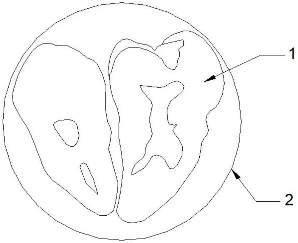
[0038] see figure 1 , this embodiment discloses a diseased tooth specimen with an ultra-clear field of view, including a diseased tooth 1, a glycerol layer and an ultra-clear epoxy resin ab crystal colloid 2 that sequentially cover the surface of the diseased tooth 1 from the inside to the outside, bacteria of the diseased tooth 1 The infection-type lesion structure is processed in at least one of full section, half section and partial section, and the non-bacterial infection type lesion structure of the diseased tooth 1 is processed by fenestration.

[0039] In an optional embodiment, the diseased tooth specimen in the ultra-clear field of view has a hemispherical structure, and the observer can observe the spherical vertical position under conventional illumination to obtain preliminary disease information. During the observation, the shape and color of the lesion structure can be seen with a magnification of about 0.5-1 times; since the distance from the spherical surface t...

Embodiment 2

[0049] A method for preparing a diseased tooth specimen with an ultra-clear field of view, comprising the following steps:

[0050] S10, selecting an isolated tooth with a bacterial infection-type diseased structure and a non-bacterial infection-type diseased structure as the diseased tooth 1, and soaking, cleaning and bleaching the diseased tooth 1;

[0051] In S10, bacterial infection-type lesions include: periodontal calculus, dental plaque; caries deep caries, superficial caries, pit and fissure caries, surface demineralization infection; dental pulp infection; restoration bonding agent residues.

[0052] In S10, non-bacterial lesions include cracked teeth, fissures, occlusal wear, dental fluorosis, non-carious dental defects, deformed teeth, secondary dentin, pulp calcification, and teeth with fillings , Restoration of crown teeth.

[0053] In S10, after the teeth are separated from the body, do not squeeze or wipe the teeth, keep the tooth tissue clean, use hydrogen pe...

Embodiment 3

[0082] Based on the diseased tooth specimen of a kind of ultra-clear visual field of embodiment 2, its observation method is as follows:

[0083] In clinical teaching, the observer wears goggles that isolate 190nm-540nm OD6, or uses an eye protection observation screen that can block blue-violet electromagnetic waves, and uses 405nm excitation light that can conically diverge beams. For example, patent number: 2019111926976, patent Name An instrument to aid in the visualization of periodontal treatment, projected into the viewing surface of the hemispherical lens specimen at an oblique angle of about 30-60 degrees.

[0084] In clinical teaching, the 405nm excitation light that forms a cone-shaped divergent beam is selected to irradiate the bacteria-infected tooth 1. After the bacteria-containing diseased tissue absorbs the purple electromagnetic wave, it produces pink fluorescence at 632 nm; after the tooth hard tissue absorbs the purple electromagnetic wave, it produces green ...


PUM


Abstract

The invention discloses a super-clear visual field diseased tooth specimen and a preparation method thereof, and the lesion tooth specimen comprises a lesion tooth, and a glycerin layer and an ultra-clear epoxy resin ab crystal colloid which sequentially cover the surface of the lesion tooth from inside to outside, and the bacterial infection type lesion structure of the lesion tooth is treated in at least one form of a full section, a half section and a local section, and the non-bacterial infection type lesion structure of the lesion tooth is subjected to windowing treatment. The in-vitro teeth which are originally planned to be discarded and destroyed can be changed into non-toxic and odorless dissection specimens which are easy to process and maintain and have super-clear view and observable lesion structures, so that students can intuitively obtain ecology and forms of corresponding lesion teeth.

Description

technical field [0001] The invention relates to the technical field of diseased tooth models in stomatology teaching, in particular to a diseased tooth specimen with an ultra-clear field of view and a preparation method and observation method thereof. Background technique [0002] The oral cavity is the entrance of the digestive organs, and there are a large number of oral microbial bacteria. Some microbial bacteria can cause a series of oral diseases, including: periodontal disease, caries, and pulp disease. However, the bacteria in dental plaque and diseased infected tissues are considered to be difficult to be directly identified by the naked eye. Another feature of oral bacterial infection is its concealment, especially the dental calculus in the deep periodontal pocket, the dentin caries with a large belly and a small mouth, and the completely invisible pulp necrosis. Electrical activity detection. However, the above detection methods are slightly harmful, especially ...

Claims


Application Information

Patent Timeline
Patent Type & Authority Applications(China)
IPC IPC(8): G09B23/28
CPCG09B23/283Y02A50/30
Inventor 傅建华傅子墨
Owner 傅建华
Why Eureka
Try Eureka