Method and device for estimating in-vivo heat source distribution based on ultrasonic imaging

An ultrasonic imaging and internal heat source technology, applied in the field of medical image processing, can solve problems such as heat source estimation error, heat source difficulty, temperature field prediction deviation, etc., and achieve good motion suppression effect and high signal-to-noise ratio

Pending Publication Date: 2022-04-08
NANJING UNIV
View PDF1 Cites 0 Cited by
  • Summary
  • Abstract
  • Description
  • Claims
  • Application Information

AI Technical Summary

Problems solved by technology

[0007] Aiming at the problem that the effective heat source in the living tissue is difficult to be accurately measured in the prior art, and the error of the heat source estimation is caused by factors such as the physiological movement of the living body, which further leads to the insufficient prediction deviation of the temperature field based

Method used


Image

  • Method and device for estimating in-vivo heat source distribution based on ultrasonic imaging
  • Method and device for estimating in-vivo heat source distribution based on ultrasonic imaging
  • Method and device for estimating in-vivo heat source distribution based on ultrasonic imaging

Examples


Embodiment 1

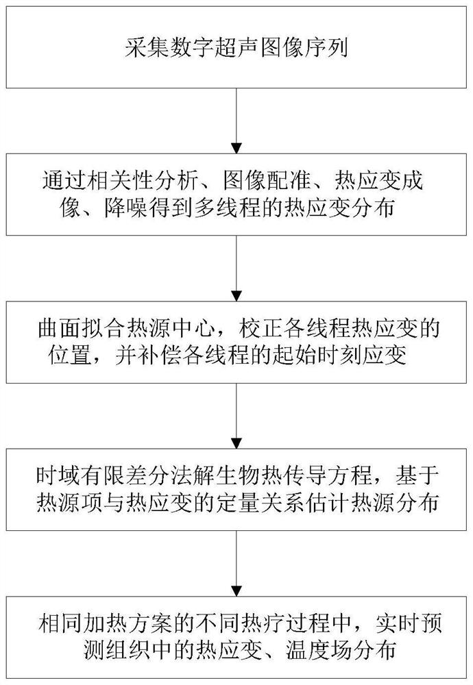
[0058] The schematic flow chart of the method of the embodiment is as figure 1 , a method and device for estimating the distribution of heat sources in the body based on ultrasound imaging of the present invention, comprising the following steps:

[0059] Acquisition steps: use hyperthermia to heat the target area, collect ultrasonic images of the hyperthermia target area in vivo, and obtain digital ultrasonic image sequence I.

[0060] Perform B-ultrasound imaging on the hyperthermia target area in vivo, and collect digital ultrasound image sequences continuously and at equal time intervals; the B-ultrasound instrument works in real-time imaging mode, adjust the angle of the probe and the parameters of the ultrasound system, so that the target area is presented in the imaging field of view; It is worth noting that in the embodiment, different beam control methods, different numbers of array elements, different geometric The imaging probe of the structure, as well as differen...

Embodiment 2

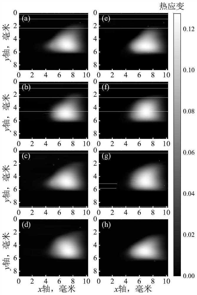
[0097] This example is based on the method of Example 1, using a microwave ablation needle to heat the visceral fat of a living pig, estimating the distribution of heat sources in the biological tissue, and predicting the thermal strain and temperature field in the living body under the same heating scheme. The specific steps of the method as follows:

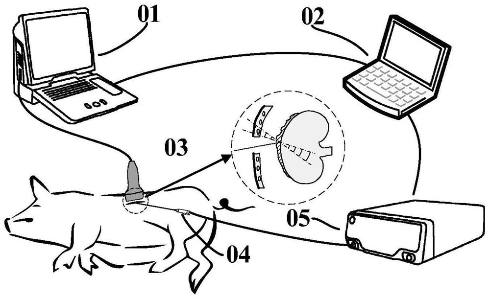
[0098] Step one, such as figure 2 As shown, in this embodiment, the portable B-ultrasound imaging device 01 with a sampling rate of 40MHz is used with the L12-5 linear array probe 03 with 128 array elements and a center frequency of 10.5MHz to image the visceral fat in the target area of ​​live pigs, continuously, etc. Ultrasonic radio frequency image data are collected at intervals, the imaging depth is set to 4cm, the linear density is set to 128, and the imaging mode is set to fundamental wave mode; thermal ablation equipment is used to heat living objects and form displacement and strain distribution in tissues. In the em...


PUM

PropertyMeasurementUnit
Densityaaaaaaaaaa

Abstract

The invention discloses a method and device for estimating in-vivo heat source distribution based on ultrasonic imaging, and belongs to the field of medical image processing. The method comprises the following steps: acquiring a digital ultrasonic image sequence in a living body, performing multi-thread division, image registration and thermal strain imaging, denoising a thermal strain image based on spatial correlation, fitting a heat source position in the thermal strain image based on a curved surface function, and correcting thermal strain distribution and compensating an initial strain value of each thread according to the heat source position. And then, estimating the heat source distribution by the thermal strain image sequence through a finite difference time domain method. And real-time prediction of thermal strain and temperature field distribution in the tissue is realized for different thermal therapy processes adopting the same heating scheme. According to the method, the spatial distribution of the heat source in the tissue can be effectively estimated without intrusive monitoring equipment, the thermal strain and temperature distribution can be predicted according to the spatial distribution, and an important dose-effect evaluation means can be provided for clinical thermal therapy.

Description

technical field [0001] The present invention relates to the field of medical image processing, in particular to a method and device for estimating the distribution of heat sources in the body based on ultrasound imaging. Background technique [0002] Currently, cancer treatment methods widely used in clinical practice, such as surgical treatment, drug chemotherapy and radiotherapy, have the disadvantages of large toxic and side effects, high recurrence rate, and poor specificity. Adhering to the purpose of reducing toxic side effects and reducing treatment costs, the development of thermal therapy has attracted much attention in recent years. Thermal dose is an important indicator related to the effectiveness and safety of thermal therapy. Therefore, in clinical practice, it is an urgent problem to be solved to dynamically plan and adjust the treatment plan through real-time temperature monitoring. [0003] Biological heat conduction equation (also known as Pennes equation)...

Claims


Application Information

Patent Timeline
IPC IPC(8): G06T7/30G06T7/32G06T5/50G06T3/40A61B5/01
Inventor 郭霞生尹楚豪章东屠娟
Owner NANJING UNIV
Why Eureka
Try Eureka