Heart three-dimensional positioning method based on MR horizontal axis image segmentation

An image segmentation and three-dimensional positioning technology, applied in image analysis, image data processing, medical science and other directions, can solve the problems of error-prone, difficult to use manual positioning method, complicated process, etc., to improve efficiency, shorten consumption time, The effect of improving accuracy

Pending Publication Date: 2022-04-29
SOUTHEAST UNIV
View PDF0 Cites 0 Cited by
  • Summary
  • Abstract
  • Description
  • Claims
  • Application Information

AI Technical Summary

Problems solved by technology

The process of this method is relatively complicated, and the general steps are "sweep axis->two-chamber heart->four-chamber heart->short axis->long axis"
The positioning accuracy of each step will affect the next process, especially the "two-chamber -> four-chamber" step is error-prone, so the manual positioning method is difficult to get started and depends on the doctor's experience

Method used


Image

  • Heart three-dimensional positioning method based on MR horizontal axis image segmentation
  • Heart three-dimensional positioning method based on MR horizontal axis image segmentation
  • Heart three-dimensional positioning method based on MR horizontal axis image segmentation

Examples


Embodiment 1

[0030] Embodiment 1: the present invention provides a kind of heart three-dimensional localization method based on MR horizontal axis image segmentation, and specific steps are as follows:

[0031] Step 1, scan the MR transverse axis, select the appropriate scanning layer thickness and number of layers, so that the transverse axis tomography covers the entire heart;

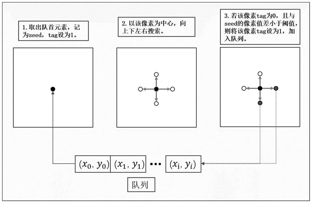
[0032] Step 2. Use the "region growing segmentation algorithm" to segment the heart region of each image. The "region growing segmentation algorithm" is a semi-automatic segmentation method. The process is as follows: figure 1 As shown, take out the first pixel from the queue, use this pixel as a "seed", search for pixels around, if the pixel tag=0, and the pixel value difference with the seed is less than the "threshold", then the The pixel is added to the queue, and the tag is set to 1;

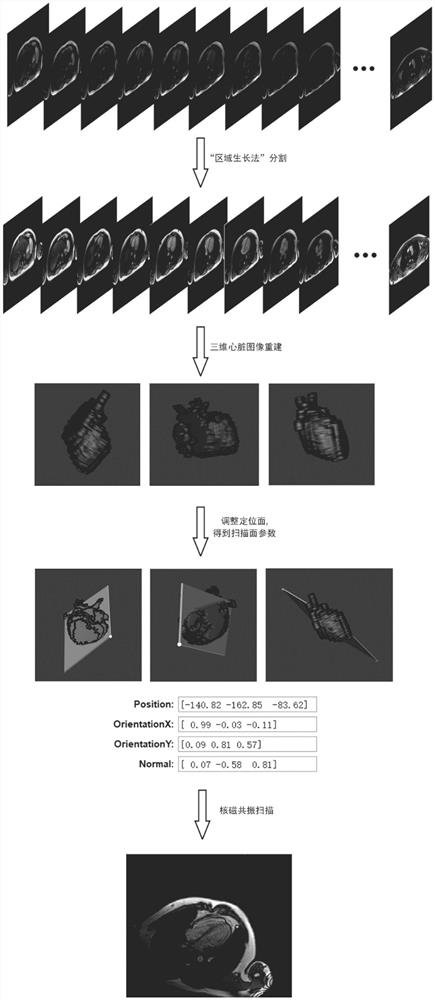
[0033] Step 3: Reconstruct the 3D heart image, and adjust the pixel interval of the heart mask obtained in step 2, and then...

specific Embodiment

[0048] Specific embodiment: a kind of heart three-dimensional localization method based on MR horizontal axis position image segmentation, concrete steps are as follows:

[0049]Step 1, scan the MR transverse axis. After the patient enters the scanner, the doctor sets the slice thickness to 8mm and the number of slices to 12, and makes the positioning line cover the entire heart, scans, and saves the obtained picture in DICOM format;

[0050] Step 2, segment the heart region of the image. Import the DICOM file obtained in step 1 into the software, adjust the image window width and window level, and use the "region growing segmentation algorithm" to segment. The segmentation process is as follows: click a certain pixel in the image with the mouse, and the pixel is regarded as " "Seed", search around, look for pixels whose pixel value difference is less than the "threshold", and classify it as the heart area. If the segmentation is not accurate, click Undo, adjust the "threshol...


PUM


Abstract

The invention discloses a heart three-dimensional positioning method based on MR horizontal axis image segmentation, which comprises the following steps: scanning multiple layers of MR horizontal axis images, segmenting a heart region by using a region growing segmentation algorithm, reconstructing a three-dimensional heart image, manually adjusting a positioning surface, calculating parameters of a scanning surface, and inputting the parameters into a scanning instrument to complete scanning of four-cavity heart, horizontal axis and long axis of the heart. Compared with a manual positioning method, the method has the advantages that scanning surfaces such as a long axis, a short axis and a four-cavity heart of the heart can be directly scanned only by scanning a thoracic cavity image at a horizontal axis position, obtaining scanning surface parameters through post-processing and inputting the scanning surface parameters into a magnetic resonance scanning system, so that the time consumed by a positioning process is shortened, and the accuracy of heart positioning is improved.

Description

technical field [0001] The invention relates to a heart three-dimensional positioning method based on MR transverse axis image segmentation, which belongs to the field of computer image processing. Background technique [0002] Magnetic resonance cardiac examination is an important means of diagnosing heart disease, and it has the advantages of other detection methods (such as CT and ultrasound). For example: the general shape of the heart can be displayed through conventional sequences; myocardial function can be evaluated through cine sequences; myocardial diseases can be diagnosed through first-pass perfusion and delayed enhancement. However, there is a necessary step before these checks - heart positioning. [0003] When analyzing anatomical structures such as the brain and liver, clinicians usually use three directions of the transverse sagittal crown for tomographic scanning. However, this method cannot be used for heart positioning scanning, because the heart is inc...

Claims


Application Information

Patent Timeline
Patent Type & Authority Applications(China)
IPC IPC(8): G06T7/11G06T7/187G06T7/55G06T7/70A61B5/055
CPCG06T7/11G06T7/187G06T7/55G06T7/70A61B5/055G06T2207/10088G06T2207/30048
Inventor 陈阳陈瑞峰朱剑锋
Owner SOUTHEAST UNIV
Why Eureka
Try Eureka