Monoclonal antibody against estrogen-stimulated leucine aminopeptidase

A monoclonal antibody, human technology, applied in the direction of antibody, enzyme, anti-receptor/cell surface antigen/cell surface determinant immunoglobulin, etc., can solve the problem of complex sample processing and so on

Inactive Publication Date: 2002-08-07
HER MAJESTY THE QUEEN & RIGHT OF CANADA REPRESENTED BY THE MIN OF HEALTH
View PDF9 Cites 0 Cited by
  • Summary
  • Abstract
  • Description
  • Claims
  • Application Information

AI Technical Summary

Problems solved by technology

[0007] However, cytology, estrogen and progesterone receptor analysis, and flow cytometry analysis all require biopsy and sample handling is complex

Method used


Image

  • Monoclonal antibody against estrogen-stimulated leucine aminopeptidase
  • Monoclonal antibody against estrogen-stimulated leucine aminopeptidase
  • Monoclonal antibody against estrogen-stimulated leucine aminopeptidase

Examples


example 1

[0051] Example 1: Isolation and purification of estrogen-dependent LAPase

[0052] Breast cancer blasts from human tumor tissue were stimulated with 100nm 17-B-estradiol for 24 hours, or culture medium as control. The cell medium is: RPMI 1640 culture medium + 10% FCS + 100U / ml penicillin + 100ug / ml streptomycin. Supernatants were collected after digestion with PBS in seamless dextran tubes (MV12,400) for 24 hours (4°C). LAP was then purified from the degraded cell supernatant using HPLC-gel permeation chromatography with DEAE-cellulose and Bestatin-Sepharose affinity chromatography. Briefly, the cell supernatant was poured onto a Bio-Sil SEC-250 column (600×7.5 mm) containing 100 mM sodium phosphate (pH 6.8), 100 mM Na 2 SO 4 , 1uM ZnCl 2 , and 10% glycerol buffer pre-equilibrated. The column was then washed with 300ml of the same buffer at a flow rate of 0.5ml / min. The protein was concentrated to 10ml by high-speed centrifugation with YM5 membrane. The concentrated so...

example 2

[0057] A) Production and purification of monoclonal antibodies

[0058] Hybridoma cell line 7B6 producing 17-B-estradiol-stimulated LAPase mouse monoclonal antibody, preparation of immunization antigen, preparation of spleen cells from immunized animals, fusion of spleen cells and myeloma cells, and the selection of fused cells in The coating methods in the media were derived from the method proposed by Campbell and the method described in detail by Lietzhe and Unsicker.

[0059] Briefly, basic immune responses were performed with purified es-LApase after desalting. Boosted with purified es-LApase at days 14, 35, and 56, BALB / C mice were screened at days 24 and 45. Mice were sacrificed on day 59 and the best responding splenocytes were fused with myeloma cells. Screening was performed using dot plot immunostaining on nitrocellulose.

[0060] Hybridoma clone 7B6 was obtained by single cell cloning by limiting dilution. Prepare four serial dilution tubes containing hybridoma...

example 4

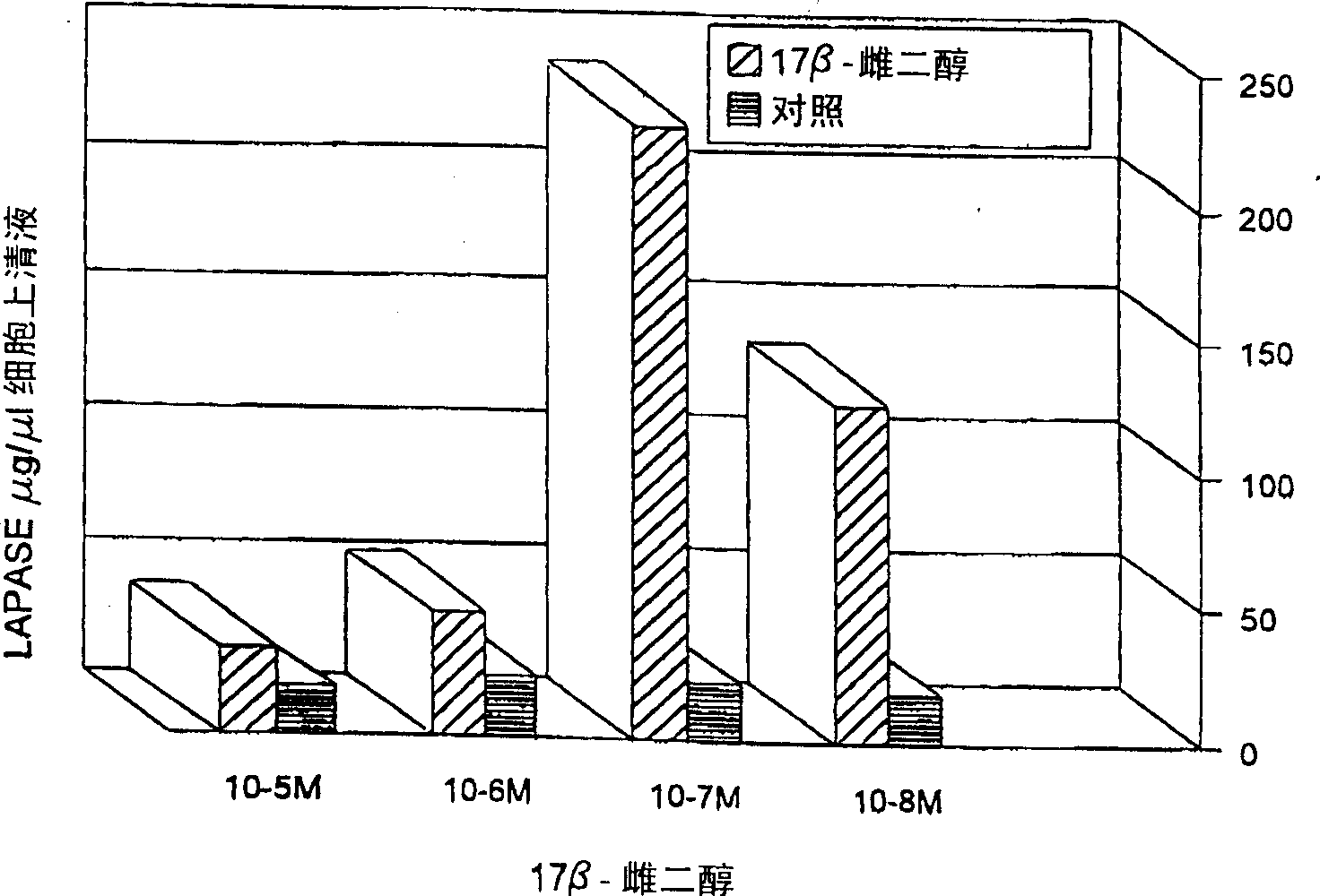
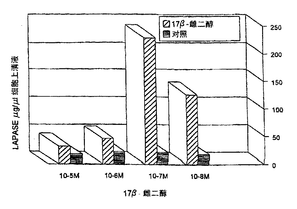
[0084] Example 4: LAPase level in cell supernatant after estrogen stimulation

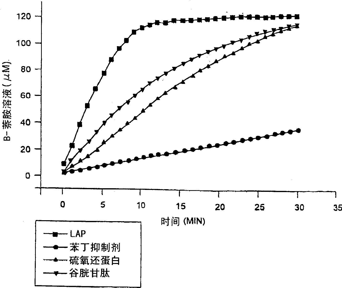
[0085] Early breast cancer mother cells were cultured with 100nM 17-B-estradiol for 24 hours, and the simple medium was used as blank. Use 7B6 IgGla) - LAPase in the supernatant isolated from protein G-LAPase beads covalently cross-linked to monoclonal antibodies. The activity of LAPase in the supernatant of LAPase immunoprecipitation was determined by fluorescence method using leucine-B-naline as substrate.

[0086] The purified enzyme preparation obtained from Sigma was incubated with 100nM 17-B-estradiol for 24 hours, and the activity of LAPase was measured by fluorescence method using leucine-B-naline as the substrate.

[0087] After estrogen stimulation, the maximum release of LAPase was at an estrogen concentration of 100 nM, after 24 hours of culture (see figure 1 ). During the same period, there was no change in LAPase in the cell supernatant of control cells. In addition, the LAPase ...


PUM


Abstract

The identification and characterization of risk factors and their molecular implications in the pathophysiology of human diseases such as cancer is essential for designing efficient diagnostic assays and therapeutic compounds. Estrogenic steroids, under normal physiological conditions, have been shown to play a critical function in several tissues. The response of such a variety of tissues to estrogen stimulation can explain in part its active role in the development and progression of different human diseases, particularly breast cancer. Searching for estrogen-responding cellular factors in parental cells of primary human breast carcinomas obtained from tumour biopsies, two cellular markers, an isoenzyme of putative leucine aminopeptidase (LAPase; EC 3.4.11.1) from parental cells of primary human breast carcinomas obtained from tumour biopsies, and cytosolic NDP-Kinase / Nm23 (EC 2.7.4.6) from HL60 cells were identified. Monoclonal antibodies against each cellular marker have been produced. Determination of the presence of these two markers, either alone or in combination, -can be used to detect breast cancer, and in particular, associated metastatis. Thus, this invention refers to the use of both LAP and NDP-Kinase / Nm23 monoclonal antibodies together in a tandem solid-matrix based immuno-assay for first line confimatory blood-based testing for breast cancer.

Description

[0001] This invention relates to monoclonal antibodies that specifically bind to the human estrogen-responsive isozyme of leucine aminoterase (es-LAPase). A myeloma cell line known as 7B6 and the monoclonal antibody produced therefrom are also discussed. A diagnostic system for detecting the level of estrogen-responsive isozymes to LAPase in blood, serum, or plasma using monoclonal antibodies from the myeloma cell line 7B6 is further discussed. This antibody is particularly useful for the rapid diagnosis of breast cancer. Background of the invention [0002] The key to effective detection and design of therapeutic drugs for human diseases such as breast cancer is the ability to identify risk factors and pathophysiological changes. Among various risk factors related to the early formation of breast cancer, estrogen and estrogen analogues with similar activities to estrogen are still the most important substances to be tested for the early occurrence and development of breast c...

Claims


Application Information

Patent Timeline
Patent Type & Authority Applications(China)
IPC IPC(8): A61K38/06A61K38/44A61K39/395A61P31/18C07K16/30C07K16/40C12N5/20C12Q1/37G01N33/569G01N33/573G01N33/574
CPCA61K39/395Y10S435/975G01N33/573G01N33/56988G01N33/57415A61K38/063A61K38/4813C07K16/40A61K2039/505C07K16/3015C12Q1/37A61K38/44A61P31/18A61K2300/00
Inventor 卡布里耶·普利多-塞胡多
Owner HER MAJESTY THE QUEEN & RIGHT OF CANADA REPRESENTED BY THE MIN OF HEALTH
Why Eureka
Try Eureka