Use of pp3774 gene in inhibiting cell growth
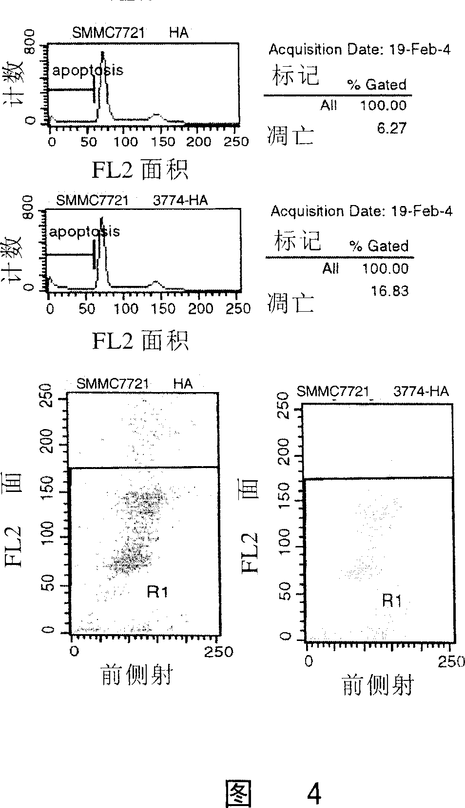
A technology of pp3774 and cells, applied in the field of functional application of pp3774 gene, can solve problems such as ignorance or understanding of functions, achieve the effect of inhibiting cell growth, enriching application prospects, and inducing cell apoptosis
- Summary
- Abstract
- Description
- Claims
- Application Information
AI Technical Summary
Problems solved by technology
Method used
Image
Examples
Embodiment 1
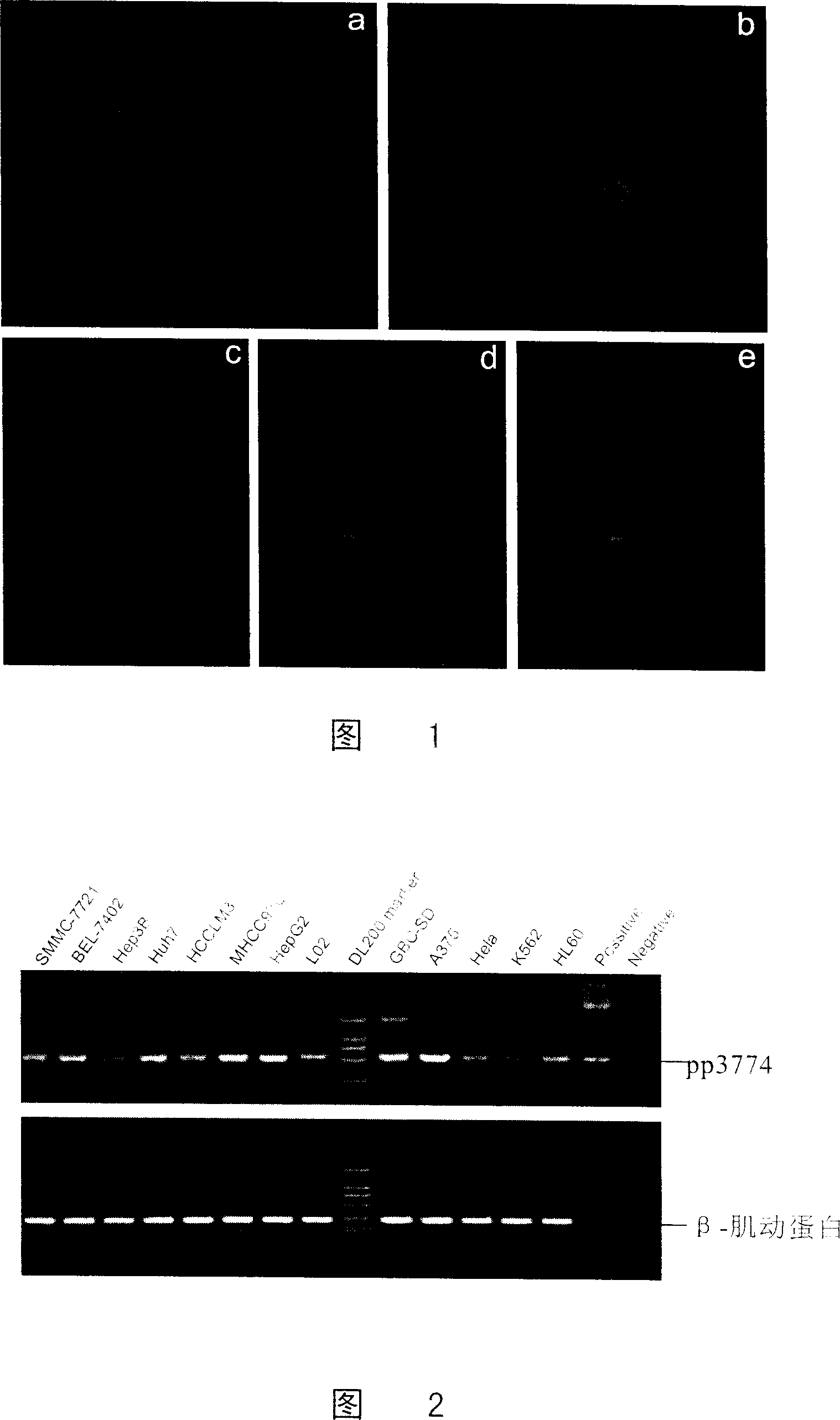
[0099] Cellular localization of pp3774 protein
[0100] Utilize specific primers containing restriction enzyme cutting sites to amplify the coding region of the pp3774 gene by the method of PCR, and load the eukaryotic expression vector EGFP-N1 (purchased from Clontech Company) with EcoRI / BamHI restriction sites, so that It can express the pp3774 fusion protein with a GFP tag at the C-terminus, which is verified by enzyme digestion and sequencing, and is constructed into a pp3774-EGFP-N1 fusion expression vector, which is transfected into SMMC-7721 cells by lipofection and observed by fluorescence Cellular localization of PP3774-GFP fusion protein.
[0101] The subcellular localization was done by transfecting live COS-7 cells with pp3774 gene and adding endoplasmic reticulum-specific fluorescent tracer ER-Tracker Blue-White DPX and then incubating with fluorescence microscope and ISIS image analysis system.
[0102] The results showed that the empty vector tagged protein GFP...
Embodiment 2
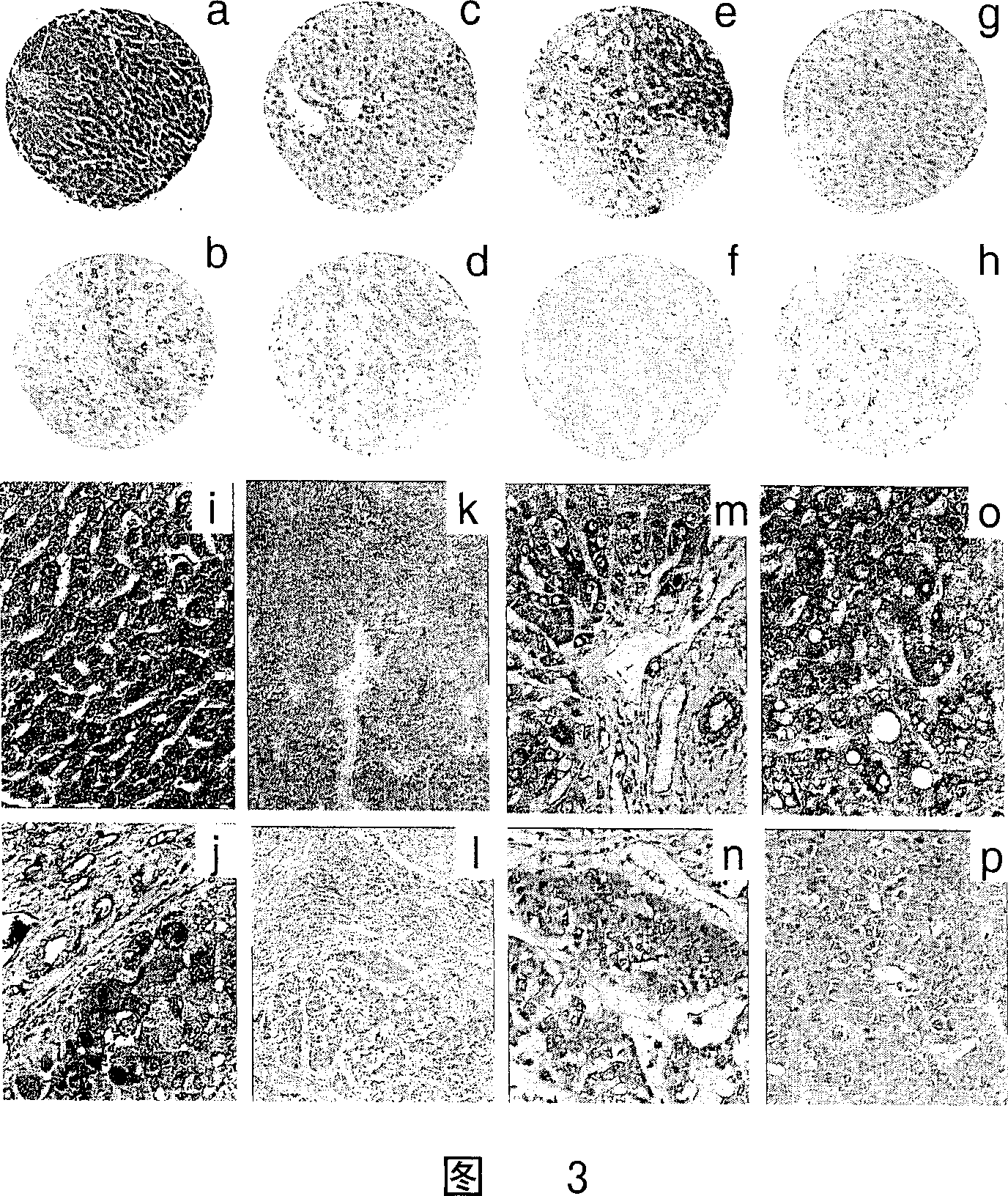
[0104] Semi-quantitative reverse transcription-polymerase chain reaction (Semi-Q RT-PCR)
[0105] a) Use Trizol reagent to extract human liver cancer cell lines SMMC-7721, BEL-7402, MHCC-LM3, MHCC-97L, Huh-7, Hep3B, HepG2 and human immortalized liver cells L-02 in good growth state, and other tumors Cell lines such as gallbladder cancer cell line GBC-SD, melanoma cell line A375, cervical cancer cell line HeLa, leukemia cell line K562, HL60, and human liver cancer tissue and paracancerous tissue total RNA were quantified and tested for purity by UV spectrophotometer, and Take a small amount of total RNA for denaturing gel electrophoresis to check whether the RNA is degraded;
[0106] b) Mix 9 μl (5 μg) of total cellular RNA treated with DNase I with 1 μl of Oligo dT, heat at 72°C for 5 minutes, and immediately place on ice for 10 minutes;
[0107] c) Add 8μl reverse transcription mixture (10×RT Buffer 2μl, 25mM MgCl 2 2 μl, 1 μl of 10 mM dNTP mix, 2 μl of 0.1 mM DTT, 1 μl of...
Embodiment 3
[0123] Immunocytochemistry and Immunohistochemical Detection of Liver Cancer Tissue Microarray
[0124] a) All monolayer cell culture sheets used for immunocytochemical detection and tissue samples used for immunohistochemical detection were fixed with 10% neutral buffered formalin;
[0125] b) Tissue samples were embedded in paraffin, serially sectioned with a thickness of 5 μm, and stained with HE for general histopathological observation, pathological grading of liver cancer tissues and positioning before preparation of liver cancer tissue chips, including 214 pairs of human liver cancer and paracancerous tissues, 18 cases The tissue chip preparation method of liver cirrhosis tissue and 10 cases of normal liver tissue is completely in accordance with the technical platform established by our laboratory;
[0126] c) Use rabbit anti-pp3774 polyclonal antiserum to isolate IgG as the primary antibody, apply EnvisionSystem-HRP (rabbit), DAB color development, Mayer's hematoxylin...
PUM
 Abstract
Description
Claims
Application Information
 - R&D
- Intellectual Property
- Life Sciences
- Materials
- Tech Scout
- Unparalleled Data Quality
- Higher Quality Content
- 60% Fewer Hallucinations
Browse by: Latest US Patents, China's latest patents, Technical Efficacy Thesaurus, Application Domain, Technology Topic, Popular Technical Reports.
© 2025 PatSnap. All rights reserved.Legal|Privacy policy|Modern Slavery Act Transparency Statement|Sitemap|About US| Contact US: help@patsnap.com


