Biofilm formation to define risk for colon cancer

a technology of colon cancer and biofilms, applied in the field of biofilm formation to define the risk of colon cancer, can solve the problems that the association between the composition of microorganisms and crc development has not been established, and achieve the effects of reducing the apoptosis of colonic epithelium, eliminating biofilms, and increasing proliferation

Active Publication Date: 2019-02-12
THE JOHN HOPKINS UNIV SCHOOL OF MEDICINE
View PDF7 Cites 0 Cited by
  • Summary
  • Abstract
  • Description
  • Claims
  • Application Information

AI Technical Summary

Benefits of technology

[0006]The present invention is based, at least in part, upon the remarkable discovery that, when colorectal cancer (CRC) subjects were examined, essentially all right-sided colon cancers and a subset of left-sided colon cancers were identified to possess a biofilm. Such biofilms were associated with increased proliferation and decreased apoptosis of colonic epithelium, both of which are important elements in transformation to colon cancer. Without wishing to be bound by theory, such biofilms would likely have allowed for an increased concentration of bacteria to contact the colonic mucosa. In contrast, only 10% of normal colonoscopy colon biopsies were identified as biofilm positive. These results indicated that detection of biofilms in normal colonoscopy specimens would identify individuals at higher risk for development of colon cancer. Furthermore, interventions such as antibiotics or probiotics that eliminate biofilms from the colon mucosa of individuals are described herein for preventing and / or diminishing development of gastrointestinal tract cancers, particularly colon cancer.
[0007]In one aspect, the invention provides a method for treating or preventing a neoplastic condition in a subject that involves (i) detecting a bacterial biofilm within the gastrointestinal tract of a subject; and (ii) administering an antimicrobial agent or a probiotic agent to the subject in an amount effective to reduce the size of the bacterial biofilm, thereby treating or preventing a neoplastic condition in the subject.
[0027]A further aspect of the invention provides a method for treating or preventing cancer in a subject that involves administering an antimicrobial agent or a probiotic agent to a subject harboring a bacterial biofilm within the gastrointestinal tract of the subject, wherein the antimicrobial agent or probiotic agent reduces or eliminates the presence of a the biofilm, thereby treating or preventing cancer in the subject.
[0053]A “growth inhibitory agent” when used herein refers to a compound or composition which inhibits growth of a cell either in vitro or in vivo. Thus, the growth inhibitory agent may be one which significantly reduces the percentage of cells in S phase. Examples of growth inhibitory agents include agents that block cell cycle progression (at a place other than S phase), such as agents that induce G1 arrest and M-phase arrest. Classical M-phase blockers include the vincas (e.g., vincristine and vinblastine), taxanes, and topoisomerase II inhibitors such as doxorubicin, epirubicin, daunorubicin, etoposide, and bleomycin. The agents that arrest G1 also spill over into S-phase arrest, for example, DNA alkylating agents such as tamoxifen, prednisone, dacarbazine, mechlorethamine, cisplatin, methotrexate, 5-fluorouracil, and ara-C. Further information can be found in The Molecular Basis of Cancer, Mendelsohn and Israel, eds., Chapter 1, entitled “Cell cycle regulation, oncogenes, and antineoplastic drugs” by Murakami et al. (WB Saunders: Philadelphia, 1995), especially p. 13. The taxanes (paclitaxel and docetaxel) are anticancer drugs both derived from the yew tree. Docetaxel (TAXOTERE®, Rhone-Poulenc Rorer), derived from the European yew, is a semisynthetic analogue of paclitaxel (TAXOL®, Bristol-Myers Squibb). Paclitaxel and docetaxel promote the assembly of microtubules from tubulin dimers and stabilize microtubules by preventing depolymerization, which results in the inhibition of mitosis in cells.
[0066]As used herein, the term “effective amount” refers to the amount of a therapy that is sufficient to result in therapeutic benefit to a patient with cancer, In one embodiment, the cancer patient has been diagnosed with a biofilm and, optionally, with CRC. In one embodiment, the effective amount is administered to a patient that has been diagnosed with cancer. The effective amount can result in the prevention of the development, recurrence, or onset of cancer and one or more symptoms thereof, to enhance or improve the efficacy of another therapy, reduce the severity, the duration of cancer, ameliorate one or more symptoms of cancer, prevent the advancement of cancer, cause regression of cancer, and / or enhance or improve the therapeutic effect(s) of another therapy. “Effective amount” also refers to the amount of a therapy that is sufficient to result in the prevention of the development, recurrence, or onset of cancer and one or more symptoms thereof, to enhance or improve the prophylactic effect(s) of another therapy, reduce the severity, the duration of cancer, ameliorate one or more symptoms of cancer, prevent the advancement of cancer, cause regression of cancer, and / or enhance or improve the therapeutic effect(s) of another therapy. In an embodiment of the invention, the amount of a therapy is effective to achieve one, two, three, or more results following the administration of one, two, three or more therapies: (1) a stabilization, reduction or elimination of the cancer stem cell population; (2) a stabilization, reduction or elimination in the cancer cell population; (3) a stabilization or reduction in the growth of a tumor or neoplasm; (4) an impairment in the formation of a tumor; (5) eradication, removal, or control of primary, regional and / or metastatic cancer; (6) a reduction in mortality; (7) an increase in disease-free, relapse-free, progression-free, and / or overall survival, duration, or rate; (8) an increase in the response rate, the durability of response, or number of patients who respond or are in remission; (9) a decrease in hospitalization rate, (10) a decrease in hospitalization lengths, (11) the size of the tumor is maintained and does not increase or increases by less than 10%, preferably less than 5%, preferably less than 4%, preferably less than 2%, (12) an increase in the number of patients in remission, (13) an increase in the length or duration of remission, (14) a decrease in the recurrence rate of cancer, (15) an increase in the time to recurrence of cancer, and (16) an amelioration of cancer-related symptoms and / or quality of life.

Problems solved by technology

However, clear associations between microbiota composition and CRC development have not been established.

Method used


Image

  • Biofilm formation to define risk for colon cancer
  • Biofilm formation to define risk for colon cancer
  • Biofilm formation to define risk for colon cancer

Examples


example 1

and Methods

Patient Selection / Sample Acquisition

[0126]CRC and paired normal tissue were collected from patients undergoing surgery at Johns Hopkins Hospital. All tissue not needed for pathologic diagnosis was rapidly preserved in formalin, Carnoy's solution and RNAlater for analysis. Patients who received pre-operative radiation and / or chemotherapy or with a personal history of CRC were excluded. For patients in this study, two bowel preparations were routinely used and recorded (mechanical bowel preparation [Miralax™], or Fleet Phospo-Soda™ enema), Pre-operative intravenous antibiotics were administered in all cases (cefotetan or clindamycin / gentamycin). No patient received pre-operative oral antibiotics.

[0127]Healthy control patients undergoing screening colonoscopy or colonoscopy for diagnostic work-up (eg, anemia) were recruited and signed informed consent. All patients underwent a standard mechanical bowel preparation (FIGS. 10A-C). Mucosal biopsies from grossly normal colon wer...

example 2

ation of Microbial Biofilms Associated with Colorectal Tumors (CRC & Adenomas Examined)

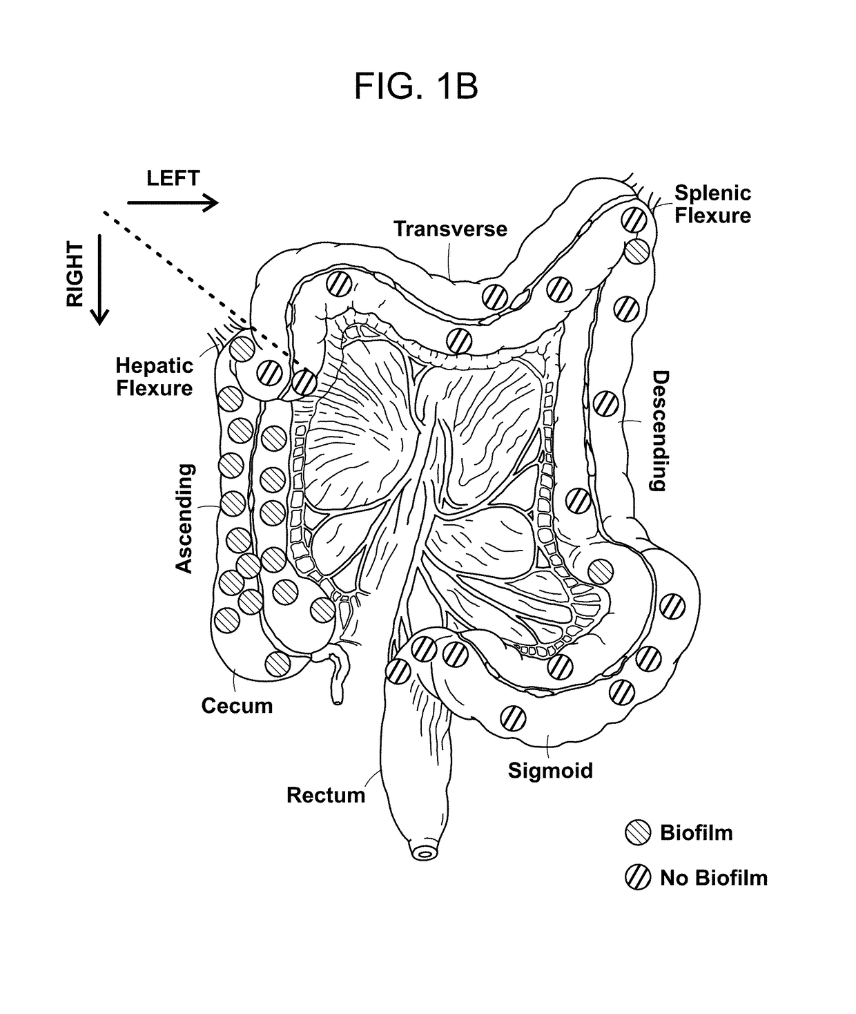
[0145]The microbial communities associated with surgically-resected colorectal tumors (CRC and adenomas) and paired normal tissue collected from the pathologically tumor-free mucosa at the surgical resection margin were systematically examined. Paired normal colon tissues were obtained from the margin of the resected specimens furthest from the site of the tumor (FIG. 4). In addition, as a healthy control population, colon biopsies obtained from individuals without colorectal tumors and without a diagnosis of inflammatory colonic disease undergoing routine screening colonoscopy were studied. FIGS. 9 and 10A-C present all patient metadata.

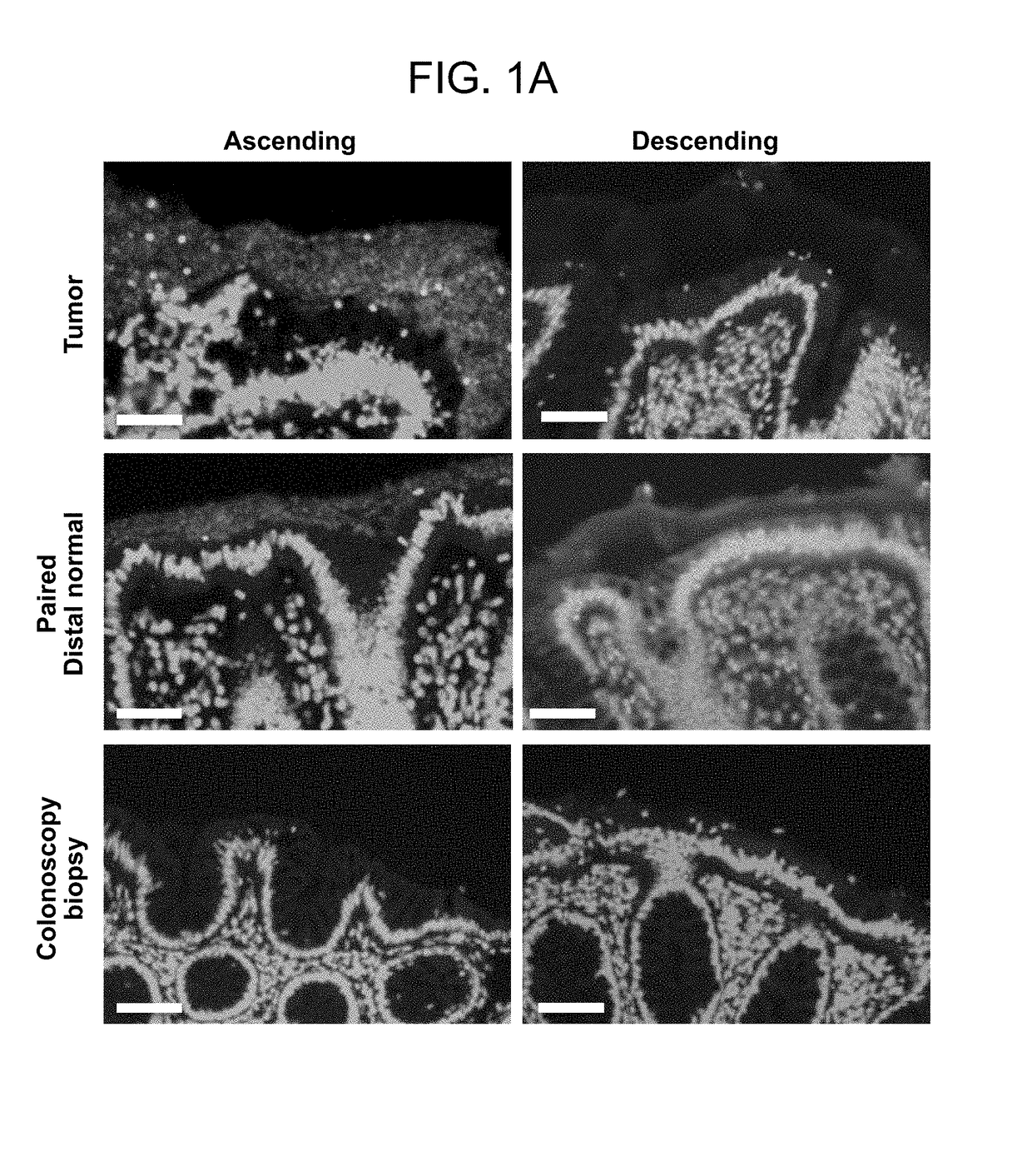
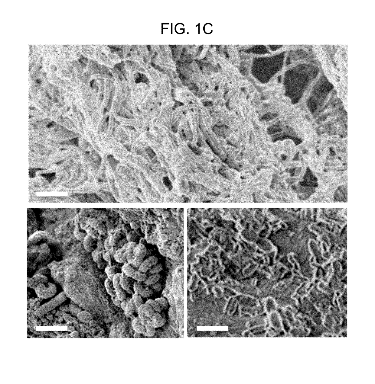
[0146]The spatial relationship of the microbiota was first compared with the host mucus layer and colonic epithelium using fluorescent in situ hybridization (FISH). Carnoy's solution-fixed, paraffin-embedded tissues known to preserve the mucus layer were employed....

example 3

ization of Microbial Biofilms Associated with Colorectal Tumors

[0147]To determine if biofilm formation was specific for the tumor microenvironment, we next used FISH to examine the paired pathologically normal colon tissues obtained from the surgical resection margin most distal from the tumor mass (FIG. 4) for biofilm formation. No biofilms were detected on the distal pathologically normal surgically-resected colon tissues paired with biofilm-negative tumors (adenomas and CRCs). In surprising contrast, all but one distal pathologically normal surgically-resected colon tissue paired with a biofilm-covered tumor were biofilm positive whether in the right or left colon; thus, 93% (14 / 15) and 100% (4 / 4) of paired pathologically normal colon tissues from the surgical resection margin of biofilm-covered CRCs and adenomas, respectively, were biofilm positive (FIGS. 1A and 1D). Of note, the single surgically-resected distal normal tissue on which we failed to detect a biofilm was fixed in ...


PUM


Abstract

Methods of identifying subjects at increased risk of cancer, based upon detection of biofilms and / or biofilm-associated microbes within a subject, are disclosed. Therapies designed to prevent formation and / or reduce the size of biofilms in a subject identified to be at increased risk of cancer based upon detection of biofilms and / or biofilm-associated microbes are disclosed. In particular embodiments, the invention provides for identification of a subject at elevated risk of developing or having colorectal cancer and / or a colorectal adenoma, based upon detection of a biofilm and / or biofilm-associated bacteria within the gastrointestinal tract of the subject (optionally, within a biopsy specimen and / or stool sample of such subject). Therapies involving administration of an antibiotic agent and / or a probiotic agent to a subject, to prevent or reduce biofilm formation within the gastrointestinal tract of the subject, optionally provided in combination with additional cancer therapy, are also disclosed.

Description

CROSS REFERENCE TO RELATED APPLICATIONS[0001]This application is a national stage application, filed under 35 U.S.C. § 371, of International Application No. PCT / US2014 / 055123, filed on Sep. 11, 2014, which claims priority to U.S. Provisional Patent Application Ser. No. 61 / 876,995, filed Sep. 12, 2013. The entire contents of each of which are hereby incorporated by reference in their entireties.STATEMENT OF RIGHTS TO INVENTIONS MADE UNDER FEDERALLY SPONSORED RESEARCH[0002]This invention was made with government support under grant no. CA151393 and CA170492 awarded by the National Institutes of Health. The government has certain rights in the invention.FIELD OF THE INVENTION[0003]The present invention relates to compounds, compositions, and methods for the study, diagnosis, and treatment of cancer and / or other proliferative diseases, disorders, or conditions. In certain embodiments, the invention specifically relates to detection, prevention and treatment of colorectal cancer (CRC) in...

Claims


Application Information

Patent Timeline
Patent Type & Authority Patents(United States)
IPC IPC(8): G01N33/574A61K31/7048A61K31/407A61K35/741A61K35/742A61K31/546A61K31/43A61K31/165A61K31/7056A61K35/745A61K35/747A61K45/06A61K35/744C12Q1/6869C12Q1/6841
CPCG01N33/57419A61K31/407A61K31/43A61K31/546A61K31/7048A61K31/7056A61K35/741A61K35/742A61K35/744A61K35/745A61K35/747A61K45/06C12Q1/6841C12Q1/6869A61K31/165G01N2800/50C12Q1/04C12Q1/689G01N2800/7028A61P35/00
Inventor SEARS, CYNTHIA L.PARDOLL, DREW M.CRAIG, CHRISTINEWICK, ELIZABETH
Owner THE JOHN HOPKINS UNIV SCHOOL OF MEDICINE
Why Eureka
Try Eureka