

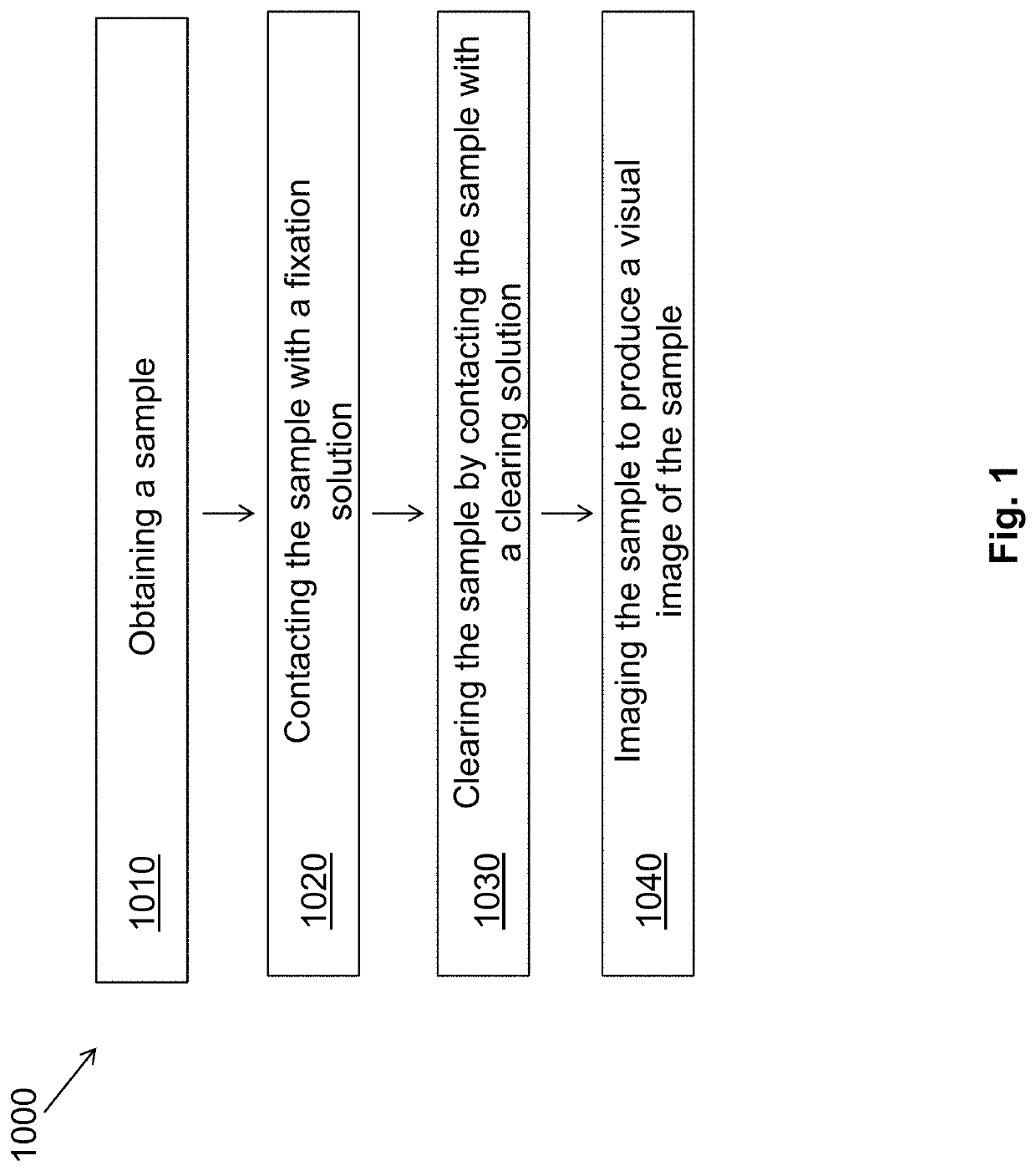
Simultaneous dehydration and staining of tissue for deep imaging
a tissue and simultaneous dehydration technology, applied in the field of simultaneous dehydration and staining of tissue for deep imaging, can solve the problems of limiting the ability to make more significant advances in the speed, quality and completeness of tissue biopsy evaluation, incomplete sample evaluation, and artifacts of preparation
- Summary
- Abstract
- Description
- Claims
- Application Information
AI Technical Summary
Benefits of technology
Problems solved by technology
Method used
Image

Examples
experimental examples
[0075]The invention is further described in detail by reference to the following experimental examples. These examples are provided for purposes of illustration only, and are not intended to be limiting unless otherwise specified. Thus, the invention should in no way be construed as being limited to the following examples, but rather, should be construed to encompass any and all variations which become evident as a result of the teaching provided herein.
example 1
High-Resolution, 2- and 3-Dimensional Imaging of Uncut, Unembedded Tissue Biopsy Samples
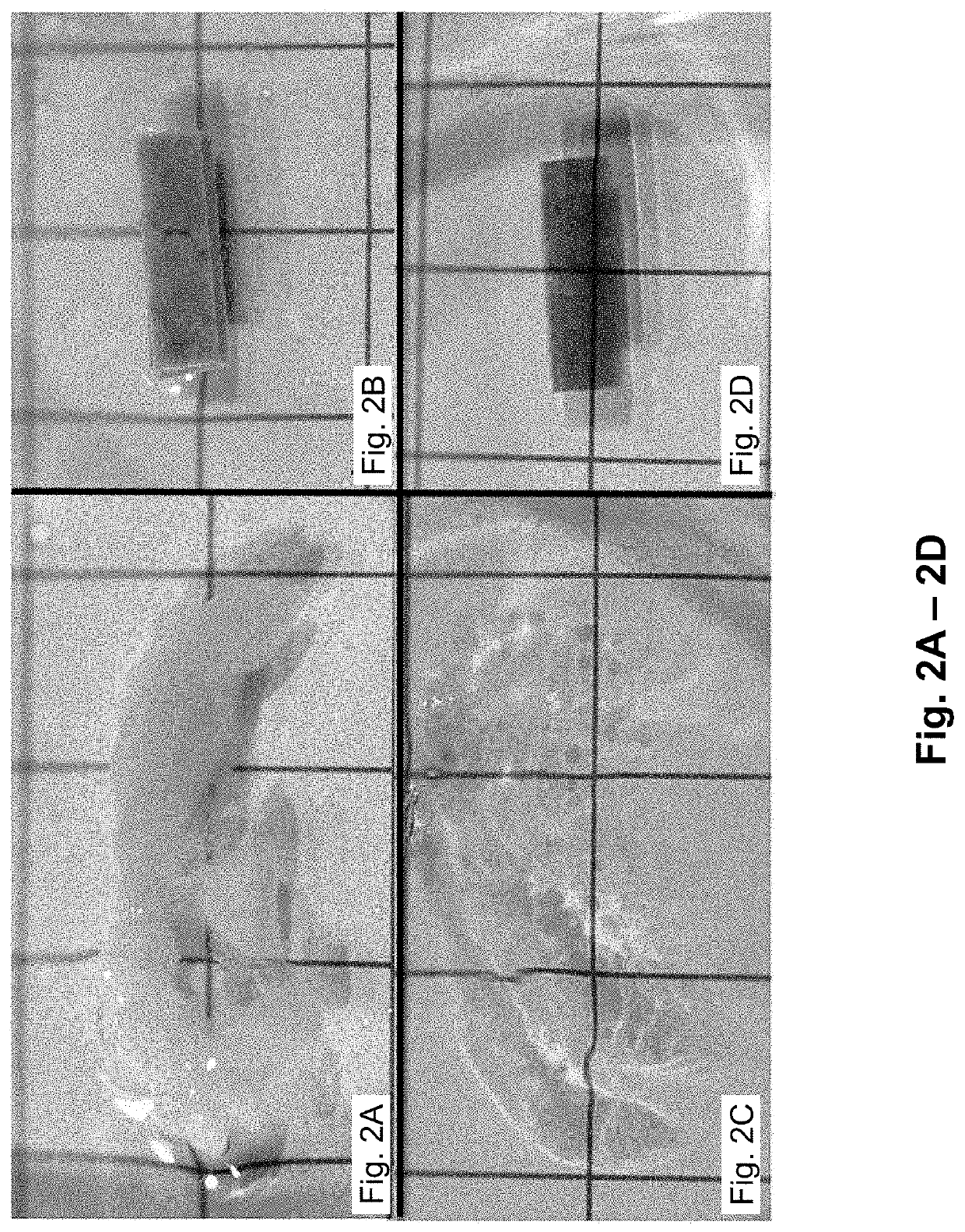
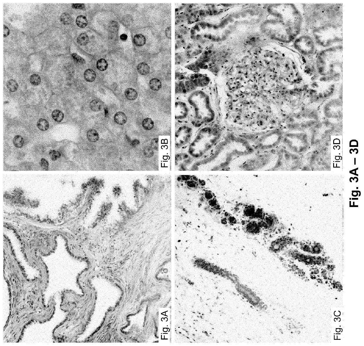
[0076]The results described herein demonstrate that the combination of clearing agents and fluorescent dyes is useful for clinical application of multiphoton imaging of complete biopsy specimens, along with added informational content from SHG. Excellent cellular contrast can be achieved from both intrinsic fluorescence and with extrinsic nucleic acid dyes. Multichannel imaging facilitated a pseudocolorization process that mimicked the appearance of traditional stains. Three-dimensional reconstructions of MPM imaging from clarified tissue may be used on complete biopsy-sized tissue specimens and may also be used to produce quantifiable characterization of collagen fibrosis.
[0077]The materials and methods employed in these experiments are now described.
Tissue Clearing and Staining
[0078]Human tissue specimens were obtained from discarded pathologic tissue of liver, kidney, breast, and prostate rese...
example 2
Exemplary Tissue Staining Protocol
[0094]A core biopsy-sized tissue specimen is fixed in formalin for a period of time from 20 minutes to 4 weeks. The specimen is then placed directly in a solution of methacarn which has 10 μM DAPI and 0.5% by volume eosin added to the solution. The specimen is incubated at 45° C. for 60 minutes. The specimen is transferred directly to a solution of 100% BABB, and incubated for 30 minutes. The specimen is imaged in a BABB bath. FIG. 15 depicts images of tissue samples prepared according to this exemplary method.
PUM
| Property | Measurement | Unit |
|---|---|---|
| depth | aaaaa | aaaaa |
| time | aaaaa | aaaaa |
| depths | aaaaa | aaaaa |
Abstract
Description
Claims
Application Information