Method for visualizing biomolecules, such as proteins or nucleic acids, with the unaided eye, without needing to use potentially toxic compounds, exposure to ultraviolet (UV) light or fluorescence

a biomolecule and visualization technology, applied in the field of biomolecule visualization, can solve the problems of difficult handling, very little use of strategies, and loss of millions of dollars in this crop

Active Publication Date: 2020-04-28
UNIVERSITY OF CHILE
View PDF0 Cites 0 Cited by
  • Summary
  • Abstract
  • Description
  • Claims
  • Application Information

AI Technical Summary

Problems solved by technology

In addition, there are pathogenic microorganisms that attack the vineyard and cause millionaires losses in this crop.
These strategies are very little used since they are not specific for DNA, since they also detect proteins and other biomolecules such as lipoproteins.
On the other hand, silver nitrate produce a long lasting stain on the surface on which is exposed, therefore is difficult to handle, dangerous if not handled properly and DNA cannot be recovered after staining.
There are no methods today for observing with the naked eye and distinguish DNA and protein in the same gel, which makes some studies difficult, such as simultaneous detection of both biomolecules.
Protein-DNA interaction studies are essential in biochemistry research, e.g., for detecting some protein specific binding sites to DNA zones through molecule crosslinking studies, whereby the silver nitrate-based staining method is not suitable for these applications because the nonspecificity thereof.

Method used


Image

  • Method for visualizing biomolecules, such as proteins or nucleic acids, with the unaided eye, without needing to use potentially toxic compounds, exposure to ultraviolet (UV) light or fluorescence
  • Method for visualizing biomolecules, such as proteins or nucleic acids, with the unaided eye, without needing to use potentially toxic compounds, exposure to ultraviolet (UV) light or fluorescence
  • Method for visualizing biomolecules, such as proteins or nucleic acids, with the unaided eye, without needing to use potentially toxic compounds, exposure to ultraviolet (UV) light or fluorescence

Examples


examples of application

Example 1

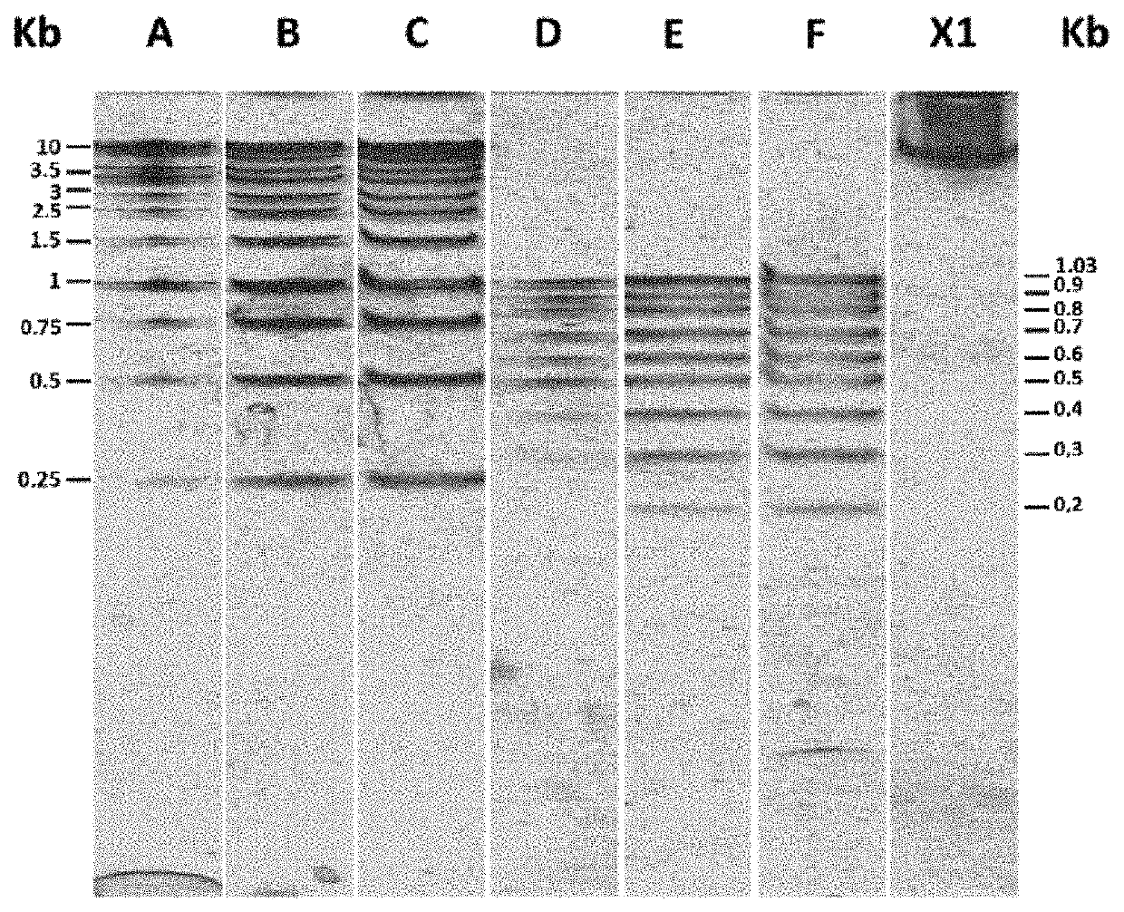
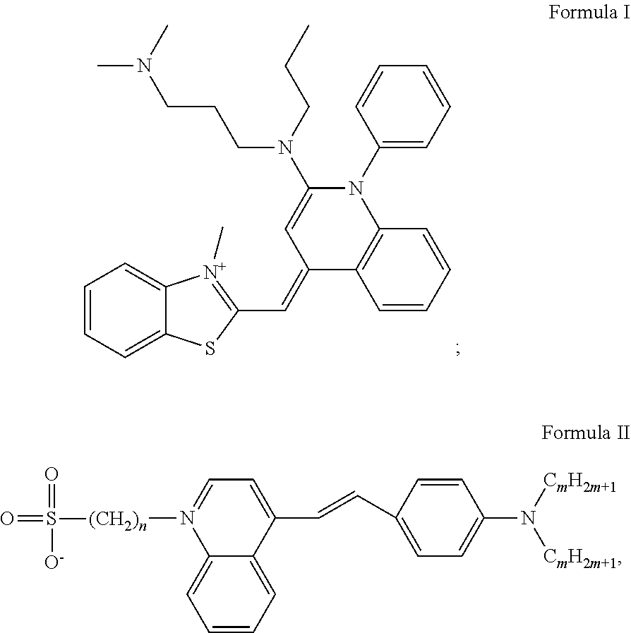
[0042]a) For separating DNA, this molecule was placed together with a loading solution (0.25% bromophenol blue, 0.25% xylene cyanol and 30% glycerol). This solution was then placed in an agarose matrix in a solution containing 1×TAE buffer (40 mM Tris, 20 mM acetate and 1 mM EDTA). By applying a voltage difference to the gel, the DNA migrates to the positive pole and DNA molecules are separated as they pass through the matrix according to their different sizes.[0043]b) The matrix was then placed in a container with cyanine-derived DNA intercalating molecule SYBR® Green I (N′,N′-dimethyl-N-[4-[(E)-(3-methyl-1,3-benzothiazol-2-ylidene)methyl]-1-phenylquinolin-1-ium-2-yl]-N-propylpropane-1,3-diamine) at a concentration of 1.96 μM (INVITROGEN®, diluted 10,000 fold, i.e., 1× final dilution) the gel completely covered in 1×TAE buffer for approx. 30 min. The container containing the gel was protected from light by covering with an aluminum foil. The gel was put under shaking at le...

example 2

[0049]a) For visualizing proteins, these molecules were placed onto a polyacrylamide matrix and the electrophoresis process was carried out.[0050]b) The gel was placed in a container containing the intercalating agent SYPRO® red (protein gel stain) at a concentration of 3.92 μM (INVITROGEN®, 5000 fold dilution) and 7.5% (v / v) acetic acid. It was covered with aluminum foil to protect the intercalating agent from light. The gel was incubated for 30 min shaking at less than 50 rpm at room temperature.[0051]c) The gel was removed and rinsed in 7.5% acetic acid to remove the excess of SYPRO® red (protein gel stain) from its surface and placed in 0.19 mM NBT for at least 90 min.[0052]d) The gel was removed from this solution and the NBT residues were removed with distilled water.[0053]e) Protein bands were visualized by the naked eye and without the need to exposure to UV light.

example 3

[0054]a) For separating RNA, this molecule was placed together with a loading solution (0.25% bromophenol blue, 0.25% xylene cyanol and 15% ficoll). This solution was then placed onto a polyacrylamide matrix in a solution containing 1×TAE buffer (40 mM Tris, 20 mM acetate and 1 mM EDTA). By applying a voltage difference to the gel, the RNA migrates to the positive pole and RNA molecules are separated as they pass through the matrix according to their different sizes.[0055]b) The matrix was then placed in a container with the cyanine-derived RNA intercalating molecule SYBR® Green II (RNA cyanine dye) at a concentration of 1.96 μM (INVITROGEN®, 10,000 fold dilution, i.e. 1× final dilution) the gel completely covered in 1×TAE buffer for approx. 30 min. The container containing the gel was protected from light by covering with an aluminum foil. It was put under shaking at less than 50 rpm and at room temperature for 30 min.[0056]c) The gel was placed in a container containing 0.198 mM o...


PUM

PropertyMeasurementUnit
temperatureaaaaaaaaaa
concentrationaaaaaaaaaa
fluorescenceaaaaaaaaaa

Abstract

The present invention relates to a method of visualizing biomolecules, having the steps of: a) providing a sample of immobilized biomolecules in a matrix and carry on the electrophoresis process; b) incubating the matrix of step a) in a solution containing a cyanine-derived molecule, for a time of 5 to 60 minutes, at room temperature, in a container preventing exposure to light, shaking the container at less of 75 rpm; c) transferring the matrix from step b) to a container with a solution having: at least one tetrazolium salt and incubating for a time of 15 to 120 minutes at room temperature under light exposure; d) removing the matrix with immobilized biomolecules from the previous step and washing with distilled water; and e) visualizing directly by the naked eye the biomolecules immobilized in the matrix.

Description

CROSS REFERENCE TO RELATED APPLICATION[0001]This Application is a 371 of PCT / IB2015 / 058363 filed on Oct. 29, 2015, which claims priority of Chilean Application No. 2014-3000 filed Nov. 5, 2014, both of which are incorporated herein by reference.[0002]The present invention describes a simple and direct method for visualizing nucleic acids and proteins by the naked eye without the need to use compounds with potential toxic effects for health, exposure to ultraviolet (UV) light or by fluorescence for their visualization.STATE OF THE ART[0003]The process for detecting biomolecules such as proteins and nucleic acids is essential in biological research, clinical diagnosis and industrial analysis services. These methods are frequently and routinely used in scientific research and basic analysis in many laboratories. In scientific research, in order to carry on experiments and analyzes it is necessary visualization and quantification of DNA and proteins on a daily basis, currently exist som...

Claims


Application Information

Patent Timeline
Patent Type & Authority Patents(United States)
IPC IPC(8): G01N33/68G01N1/30G01N33/52C09B23/04C12Q1/68G01N27/447G06T7/00G01N21/64
CPCC12Q2563/173C12Q2565/125G01N1/30G01N33/6803G06T7/0012C09B23/04C12Q1/68G01N33/52C12Q2523/115G01N27/447G01N2021/6439G06T2207/30242G01N27/44726C09B23/105C09B23/145
Inventor WILSON MOYA, CHRISTIAN ANDRES MARCELOBABUL CATTAN, JORGECONTRERAS ARRIAGADA, GABRIELA ISABEL
Owner UNIVERSITY OF CHILE
Why Eureka
Try Eureka