Method of DNA analysis using micro/nanochannel

a micro-channel and nanochannel technology, applied in the field of presenting polymeric biomolecules, can solve the problems of difficult control of chemical interactions with dna, cleaved dna may not be suitable for subsequent use, etc., and achieve the effects of increasing the stiffness of the molecul

Active Publication Date: 2011-06-14
WISCONSIN ALUMNI RES FOUND
View PDF23 Cites 61 Cited by
  • Summary
  • Abstract
  • Description
  • Claims
  • Application Information

AI Technical Summary

Benefits of technology

[0008]The invention also relates to methods for providing the nicked, polymeric nucleic acid molecules in a low ionic strength buffer to increase the stiffness of the molecules, thereby facilitating use of devices having convenient geometries for characterization.
[0009]The third technique, rendered possible by the increased stiffness, is to use a channel having a nanometer scale in height, but a micrometer scale in width, relying on the greater stiffness of the elongated DNA molecule to remain properly aligned within the larger channels. The larger channel simplifies loading of the DNA, the introduction of reagents, and greatly simplifies channel construction, thereby reducing costs and making disposable channels practical.
[0010]These simplified processes for preparing the individual DNA molecules (which can include multiple copies of particular individual molecules, where, e.g., multiple genome-equivalents are analyzed) away from a surface and before introduction into the nanochannel, also simplify marking of the DNA molecule with multiple fluoroscopic materials. The use of fluorescence resonance energy transfer (FRET), in which one fluorescent material produces light exciting the other, allows dual color imaging and reduces the effects of unincorporated dyes because of the limited range of this effect. The profile of fluorescent labels on each imaged molecule, referred to as its ‘barcode,’ is characteristic of that molecule. From the characteristic barcode, the location of the molecule can be ascertained from an existing database of nucleic acid sequences of the source organism, tissue or cell type, in a manner known to the art. Individual molecules of interest can be flagged according to a user-specified set of parameters for further collection, analysis or other use.
[0012]After capture on one or a plurality of such electrodes, identical or related captured molecules can be released from the electrodes in an ordered manner by reversing charge on the electrodes. Again, the release can be separately or coordinately controlled by the controller or controllers. Upon release, the molecules can be conveyed for subsequent analysis, such as sequencing analysis. The powerful labeling and identification features of the invention, coupled with the ability to selectively capture or discard identified molecules, yield a system for high-throughput analysis of nucleic acid molecules. One appropriate use for such released molecule(s) is a conventional nucleotide-level sequence analysis of the molecules to ascertain whether the obtained molecule(s) are identical to or different from a reference sequence in a pre-existing database. In a related embodiment, after the profile of each molecule is determined in a nanochannel and its value assessed, the molecule flows in a stream from the nanochannel into a sorting microchannel-sized chamber whereupon the controller instructs a microvalve in the chamber either (1) to open to reject a molecule of low interest from the chamber into a waste chamber or (2) to close so that a molecule of high interest is drawn (e.g., by charge) into a downstream system, such as a sequencing platform or amplification system. It will be understood that high throughput can be achieved in any of these embodiments by providing a plurality of nanochannels for sorting multiple molecules in parallel from a single inlet source along with a controller network and software having adequate facility to manage the parallel sorting and disposition of the tagged molecules.

Problems solved by technology

Although fixing the DNA to the substrate simplifies the analysis by preserving the elongated state, stabilizing the position of the molecule, and preventing fragment shuffling after cleavage, it is difficult to control chemical interactions with the DNA because of its close proximity to the substrate.
Also, cleaved DNA may not be not suited for subsequent use, especially where fragment shuffling diminishes information content that can be obtained from an uncleaved molecule.

Method used


Image

  • Method of DNA analysis using micro/nanochannel
  • Method of DNA analysis using micro/nanochannel
  • Method of DNA analysis using micro/nanochannel

Examples


example 1

Construction of Nanochannel Devices

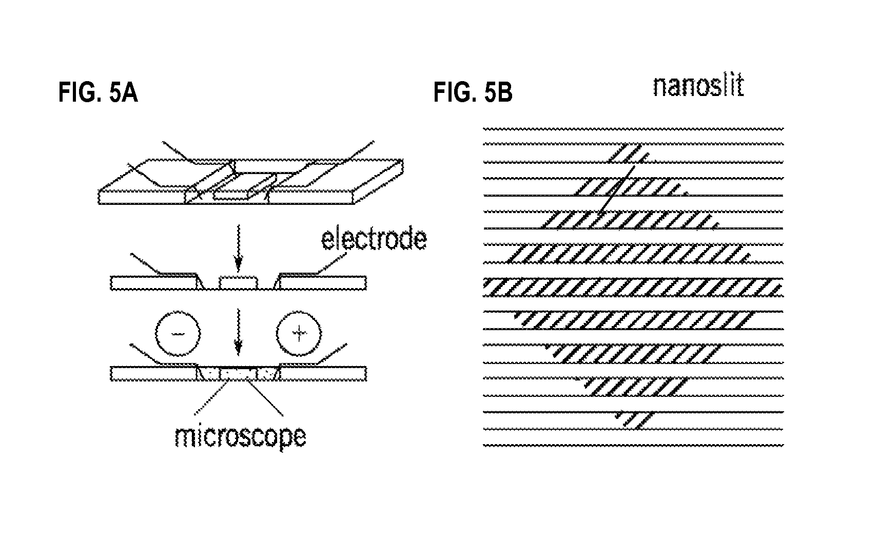
[0035]Fabrication of nanochannel devices followed standard soft lithography techniques known to one skilled in the art. Whitesides G, et al., “Soft lithography in biology and biochemistry,” Annu. Rev. Biomed. Eng. 3:335-373 (2001). To begin, a chrome mask, created with e-beam lithography at the University of Wisconsin Center for nanotechnology, was used as a mask in photolithography on negative photoresist spin-coated onto silicon wafers, creating an array of 1 μm channels. The wafer was etched to a depth of 100 nm by CF4 gas. Photoresist was lifted off by piranha solution. The height of the pattern was determined by an alpha step profilometer and the width of the pattern was measured under scanning electron microscope. After fabrication of the nanochannels, a microchannel array was overlaid on the nanopatterned wafer. SU-8 2005 photoresist was used to create channels 5 μm high and 100 μm wide. FIG. 5 shows the nanochannels (diagonal lines; 100 nm ...

example 2

Sequence-Specific Labeling of Nicked DNA

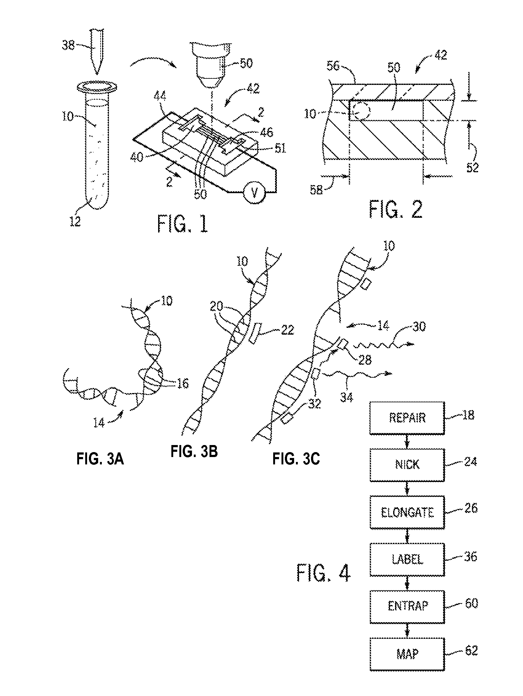
[0037]Sequence-specific labeled DNA was the result of the following steps: (1) linearization, (2) ligation, (3) nick site blocking, (4) nick translation, and (5) protein digestion. Linearization is required only when using circular DNA. Regardless of whether linearization was required, DNA ligase was used to remove inherent nicks in DNA. A DNA ligase reaction with T4 DNA ligase was performed at room temperature overnight. An overnight reaction ensures that ligation is complete and kills DNA ligase activity. To further ensure that and all inherent nicks were removed, DNA polymerase I (10 units) was added with dideoxy nucleotides (ddNTPs; 0.2 μM each) for 30 minutes at 37° C. ddNTPs incorporated into nicks block additional polymerase reaction and avoid random labeling. A nicking enzyme (Nb.BbvCI, 20 units; GC^TGAGG) was then added with deoxynucleotides (dNTPs), such as ALEXA FLUOR 647-aha-dCTP fluorescent marker (2 μM), ALEXA FLUOR 647-aha-dUTP ...

example 3

Elongation of Labeled DNA

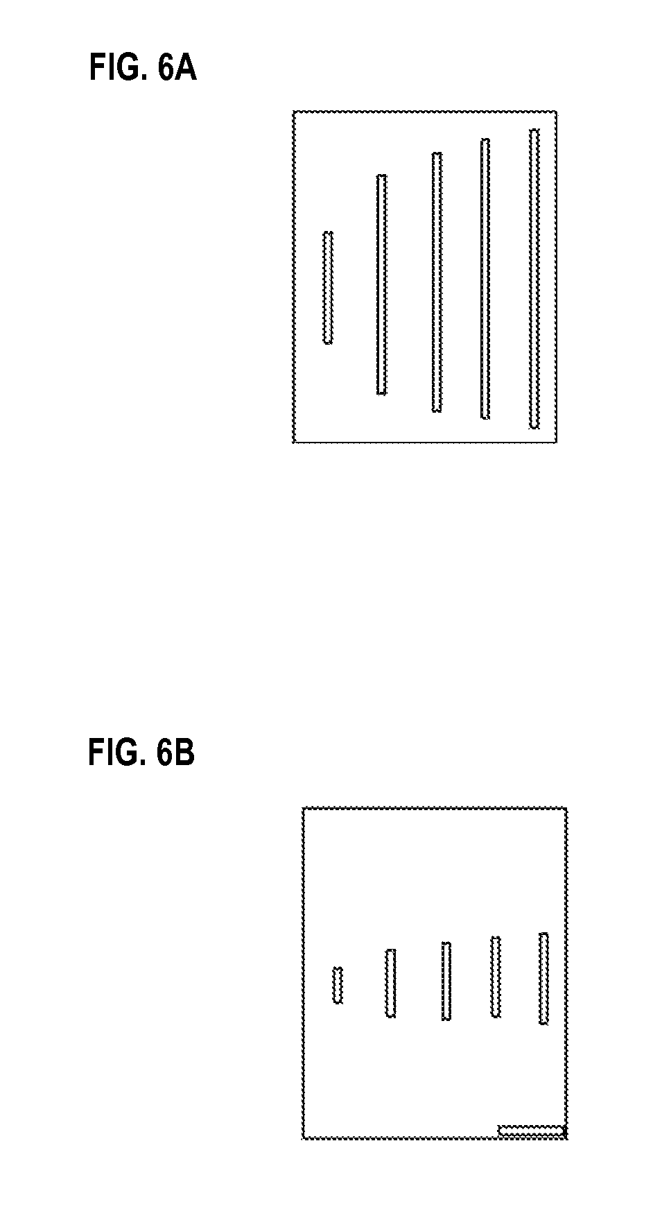
[0040]As shown in FIGS. 6a and 6b, diminished ionic strength buffer conditions increased intrachain electrostatic repulsion of DNA in the nanochannels, thereby increasing persistence length. Specifically, FIGS. 6a and 6b show typical images of T4 DNA (166 kbp; polymer contour length, 74.5 μm) and λDNA (48.5 kbp; polymer length, 21.8 μm) elongated within nanochannels under low ionic strength buffer conditions (0.05×TE and 0.01×TE) showing apparent lengths of 40.9±8.4 μm (T4 DNA) and 13.0±2.4 μm (λDNA).

[0041]As shown in FIG. 7, DNA elongation is a function of the dilution of TE (1 / TE) reported as “stretch,” and defined as the average apparent length, X, divided by the dye adjusted polymer contour length, L (e.g., L=21.8 for λDNA); a stretch value of 1.0 indicates complete elongation. FIG. 7 also shows that the degree of DNA elongation is size independent and inversely related to salt concentration or ionic strength used for the calculation of persistence by th...


PUM

PropertyMeasurementUnit
widthaaaaaaaaaa
widthaaaaaaaaaa
heightaaaaaaaaaa

Abstract

Methods are provided for tagging, characterizing and sorting double-stranded biomolecules while maintaining the integrity of the biomolecules.

Description

CROSS-REFERENCE TO RELATED APPLICATIONS[0001]This application claims the benefit of U.S. Provisional Patent Application Nos. 60 / 740,583, filed Nov. 29, 2005; and 60 / 740,693, filed Nov. 30, 2005, each of which is incorporated herein by reference as if set forth in its entirety.STATEMENT REGARDING FEDERALLY SPONSORED RESEARCH OR DEVELOPMENT[0002]This invention was made with United States government support awarded by the following agencies: NIH HG000225 and NSF 0425880. The United States has certain rights in this invention.BACKGROUND OF THE INVENTION[0003]The present invention relates to methods of presenting polymeric biomolecules such as nucleic acids for analysis, and in particular, to methods for tagging and elongating the biomolecules for characterization, sorting and analysis or other use.[0004]A single DNA molecule can be elongated, for example, by shear forces of liquid flow, by capillary action or by convective flow, and then fixed in an elongated form to a substrate by elec...

Claims


Application Information

Patent Timeline
Patent Type & Authority Patents(United States)
IPC IPC(8): C12Q1/68C12M1/34C07H21/04G01N33/53
CPCB82Y15/00B82Y30/00C12Q1/6816C12Q1/6837C12Q1/6869B01L3/5027C12Q2537/143C12Q2565/631C12Q2527/137C12Q2521/307C12Q2565/601Y10S436/80
Inventor SCHWARTZ, DAVID C.JO, KYUBONGDHINGRA, DALIA M.
Owner WISCONSIN ALUMNI RES FOUND
Why Eureka
Try Eureka