Nuclear magnetic resonance screening method
a technology of nuclear magnetic resonance and screening method, which is applied in the direction of nmr measurement, nmr spectroscopy measurement, instruments, etc., can solve the problems of limiting experimental work, requiring several months of experimental work and data analysis, and a lot of experimental work and data achieved
- Summary
- Abstract
- Description
- Claims
- Application Information
AI Technical Summary
Benefits of technology
Problems solved by technology
Method used
Image


Examples
example 1
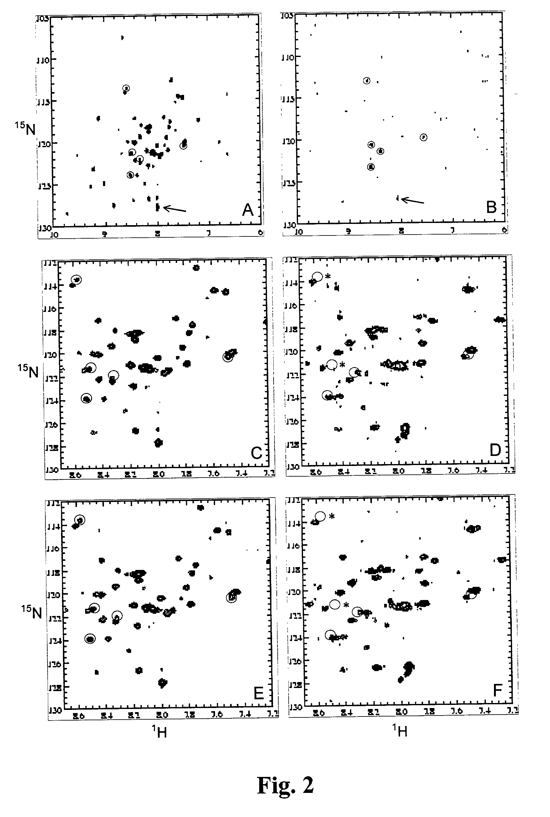
[0055] Screening for Binding to the Y-Loop of Protein Tyrosine Phosphatase-1B (PTP1B) (SEQ ID NO:1)
[0056] PTP1B (35 kD, single domain protein] is a protein that dephosphorylates phosphotyrosines. The active site binding cleft is centered around an active cysteine residue. It has been speculated that the so-called Y-loop of PTP1B is important for binding of some ligands. The Y-loop contains a sequence motif that is unique in the sequence, i.e. Arg47-Asp48 (FIG. 1). A selective labeled sample of PTP1B was prepared to monitor binding to this site.
[0057] Site specific labeled PTP1B (residues 1-298) was prepared from transformed Escherichia coli strain BL21 (DE3) cells. Bacteria were grown in rich medium containing all amino acids and 5 nucleotides according to the protocol described by Muchmore et al. [Muchmore, 1989] Aspartate and arginine were supplied .sup.15N-enriched and .sup.13C-enriched, respectively. It should be noted that this protocol (i.e. the use of a prototrophic bacterial...
example 2
[0065] Site Selective Screening of Human Muscle Fatty Acid Binding Protein (M-FABP)(SEQ ID NO:2).
[0066] The principle of the site-selective screening method is demonstrated for the human muscle fatty acid binding protein M-FABP. A 1.4 .ANG. X-ray crystal structure of M-FABP in complex with a ligand, oleic acid, is available (PDB ID code: 1 HMS). [Young, 1994] The structure consists of 10 anti-parallel .beta.-strands and two .alpha.-helices that connect .beta.-strands 1 and 2. The .beta.-strands form two nearly orthogonal .beta.-sheets. The fatty acid binding site is situated in a cavity between the two .beta.-sheets. The cavity is also lined by residues from helices 1 and 2. The carboxylate group of the fatty acid forms hydrogen bonds (direct and solvent mediated) to the protein. The aliphatic tail of the fatty acid adopts a u-shaped conformation in the highly hydrophobic cavity. Alanine 33 makes contacts with carbons C12, C13 and C14 of the oleic acid. [Young, 1994] Valine 32 and a...
PUM
 Abstract
Description
Claims
Application Information