
Method for identifying biologically active structures of microbial pathogens

Inactive Publication Date: 2004-07-08
SAHIN UGUR +2
View PDF3 Cites 11 Cited by
  • Summary
  • Abstract
  • Description
  • Claims
  • Application Information

AI Technical Summary

Benefits of technology

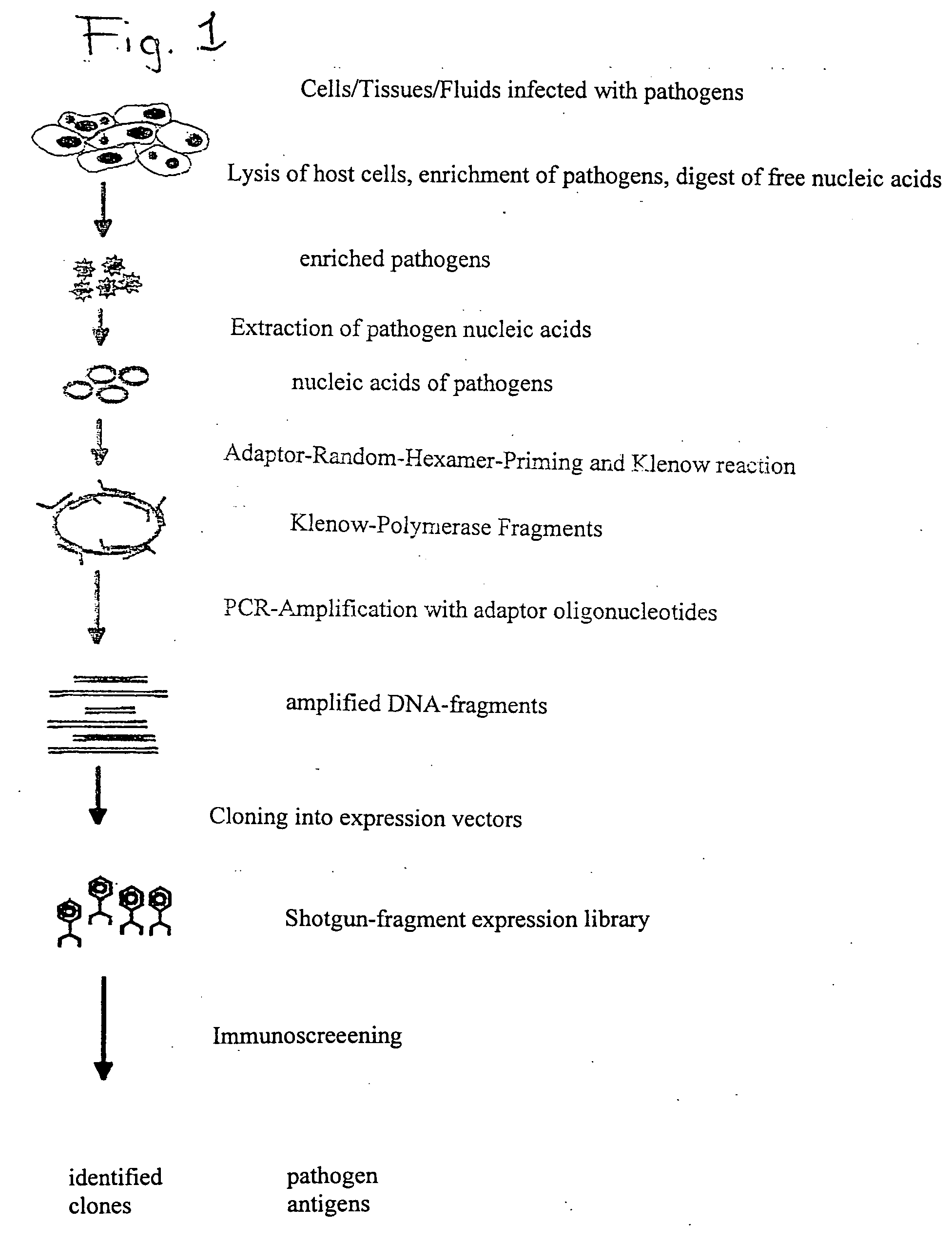
[0019] The method according to the invention has the decisive advantage that a comprehensive identification of pathogen antigens recognized by the host organism (microbial immunone) is possible even for very small amounts of pathogens.
[0092] Vaccination against variola major with sub-unit vaccines. Vaccination with the vaccinia virus provides good protection against the smallpox pathogen. Individual vaccinia virus antigens are therefore potential candidates for the induction of immunological protection against the smallpox pathogen.

Problems solved by technology

Although most relevant antigens have been identified molecularly in the case of low-complex infection pathogens with a known genome (e.g. simple viruses), it is still not clear which antigens are immunologically relevant in the case of more complex infection pathogens (e.g. bacteria).
The reason for this is that the complex genomes of these pathogens contain a large number of genes (1000 to over 4000), which makes a quick identification of the relevant antigens difficult.
This method has a number of limitations and disadvantages, since large amounts of pathogenic material are needed for the analyses.
Analysis is not possible directly from the primary lesion, but only after often very time-consuming culturing (e.g. in the case of mycobacteria).
Some pathogens cannot be cultured in a simple manner; for unknown pathogens, culturing conditions are often not defined.
Another disadvantage of the 2-D gel technology is that the gene expression status of a pathogen in a cell culture is clearly different from that in vivo.
The disadvantage of this method is that only proteins which are known to be expressed in the pathogen of interest can be analyzed.
Since the aforementioned analytical technologies require a great amount of material, time, staff and costs, they are reserved for only a few large centres.
Consequently, the method has hitherto been limited to pathogens whose culturing and purification modalities are known and established.
One problem is to identify infection pathogens which have so far not been characterized or only insufficiently characterized.
1) Inflammatory diseases whose cause, based on epidemiology and their clinical course, is likely to be an infection whose pathogens cannot be defined and / or only insufficiently characterized by means of known methods. This includes diseases such as multiple sclerosis, Kawasaki's disease, sarcoidosis, diabetes mellitus, morbus Whipple, pityriasis rosea, etc. It would be desirable to have a method for these diseases that allows a systematic analysis for determining unknown infection pathogens from primary patient material such as lymph node biopsies.
2) Newly emerging infectious diseases. This includes infectious diseases caused by hitherto unknown or not well-characterized pathogens (e.g. HIV in the 80s) und which, for example by reason of a change in epidemiology, are suddenly the focus of clinical interest. From a medical and socioeconomical point of view, rapid pathogen identification, the development of corresponding diagnostics and, possibly, the production of vaccines are essential. Since, in general, establishing culturing conditions for non-characterized pathogens may take up to several years, it is highly desirable, in this case as well, to identify pathogens and pathogen antigens directly from the infected tissue; however, this cannot be done by using the known methods.

Method used


Image

  • Method for identifying biologically active structures of microbial pathogens
  • Method for identifying biologically active structures of microbial pathogens
  • Method for identifying biologically active structures of microbial pathogens

Examples


example 1

[0114] Isolation of Pathogen Nucleic Acids from Virus-infected Cells:

[0115] BSC40 cells were infected with 2.times.10.sup.6 pfu of vaccinia viruses. The infected cells were incubated for 24 hours at 37.degree. C. in a CO.sub.2 incubator and then harvested. The harvested cells were then homogenized and absorbed in a buffered medium. To separate virus particles from host cell fragments, the cell lysate absorbed by the medium was then treated with ultrasound. Coarse particulate structures were pelleted by centrifugation for 15 min at 3000 rpm. Following centrifugation, the supernatant was removed and the pellet discarded. In order to precipitate corpuscular particles in the supernatant, 2 mL of cooled supernatant were precipitated with 6% PEG6000 / 0,6M NaCl (1 h incubation on ice); the precipitate was then pelleted for 10 min by centrifugation at 10000.times.g. As the preceding steps should have yielded both a separation and a lysis of contaminating host cells, the precipitate was then ...

example 2

[0116] Infection with Vaccinia Virus and Obtaining Serum

[0117] Recombinant vaccinia virus with the glycoprotein of the vesicular stomatitis virus (VaccG) (Mackett et al. (1985), Science 227, 433-435.) was cultured on BSC40 cells and the virus concentration determined in a plaque assay. C57BL / 6 mice (Institute of Laboratory Animal Science, University of Zurich) were infected intravenously with 2.times.10.sup.6 pfu of VaccG. Blood samples of 200-300 .mu.l were taken from the mice on days 8, 16 and 30 following infection, and serum was obtained through centrifugation and stored at -20.degree. C.

[0118] After the mice had been immunized, the successful induction of anti-vaccinia antibodies was tested against VSV in a neutralization assay (Ludewig et al. 2000. Eur J Immunol. 30:185-196) to determine the best time to extract serum for the planned analyses. As shown in Table 1, the increase of both the total immune globulin and the IgG class reached its maximum as of day 16, so that this se...

example 3a

[0119] Global Amplification of Minimai Amounts of Pathogen Nucleic Acids

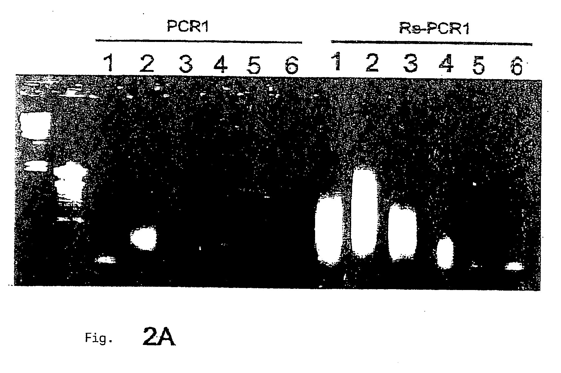
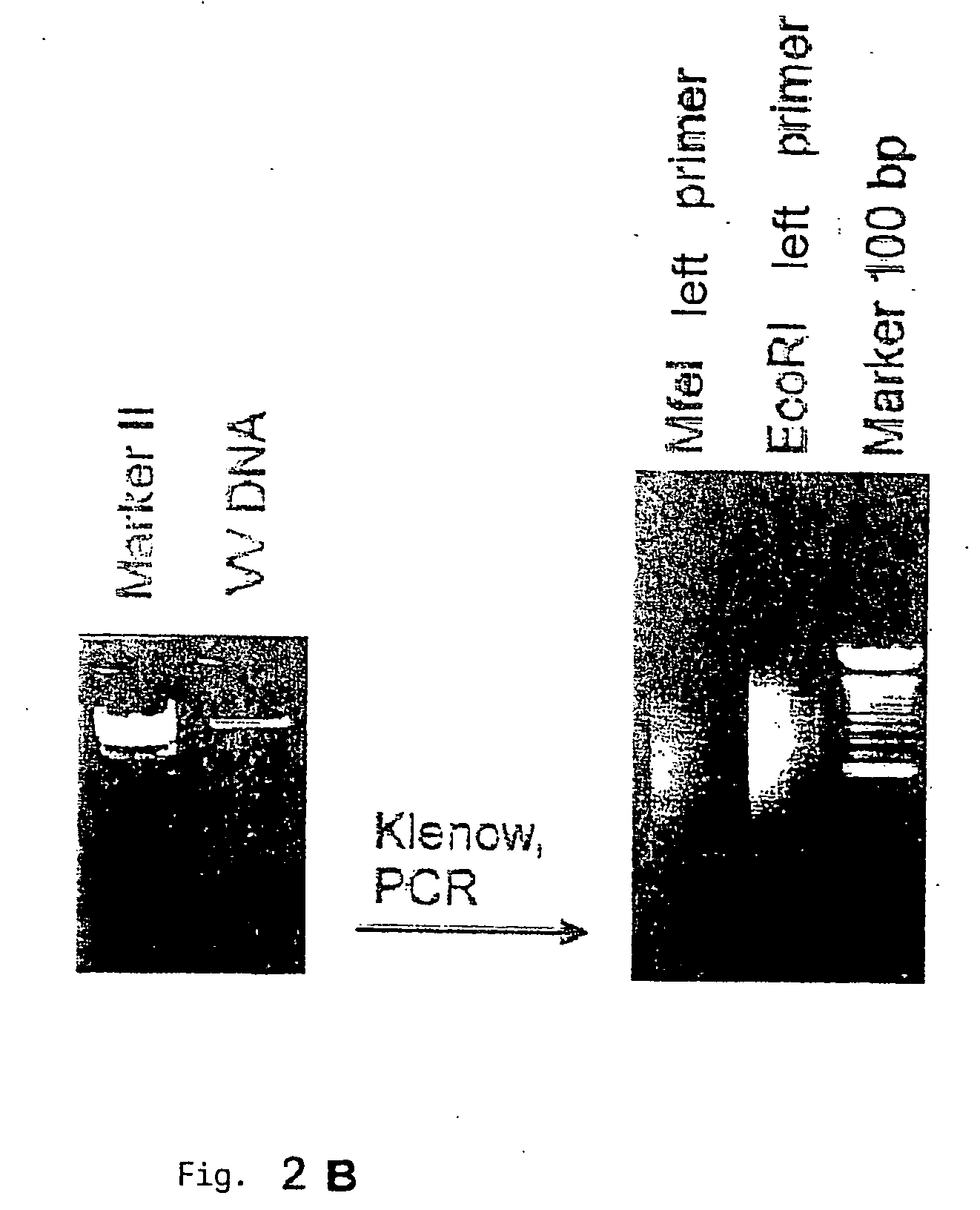
[0120] The global amplification of genomic nucleic acids of the pathogen is an essential step in the procedure according to the invention. The main challenge in this is to amplify in a comprehensive manner (i.e. including all the segments of the genome, if possible) the very small amount of genomic germinal nucleic acids which are isolated (without pre-culturing) from infected tissue. The amplified DNA must also be expressable and clonable for subsequent screening. While PCR-amplified cDNA-expression libraries are often described, produced and used (e.g. Edwards et al., 1991) and are sometimes commercialized as kits (e.g. SMART-cDNA library construction kit, Clontech), the establishment of comprehensive genomic libraries based on small amounts of pathogen nucleic acids (<10 ng) has hitherto not been described. It was therefore necessary to develop a DNA amplification module for the method according to the invent...


PUM

PropertyMeasurementUnit
Fractionaaaaaaaaaa
Fractionaaaaaaaaaa
Fractionaaaaaaaaaa

Abstract

The present invention concerns a method for identifying biologically active structures which are coded by the genome of microbial pathogens, using genomic pathogen nucleic acids.

Description

[0001] The invention described below concerns a procedure for identifying biologically active structures which are coded by the genome of microbial pathogens, on the basis of genomic pathogenic nucleic acids.[0002] A requirement for the development of molecularly defined serodiagnostic agents and vaccines is the molecular knowledge and availability of the antigens of the pathogenic agent (the microbial immunome) recognized by the immune system of an infected host. Serodiagnosis of infectious diseases is based on the detection of antibodies circulating in the blood, which are directed specifically against immunogenic components (antigens) of the pathogen and thus indicate an existing or recent infection. Knowledge of these antigens makes it possible to produce antigens through recombination as molecularly defined vaccines. These vaccines can give an organism protection against an infection caused by the pathogen in question (prophylactic immunization), but also, in the case of persis...

Claims


Application Information

Patent Timeline
IPC IPC(8): C07K14/07C12N15/09G01N33/50C12N15/10C12N15/39C12Q1/06C12Q1/68G01N33/15G01N33/569
CPCC07K14/005C12N2710/24122C12N15/1034
Inventor SAHIN, UGURTUERECI, OZLEMLUDEWIG, BURKHARD
Owner SAHIN UGUR
Learn More