
Oxidation reduction sensitive green fluorescent protein variants

a green fluorescent protein and variant technology, applied in the field of gene engineering, can solve the problems of inability to monitor redox changes within the same cell with this technique, and the current methods of determining in vivo redox status have many limitations, and can not be used in tissue culture, etc., to achieve the effect of reducing the number of redox changes, and improving the redox ra

Inactive Publication Date: 2004-09-02
STATE OF OREGON ACTING BY & THROUGH THE OREGON STATE BOARD OF HIGHER EDUCATION ON BEHALF OF THE OREGON HEALTH SCI UNIV
View PDF1 Cites 16 Cited by
  • Summary
  • Abstract
  • Description
  • Claims
  • Application Information

AI Technical Summary

Benefits of technology

0008] To overcome disadvantages of available methods for determining redox status in cells, GFP mutants (also referred to as redox sensitive GFP (rosGFP) variants) have been designed and are described herein, which

Problems solved by technology

Current methods of determining in vivo redox status have many limitations.
This type of procedure is not only very invasive but is also not a very accurate measure of the in vivo state of the cells.
Moreover, it would be impossible with this technique to monitor redox changes within the same cell over a period of time.
447:135-138, 1999) have developed an indicator of redox state in which glutathione reductase crystals were microinjected into the cytosol of human fibroblasts, and by detecting a color change of the crystals, they were able to determine the redox potential of the cytosol to be more reducing than -0.270 V. While this method may allow redox determination within single living cells, the cumbersome nature of the technique is still a major drawback.
However, is method still required harvesting of the cells and like all the other methods, it is very labor intensive.
Moreover, this technique determined redox potentials based only upon the ratio of reduced glutathione (GSH) to oxidized glutathione (GSSG), potentially ignoring other redox buffering components.

Method used


Image

  • Oxidation reduction sensitive green fluorescent protein variants
  • Oxidation reduction sensitive green fluorescent protein variants
  • Oxidation reduction sensitive green fluorescent protein variants

Examples


example 1

Selection and Design of Redox-Sensitive GFPs

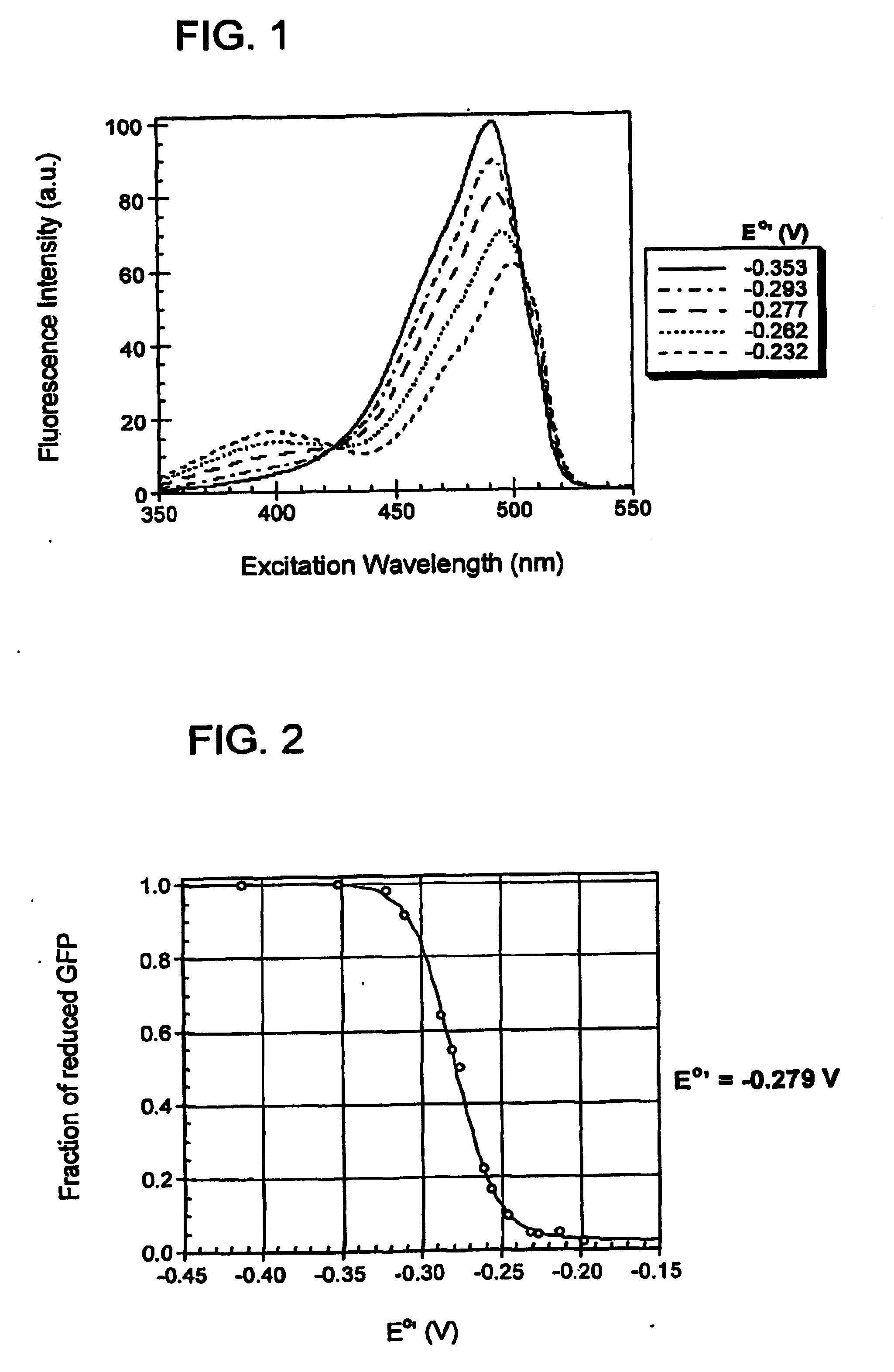
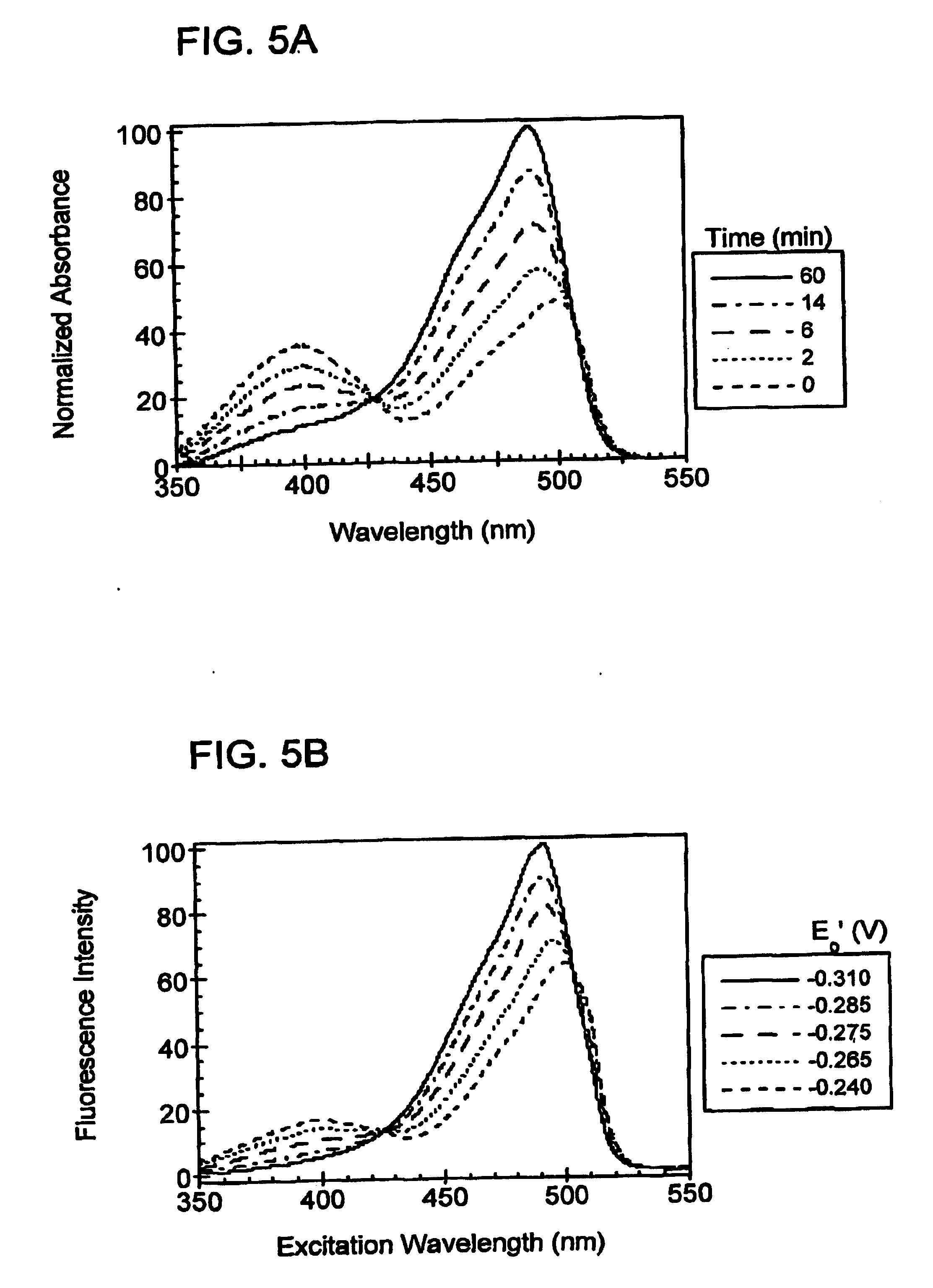
[0169] In order to create redox-sensitive GFPs, two cysteine residues were introduced into GFP that would be within disulfide bonding distance of each other, near the chromophore, and on the surface of the protein. The cysteine residues bring about fluorescence changes based upon whether they are reduced or oxidized. In addition, by being on the surface of the protein, the cysteines are solvent accessible, which enables them to reversibly respond to redox changes in the surrounding environment.

[0170] After close examination of the crystal structure of the S65T variant of GFP, two borderline suitable sites for the introduction of a pair of cysteines were chosen. The first site chosen was positions 147 and 204, while the second site chosen was positions 149 and 202. All four amino acid side-chains at these positions pointed away from the protein's interior. The distance between the C.sub..alpha.-C.sub..alpha.and C.sub..beta.-C.sub..beta. pos...

example 2

Construction and Expression of Redox Sensitive GFPs

[0171] Wild-type GFP contains two cysteine residues at positions 48 and 70. To avoid possible thiol / disulfide interchange reactions with the newly engineered cysteines, cysteine 48 and cysteine 70 were replaced with serine and alanine, respectively. Although the substitution C48S did not alter the properties of GFP, C70A appeared to be deleterious to obtaining soluble, fluorescent protein. Since position 70 is located within the interior of GFP and is in close proximity to the chromophore, it was deemed a mutation-sensitive position and was therefore left as a cysteine.

[0172] The C48S mutation was introduced into a histidine-tagged version of the S65T variant of GFP in the plasmid pRSET.sub.B. This construct served as the template for introduction of the cysteines at sites one (S147C / Q204C) and two (N149C / S202C). All mutations were introduced via the QuikChange.TM. Site-Directed Mutagenesis Kit (Stratagene, La Jolla, Calif.), follow...

example 3

Disulfide-Bond Formation and Redox Sensitivity of rosGFPs

[0176] Samples of rosGFP2 and GFP S65T (control) were treated with 1 mM DTT or 1 .mu.M CuCl.sub.2 and incubated at room temperature for 3-4 hours, then 2 mM N-ethyl maleimide was added to prevent disulfide exchange reactions. Molecular weights were determined by comparison to BenchMark.TM. protein ladder (InVitrogen, Carlsbad, Calif.). The gel was visualized with Coomassie blue stain.

[0177] Results

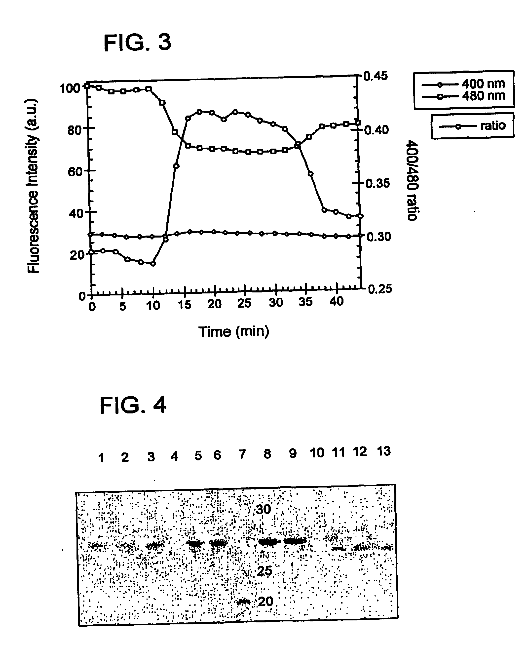
[0178] To verify that the introduced cysteines formed disulfide bonds and to show that the disulfide bonds were intramolecular, SDS-PAGE was run on redox-sensitive GFP #2 (rosGFP2), harboring the mutations C48S / S65T / S147C / Q204C, and on a control GFP (C48S / S65I) under reducing and non-reducing conditions. If an intramolecular disulfide bond forms, the resulting polypeptide would be expected to migrate further based on its slightly more compact structure in the denatured form. However, if an intermolecular disulfide bridge forms, then ...


PUM

PropertyMeasurementUnit
Sensitivityaaaaaaaaaa
Fluorescenceaaaaaaaaaa
Spectrumaaaaaaaaaa

Abstract

The disclosure provides proteins that can be used to determine the redox status of an environment (such as the environment within a cell or subcellular compartment). These proteins are green fluorescent protein (GFP) variants (also referred to as redox sensitive GFP (rosGFP) mutants), which have been engineering to have two cysteine amino acids near the chromophore and within disulfide bonding distance of each other. Also provided are nucleic acid molecules that encode rosGFPs, vectors containing such encoding molecules, and cells transformed therewith. The disclosure further provides methods of using the rosGFPs (and encoding molecules) to analyze the redox status of an environment, such as a cell, or a subcellular compartment within a cell. In certain embodiments, both redox status and pH are analyzed concurrently.

Description

FIELD[0002] The present disclosure relates to the field of genetic engineering, and in particular to green fluorescent protein (GFP) mutants that can be used to detect oxidation-reduction state, or a change in oxidation-reduction state.[0003] The green fluorescent protein (GFP) from the Pacific Northwest jellyfish, Aequorea victoria, has been used extensively in molecular and cell biology as a fluorescent marker. It is a 238 amino acid protein that generates its own fluorescent chromophore. The spontaneous generation of the chromophore is achieved by cyclization of the internal Ser65-Tyr66-Gly67 sequence followed by oxidation of Tyr 66 in the presence of molecular oxygen (Heim et at, Proc. Natl. Acad. Sci. USA 91:12501-12504, 1994). The overall fold of the protein consists of an 11-stranded .beta.-barrel capped by .alpha.-helices at both ends and contains a coaxial .alpha.-helix from which the chromophore is generated (Brejc et al., Proc. Natl. Acad Sci. USA 94:2306-2311, 1997; Orm{...

Claims


Application Information

Patent Timeline
IPC IPC(8): C07K14/435C12N5/02
CPCC07K14/43595
Inventor REMINGTON, S. JAMESHANSON, GEORGE T.
Owner STATE OF OREGON ACTING BY & THROUGH THE OREGON STATE BOARD OF HIGHER EDUCATION ON BEHALF OF THE OREGON HEALTH SCI UNIV
Learn More