
Use of protein biomolecular targets in the treatment and visualization of brain tumors

Inactive Publication Date: 2005-04-07
MEDAREX LLC
View PDF17 Cites 4 Cited by
  • Summary
  • Abstract
  • Description
  • Claims
  • Application Information

AI Technical Summary

Benefits of technology

[0050] The present invention provides novel methods and reagents for specifically targeting brain tumor neoplastic cells for both therapeutic and imaging purposes, by targeting brain tumor protein targets (TBT). These targets have been identified by the applicants as being overexpressed in brain tumors, and thus allow for the selective inhibition of cell function or selective marking for visualization with therapeutic or visualizing compositions which have a specific affinity for thes eprotein targets. Each of angiopoietin related protein 2 (ARP-2,) secreted protein acidic, rich in cysteine (SPARC,) c-met

Problems solved by technology

First and foremost, the intracranial space and physical layout of the brain create significant obstacles to treatment and recovery.
Because of the relatively small volume of the skull cavity, minor changes in the volume of tissue in the brain can dramatically increase intracranial pressure, causing damage to the entire organ (i.e., “water on the brain”).
Thus, even small tumors can have a profound and adverse affect on the brain's function.
The cramped physical location of the cranium also makes surgery and treatment of the brain a difficult and delicate procedure.
However, because of the dangers of increased intracranial pressure from the tumor, surgery is often the first strategy of attack in treating brain tumors.
However, it also prevents therapeutic molecules, including many chemotherapeutic agents that are useful in other types of tumors, from crossing into the brain.
However, the amount of radiation necessary to completely destroy potential tumor-producing cells also produce unacceptable losses of healthy brain tissue.
The retention of patient cognitive function while eliminating the tumor mass is another challenge to brain tumor treatment.
Thus, it is impossible to define a true “tumor margin,” unlike, for example, in lung or bladder cancers.
Unlike reproductive (ovarian, uterine, testicular, prostate, etc.), breast, kidney, or lung cancers, the entire organ, or even significant portions, cannot be removed to prevent the growth of new tumors.
This pervasive and variable nature greatly adds to the difficulty of treating brain tumors while preserving the health and function of normal brain tissue.
Although current surgical methods offer considerably better post-operative life for patients, the current combination therapy methods (surgery, low-dosage radiation, and chemotherapy) have only improved the life expectancy of patients by one month, as compared to the methods of 30 years ago.
Without effective agents to prevent the growth of brain tumor cells that are present outside the main tumor mass, the prognosis for these patients cannot be significantly improved.
Although some immuno-affinity agents have been proposed and tested for the treatment of brain tumors, see, e.g., the tenascin-targeting agents described in U.S. Pat. No. 5,624,659, these agents have not proven sufficient for the treatment of brain tumors.
Some tyrosine kinases (PTK's) and phosphatases (PTPase's) have been theorized to have a role in some types of oncogenesis, which is thought to result from an imbalance in their activities.
Results suggested that PTN signals through “ligand-dependent receptor inactivation” of PTPζ and disrupts its normal roles in the regulation of steady-state tyrosine phosphorylation of downstream signaling molecules.

Method used


Image

  • Use of protein biomolecular targets in the treatment and visualization of brain tumors
  • Use of protein biomolecular targets in the treatment and visualization of brain tumors
  • Use of protein biomolecular targets in the treatment and visualization of brain tumors

Examples


example 1

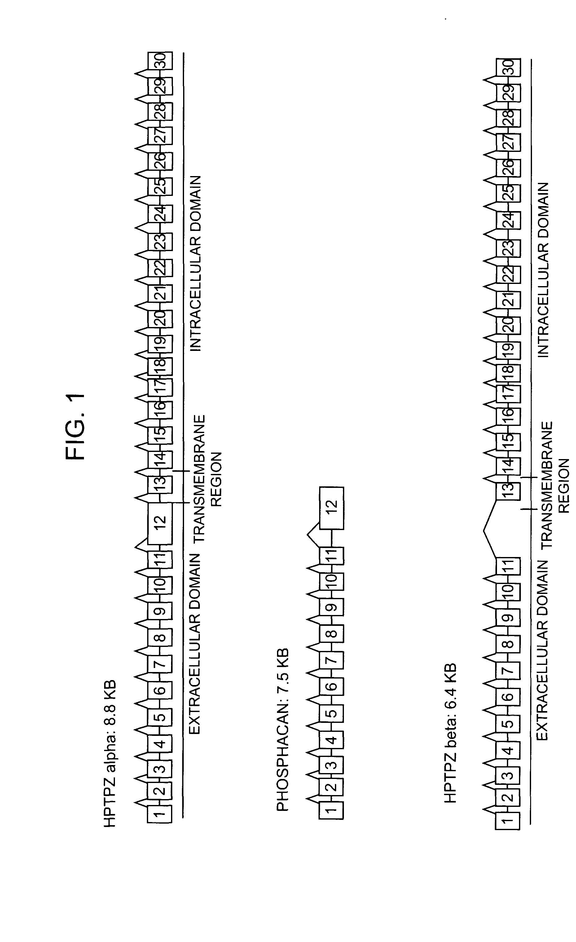
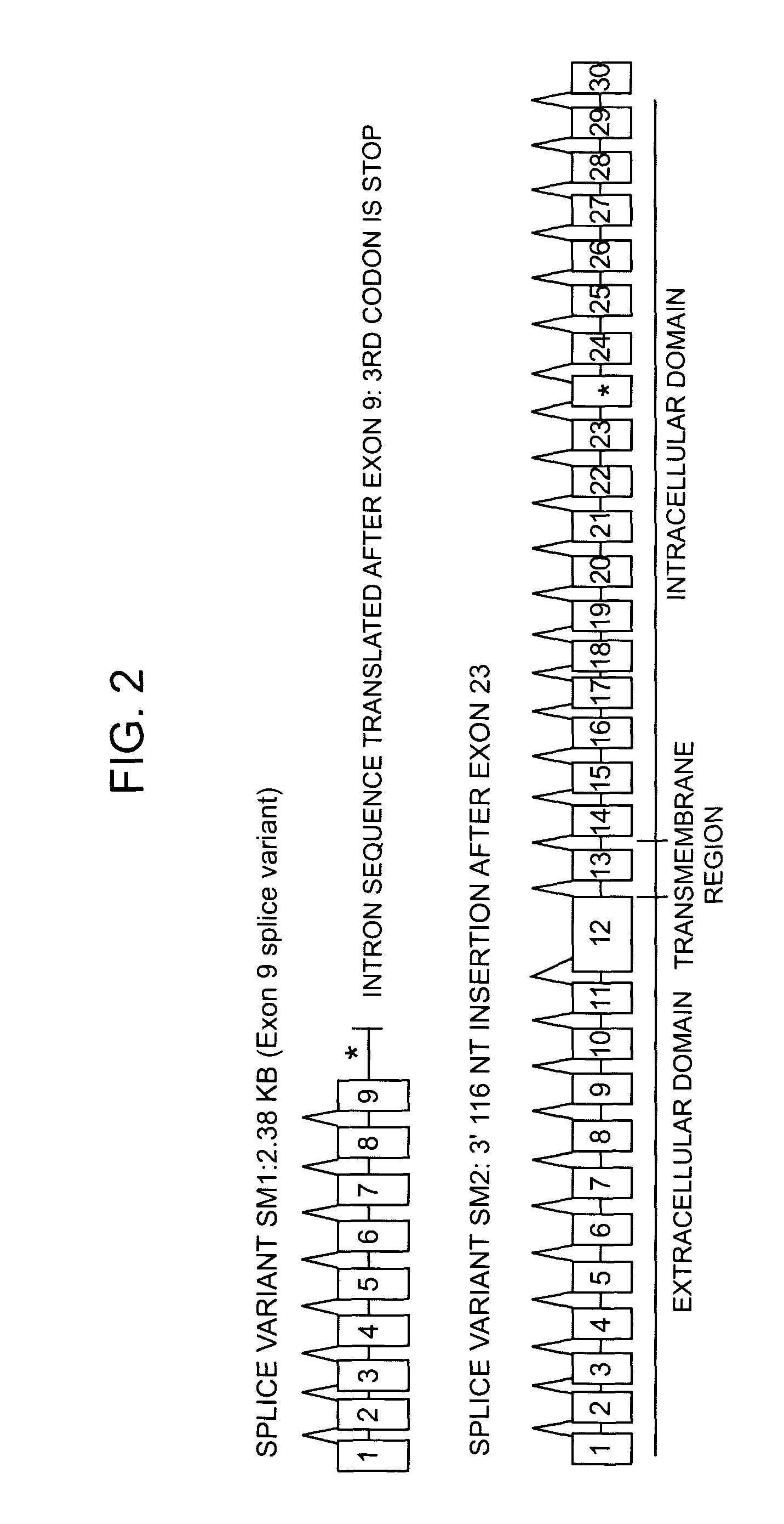
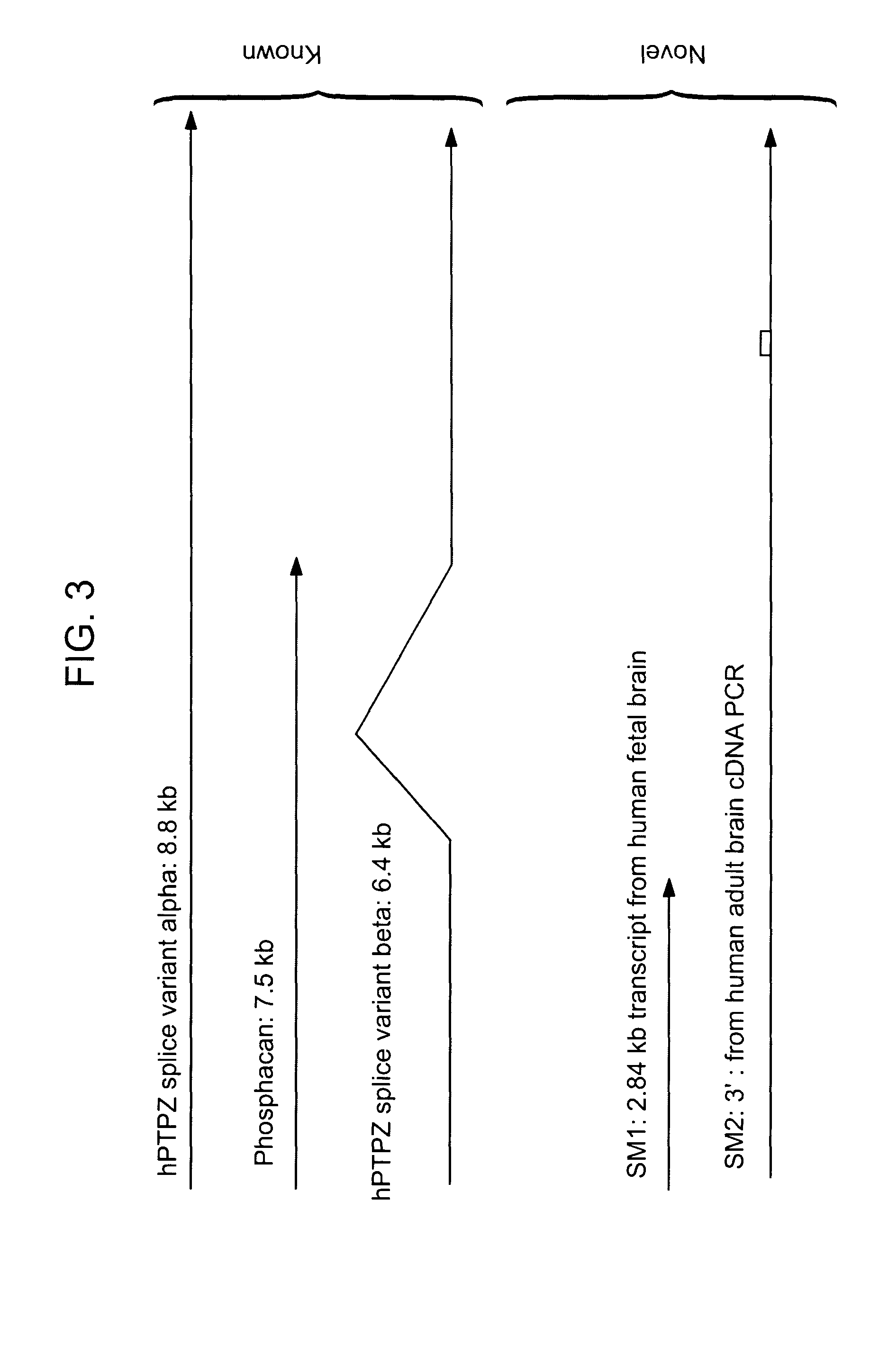
Identification of Two New Splicing Variant Isoforms of PTPζ: PTPζ SM1 and SM2

[0276] The mRNA nucleotide sequence for PTPζ SM1 was identified in a human fetal brain phage cDNA library by sequencing.

[0277] The mRNA nucleotide sequence for PTPζ SM2 was identified by PCR amplification of adult human brain cDNA, and sequencing of the resulting nucleic acids.

[0278] For the RT-PCR analyses performed below, total RNA was isolated from either cells (glioblastoma cultured lines) or tissue using Trizole (Gibco Life Technologies, Inc.), following the manufacture's protocol. cDNA was generated from total RNA using the 1st Strand synthesis kit from Gibco Life Technologies, Inc., and an oligo dT30 anchored primer. For each RT-PCR reaction, 1 μl of cDNA was utilized. The PCR reaction was carried out using an Advantage 2 kit (Clontech) under standard conditions. The products of the PCR reactions were confirmed via sequencing.

[0279] Both clones were verified by RT-PCR analysis of glioblastoma cel...

example 2

HUVEC(Human Umbilical Vein Endothelial Cells) Endothelial Sprouting assay For Determining Antibody Activity on Protein Targets

[0288] Cell-sprouting morphology can be utilized as an easily visualized assay to determine the inhibitory effect of a candidate antibody on the protein target function for protein targets which stimulate endothelial cell sprouting, such as ARP2. Such assays have been described extensively in the literature (Nehls, V., et al., Histochem. Cell Biol. 104: 459-466 (1995); Koblizek, T. I., et al., Curr. Biol. 8: 529-532 (1988); and Kwak, H. J., et al., FEBS Lett. 448: 249-253). Briefly, a endothelial cells from a suitable source, such as HUVECs or PPAECs (porcine pulmonary artery endothelial cells) are grown to confluence on microcarrier (MC) beads (diameter 175 μm, available from Sigma) and placed into a 2.5 mg / ml fibrinogen gel containing the protein target at an appropriate effective concentration (200 ng / ml is an suitable starting concentration, which the sk...


PUM

PropertyMeasurementUnit
Fractionaaaaaaaaaa
Pharmaceutically acceptableaaaaaaaaaa
Affinityaaaaaaaaaa

Abstract

The present invention relates to the use of proteins which are differentially expressed in primary brain tumor tissues, as compared to normal brain tissues, as biomolecular targets for brain tumor treatment therapies. Specifically, the present invention relates to the use of immunotherapeutic and immunoimaging agents which specifically bind to one or more of human proteins angiopoietin related protein 2 (ARP-2,) secreted protein acidic, rich in cysteine (SPARC,) c-met proto-oncogene (C-MET,) brevican (BEHAB,) CD-44 antigen (CD-44,) tetraspanin 3 (TSPN3,) pleiotrophin (PTN,) osteopontin (OPN,) vasoactive intestinal peptide receptor-2 (VIPR-2,) and receptor protein tyrosine phosphatase zeta (PTPζ) for the treatment and visualization of brain tumors in patients. The present invention also provides compounds and pharmaceutically acceptable compositions for administration in the methods of the invention. The present invention also provides novel splice variants of protein PTPζ, PTPζ SM1 and PTPζ SM2. Nucleic acid probes specific for the spliced mRNA encoding these variants and affinity reagents specific for the novel proteins are also provided.

Description

FIELD OF USE [0001] The present invention relates to the use of proteins which are differentially expressed in primary brain tumor tissues, as compared to normal brain tissues, as biomolecular targets for brain tumor treatment therapies. Specifically, the present invention relates to the use of immunotherapeutic and immunoimaging agents which specifically bind to one or more of angiopoietin related protein 2 (ARP-2,) secreted protein acidic, rich in cysteine (SPARC,) c-met proto-oncogene (C-MET,) brevican (BEHAB,) CD-44 antigen (CD-44,) tetraspanin 3 (TSPN3,) pleiotrophin (PTN,) osteopontin (OPN,) vasoactive intestinal peptide receptor-2 (VIPR-2,) and receptor protein tyrosine phosphatase zeta (PTPζ) for the treatment and visualization of brain tumors in patients. The present invention also provides compounds and pharmaceutically acceptable compositions for administration. BACKGROUND OF THE INVENTION [0002] Brain Tumor Biology and Etiology [0003] Brain tumors are considered to have ...

Claims


Application Information

Patent Timeline
IPC IPC(8): A61K49/00
CPCA61K49/0002
Inventor MUELLER, SABINEMELCHER, THORSTENCHIN, DANIEL
Owner MEDAREX LLC
Learn More