Methods and systems for assessing biological material using optical detection techniques

a biological material and optical detection technology, applied in the field of optical or spectroscopic detection, can solve the problems of insufficient high throughput screening of biological materials, inability to accurately predict the response and side effects observed in human clinical trials, and inability to scale up to provide the high throughput screening necessary to test the numerous candidate compounds generated by traditional and computational means. , to achieve the effect of high throughpu

Inactive Publication Date: 2005-06-02
CYTOSCAN SCI
View PDF6 Cites 19 Cited by
  • Summary
  • Abstract
  • Description
  • Claims
  • Application Information

AI Technical Summary

Benefits of technology

[0021] An important application of the methods and systems of the present invention involves screening cell populations to assess the effect(s) of exposure to various types of test agents or test conditions, including drugs, hormones and other biological agents, toxins, infectious agents, physiological stimuli, radiation, chemotherapy, and the like. The effect of various test agents and conditions may be evaluated on both normal and pathological sample populations. Safety and cytotoxicity testing is conducted by exposing a sample population to a test agent or test condition and assessing the physiological state of the sample population using optical techniques at one or more time points following administration of the test agent or test condition. Such testing may be conducted on various sample populations to determine how a test agent or condition affects a desired target sample population, as well as to predict whether a test agent or condition produces physiological side effects on sample populations that are not the target of the test agent or condition.
[0022] According to a preferred embodiment, a disease state or compromised condition is simulated in biological materials prior to administration of a test agent or test condition to assess the suitability of the test agent or condition for treating the disease state or compromised condition. Exposure of sample populations to a physiological challenge, such as a change in extracellular osmolarity or ion concentration, altered oxygen or nutrient or metabolite conditions, drugs or diagnostic or therapeutic agents, a disturbance in ion homeostasis, electrical stimulation, inflammatio

Problems solved by technology

Screening methods currently used are generally difficult to scale up to provide the high throughput screening necessary to test the numerous candidate compounds generated by traditional and computational means.
Moreover, studies involving cell culture systems and animal model responses frequently don't accurately predict the responses and side effects observed during human clinical trials.
Conventional methods for assessing the effects of various agents or physiological activities on biological materials, in both in vitro and in vivo systems, generally are not highly sensitive or informative.
Cytotoxicity assays ge

Method used


Image

  • Methods and systems for assessing biological material using optical detection techniques
  • Methods and systems for assessing biological material using optical detection techniques
  • Methods and systems for assessing biological material using optical detection techniques

Examples


example 1

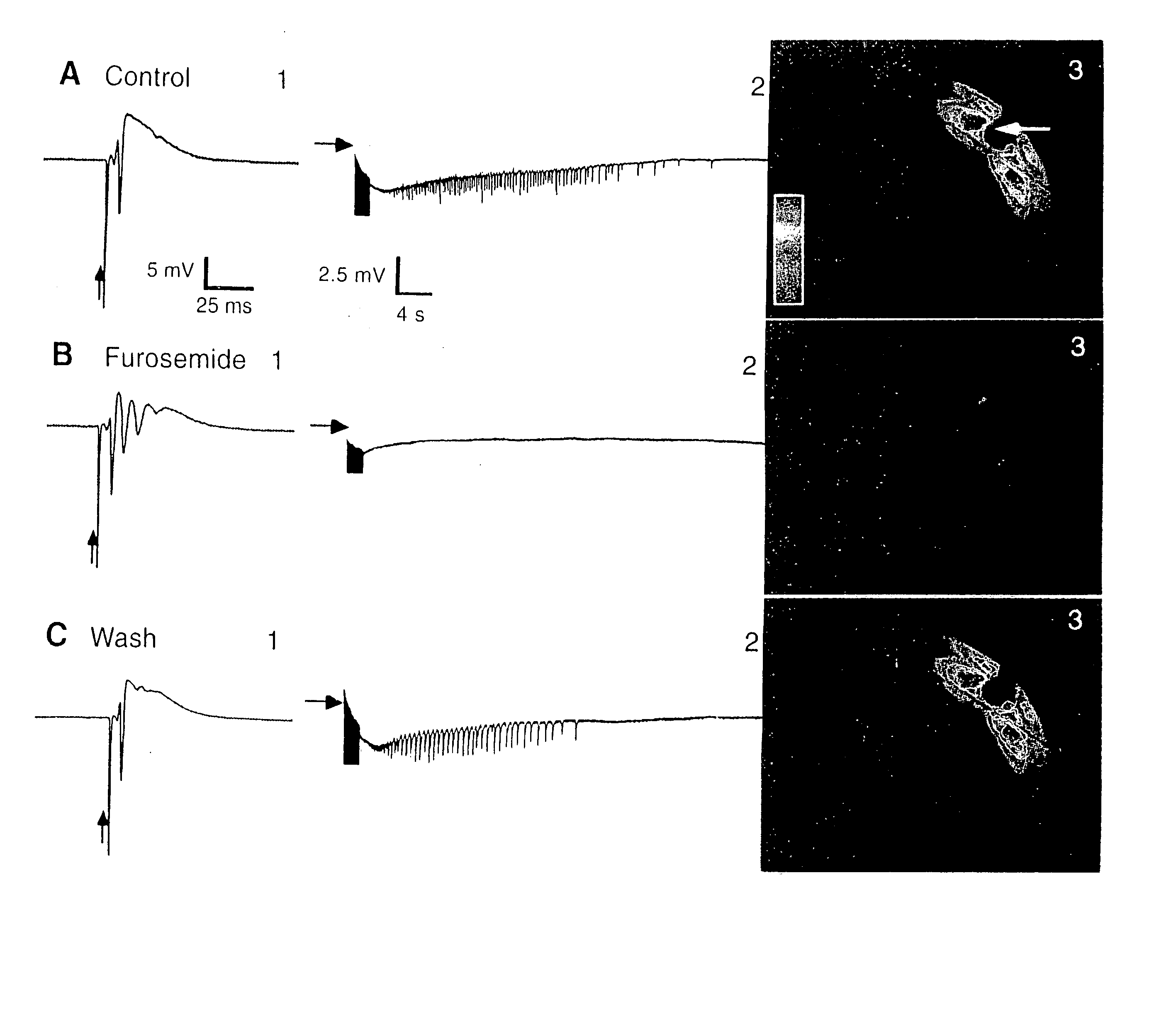
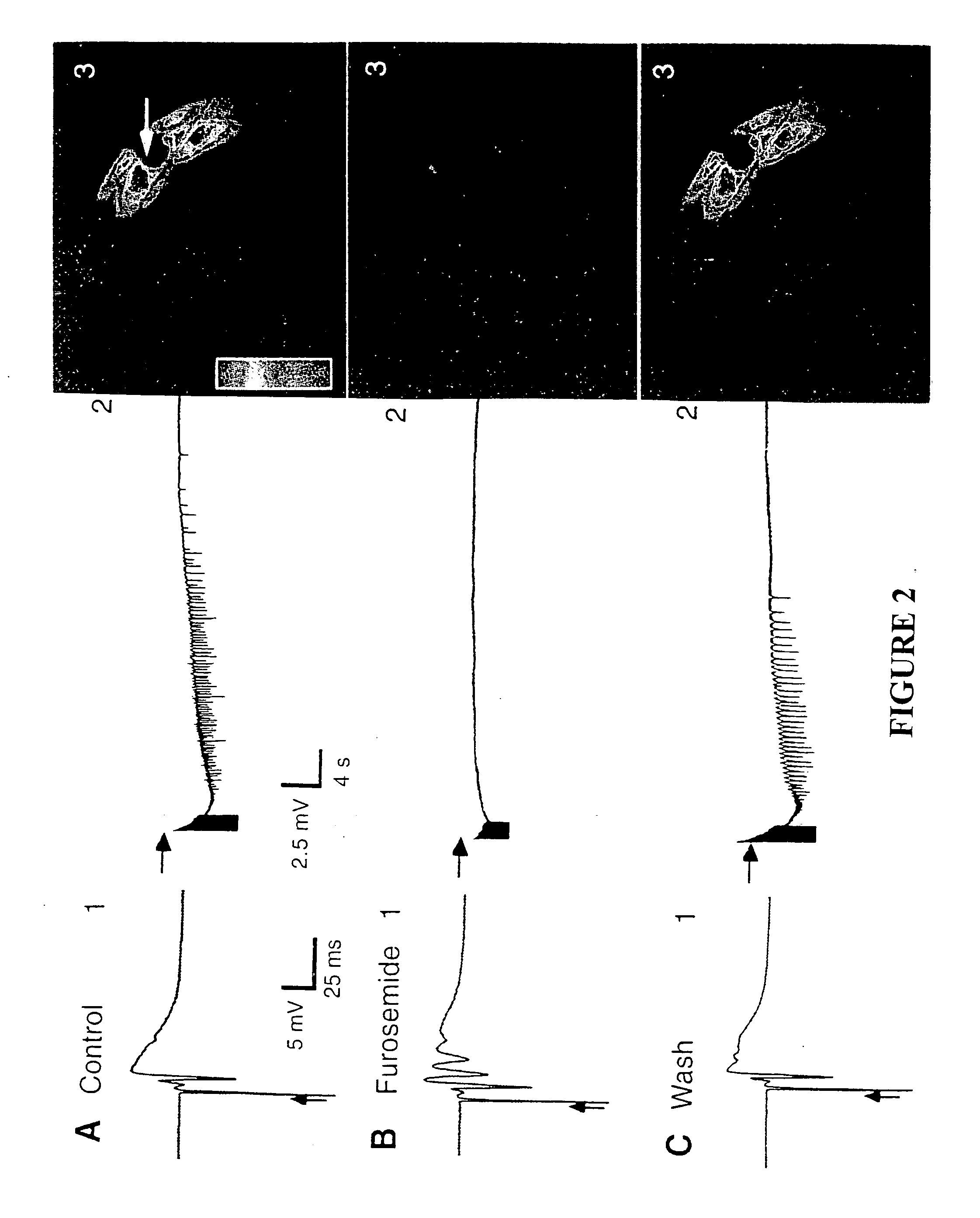
[0100] Sprague-Dawley rats (male and female; 25 to 35 days old) were prepared as described in Aghajanian, A. K. and Rasmussen, K., Synapse 31:331, 1989; and Buckmaster, P. S., Strowbridge, B. W., Schwartzdroin, P. A., J. Neurophysiol. 70:1281, 1993. In most hippocampal slice experiments, simultaneous extracellular field electrode recordings were obtained from CA1 and CA3 areas. For stimulation-evoked afterdischarge (13 slices, 8 animals), the concentration of Mg2+ in the bathing medium was reduced to 0.9 mM. A bipolar tungsten stimulating electrode was placed on the Schaffer collaterals to evoke synaptically driven field responses in CA1; stimuli consisted to 100 to 300-μs-duration pulses at an intensity of four times population-spike threshold. Afterdischarges were evoked by a 2-s train of such stimuli delivered at 60 Hz. Spontaneous interictal-like bursts were observed in slices treated with the following modifications or additions to the bathing medium: 10 mM K+ (6 slices; 4 anim...

example 2

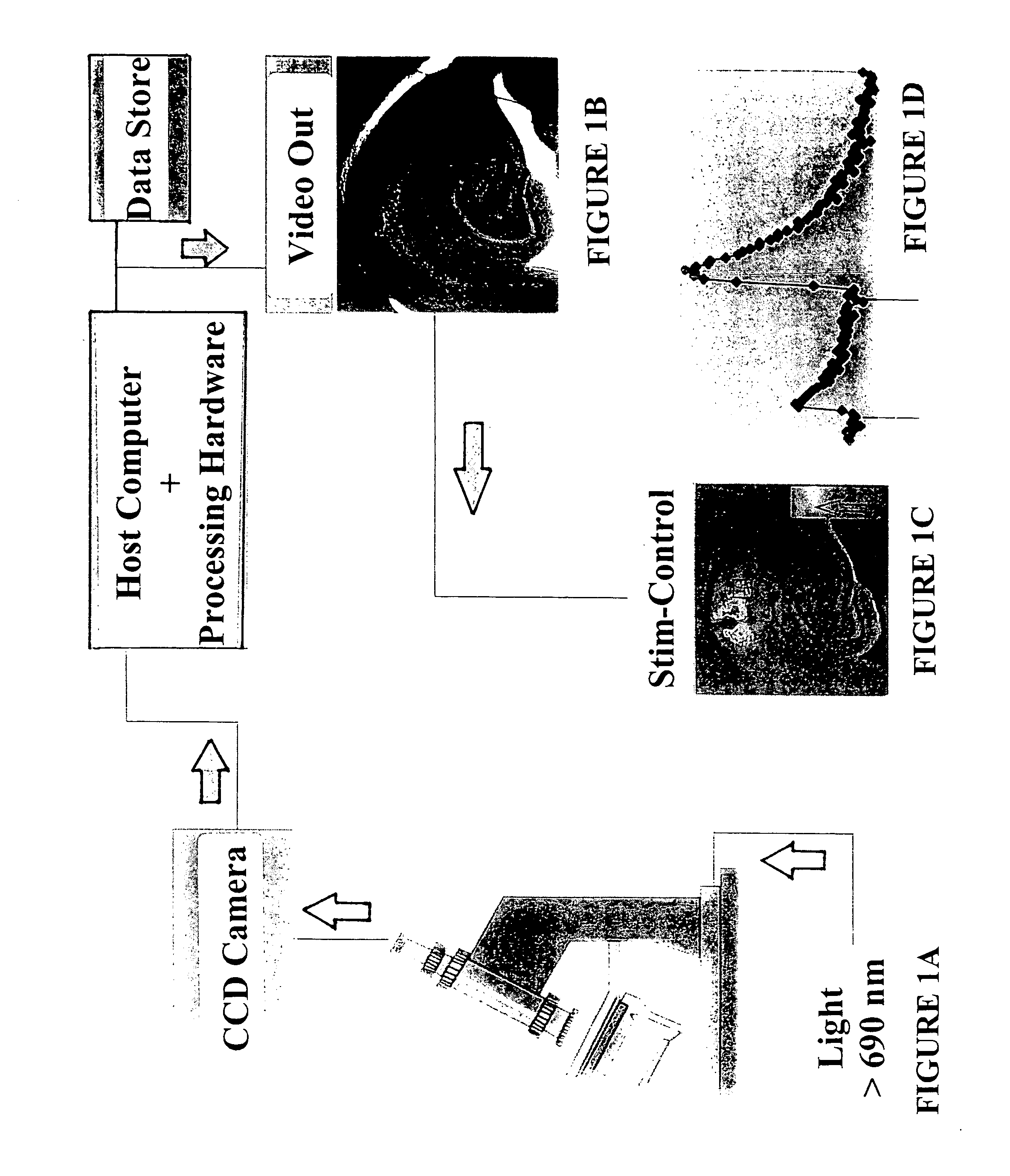
[0105] This example illustrates optical changes indicative of neuronal activity in a human subject by direct cortical electrical stimulation. Surface electrical recordings (surface EEG, ECOG) were correlated with optical changes. Intrinsic optical changes were evoked in an awake patient during stimulating-electrode “calibration.” Four stimulation trials were sequentially applied to the cortical surface, each stimulation evoking an epileptiform afterdischarge episode. A stimulation trial consisted of: (1) monitoring resting cortical activity by observing the output of the recording electrodes for a brief period of time; (2) applying an electric current via the stimulation-electrodes to the cortical surface at a particular current for several seconds; and (3) monitoring the output of the recording electrodes for a period of time after stimulation has ceased.

[0106] The cortex was evenly illuminated by a fiber optic emr passing through a beam splitter, controlled by a D.C. regulated po...

example 3

[0117] Stimulation mapping of the cortical surface was performed on awake human patients under local anesthesia to identify sensory / motor cortex and Broca's areas. The illumination source and optical detection device and processing techniques used were the same as those described in Example 2. During three “tongue wiggling” trials, images were averaged (32 frames, 1 sec) and stored every 2 seconds. A tongue wiggling trial consisted of acquiring 5-6 images during rest, then acquiring images during the 40 seconds that the patient was required to wiggle his tongue against the roof of his mouth, and then to continue acquiring images during a recovery period. The same patient was then required to engage in a “language naming” trial. A language naming trial consisted of acquiring 5-8 images during rest (control images—the patient silently viewing a series of blank slides), then acquiring images during the period of time that the patient engaged in the naming paradigm (naming a series of o...


PUM


Abstract

Optical detection techniques for the assessment of the physiological state, health and/or viability of biological materials are provided. Biological materials which may be examined using such techniques include cells, tissues, organs and subcellular components. The inventive techniques may be employed in high throughput screening of potential diagnostic and/or therapeutic agents.

Description

REFERENCE TO PRIORITY APPLICATIONS [0001] This application is a divisional application of U.S. patent application Ser. No. 09 / 326,244, filed Jun. 4, 1999, issuing as U.S. Pat. No. 6,834,238 on Dec. 21, 2004, which claims priority from U.S. Provisional Patent Application No. 60 / 088,494, filed Jun. 8, 1998, entitled METHODS AND APPARATUS FOR ASSESSING BIOLOGICAL MATERIALS USING OPTICAL DETECTION TECHNIQUES, which are hereby incorporated herein by reference.FIELD OF THE INVENTION [0002] The methods and systems of the present invention employ optical, or spectroscopic, detection techniques for assessing the health, physiological condition, and viability of biological materials such as tissues, cells, and subcellular components, and may be used in both in vitro and in vivo systems. One important application of the methods and apparatus of the present invention is high throughput screening of candidate agents and conditions to evaluate their suitability as diagnostic or therapeutic agents...

Claims


Application Information

Patent Timeline
IPC IPC(8): A61K31/00A61K31/635A61K45/06
CPCA61K31/00A61K31/635A61K45/06A61K2300/00
Inventor HOCHMAN, DARYL W.
Owner CYTOSCAN SCI
Why Eureka
Try Eureka