Eureka AIR delivers breakthrough ideas for toughest innovation challenges, trusted by R&D personnel around the world.

Device and method for controlled electroporation and molecular delivery into cells and tissue

a technology of molecular delivery and electroporation, which is applied in the field of devices and methods for controlling electroporation and molecular delivery into cells and tissues, can solve the problems of significant cell death, low transport efficiency of tissue-derived cells and other primary cells, and low overall efficiency of traditional electroporation, etc., and achieve accurate measurement and precise control of voltage applied.

Inactive Publication Date: 2005-08-04
HUANG YONG +2
View PDF6 Cites 59 Cited by
  • Summary
  • Abstract
  • Description
  • Claims
  • Application Information

AI Technical Summary

Benefits of technology

[0009] In the present invention, electrical current is directed to flow through biological cells, making it possible to accurately measure, and thus precisely control, the voltage over the cells—ensuring a uniform, homogeneous field applied to the cell or cell population. Since the present invention allows more precise control of the voltage applied to cells, it provides a means of ensuring that cells are not killed during electroporation.
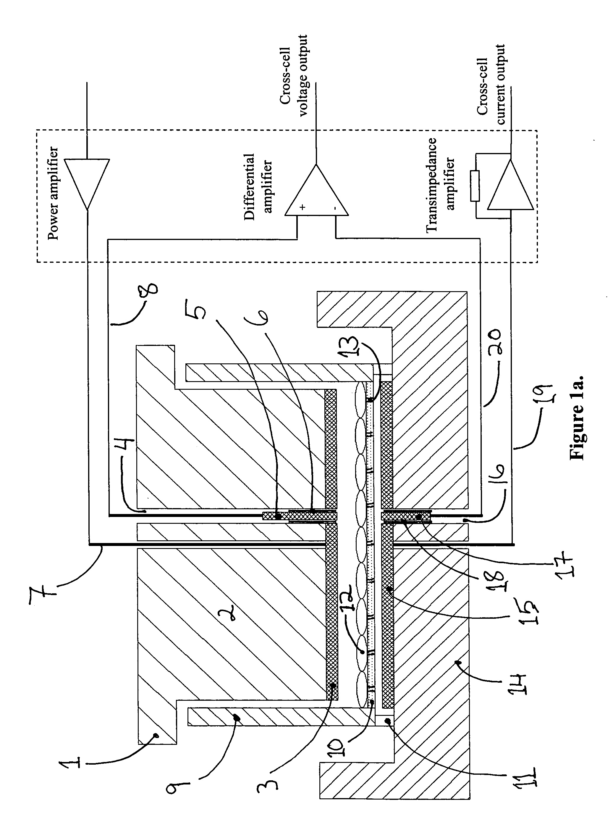
[0010] Furthermore, the voltages applied to the electroporation electrodes (3 and 15) in the present invention can be more than two orders of magnitude lower than those used in electroporation systems where the cells are in suspension. Since the cells present a large electrical impedance, the bulk impedance of the electrolyte becomes negligible, and most of the voltage applied to the electroporation electrodes (3 and 15) drops over the cells. This ‘focusing’ of the electric field permits application of electroporation voltages to the electroporation electrodes (3 and 15) that is very close to the actual cross-cell voltage required to initiate electroporation.(roughly between 0.3V and 1.0V). Lower voltages in turn reduce the complexity, size and cost of the power amplifier (21), and also allow electroporation pulses of arbitrary shape and duration without adding complexity to the power amplifier (21).
[0012] The traditional methods of mechanical cell delivery (electroporation, ballistics and microinjection) generally cause significant cell death due to irreversible membrane rupture. In the case of traditional electroporation (FIG. 18: EP1-EP4 indicate typical results), generally less than 50% of the cells survive the process and the transport efficiency is less than 50% for the remaining cells. For this case, traditional electroporation often has less than 25% overall efficiency. Chemical methods of cell delivery such as lipofection (FIG. 18: LF1-LF4 indicate typical results) show little immediate harm to cell viability, but these methods often have very low transport efficiency with tissue-derived cells and other primary cells, typically less than 30% overall efficiency. The controlled electroporation process typically produces greater than 80% overall efficiency, and in many cases greater than 90% overall efficiency—produced by the >90% cell viability and >80% transport efficiency of this process. This very high overall efficiency provides a new utility for cell engineering, particularly important for delicate cells and primary cells, in that the engineered cells can be studied without tedious and time consuming cell sorting to remove dead or unprocessed cells. Particularly because of the high cell viability of the controlled electroporation process, this method and device enables new research in areas where cells are precious or rare, or where speed and efficiency in processing is critical.
[0013] The controlled electroporation process and apparatus provides great benefit in the ability to observe transient transfection in hard-to-transfect cells, as the researcher can now observe gene expression within a few hours the transfection event. Without this technique, researchers may spend more than a month developing each stably-transfected cell type to ensure a consistent signal for cell detection; the other possibility is to spend days sorting the cells to isolate the transfected cells, racing against time to take data before the expression levels fade.
[0014] Primary cells greatly benefit from the process of controlled electroporation. For example, in the area of regenerative medicine, this process and device can be used to import genes, proteins and other material to induce differentiation or selective regeneration of stem cells, nerve cells and other critical primary cells of interest. These cells are useful both for research and development, but also may be used as a source for tissue generation for re-implantation and regeneration. As another example, blood products may be infused with drugs, proteins and other therapeutic compounds—then infused back into the body as cell-based drug carriers. In the case of blood products (including, but not limited to platelets, white blood cells and red blood cells), these cells often aggregate at the sites of trauma, tumors or blood clots. In this way, one may infuse the blood cells with therapeutics for targeted drug delivery, using the cells as natural targeting agents. Using the described process of controlled electroporation, the high level of efficiency in molecular delivery allows a robust and reliable process for targeted cell therapeutics, without significant loss in cells and without the need to sort or screen cells before use.

Problems solved by technology

The traditional methods of mechanical cell delivery (electroporation, ballistics and microinjection) generally cause significant cell death due to irreversible membrane rupture.
For this case, traditional electroporation often has less than 25% overall efficiency.
Chemical methods of cell delivery such as lipofection (FIG. 18: LF1-LF4 indicate typical results) show little immediate harm to cell viability, but these methods often have very low transport efficiency with tissue-derived cells and other primary cells, typically less than 30% overall efficiency.

Method used


Image

  • Device and method for controlled electroporation and molecular delivery into cells and tissue
  • Device and method for controlled electroporation and molecular delivery into cells and tissue
  • Device and method for controlled electroporation and molecular delivery into cells and tissue

Examples


Embodiment Construction

Device Configuration

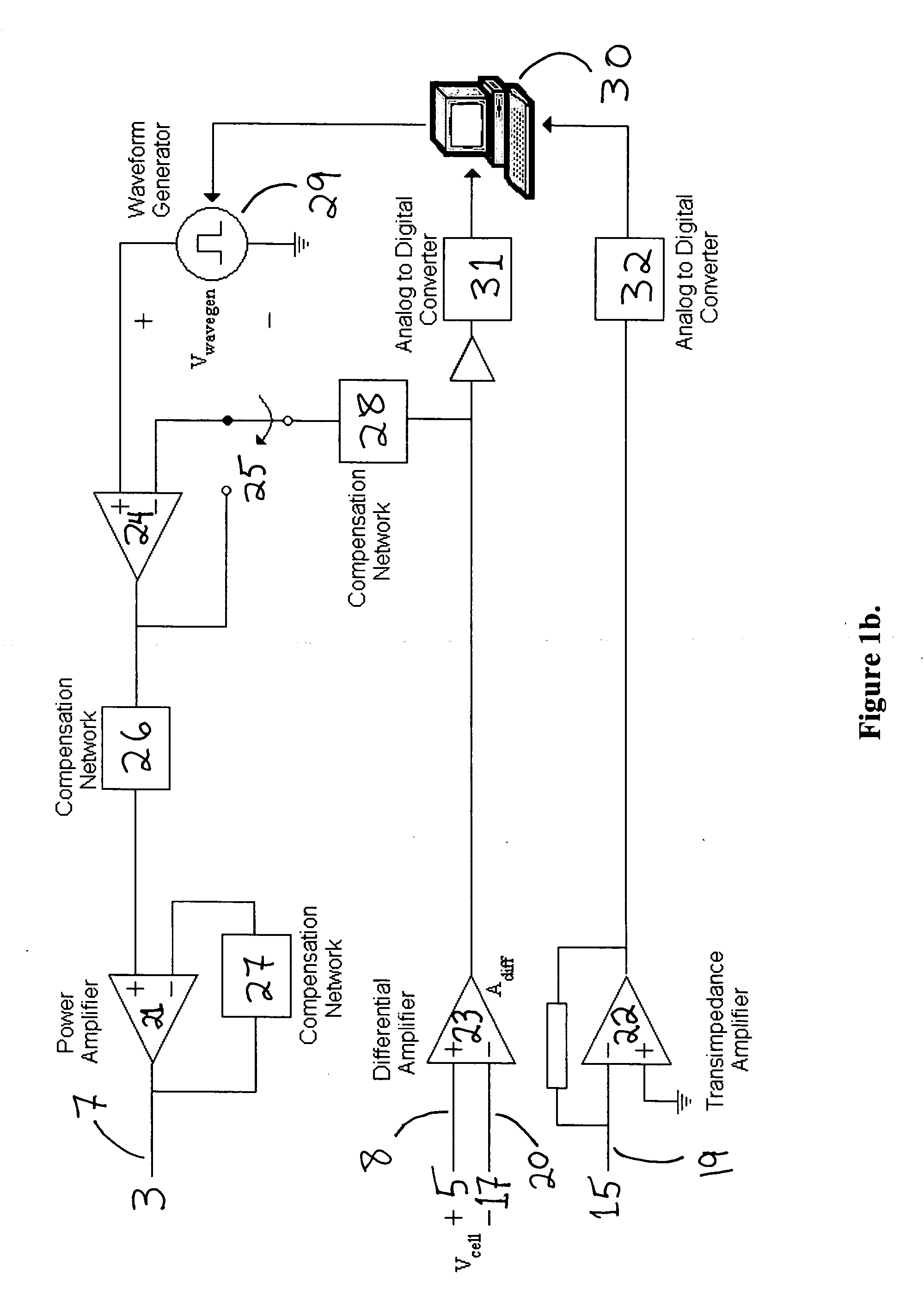
[0033]FIG. 1a shows the cross-section schematic of a device as well as the electronic configuration for monitoring and controlling electroporation of cells (including but not limited to biological cells, lipid vesicles, cell cultures, cell monolayers, spheroids, biological tissue and tissue slices and any combination thereof) on porous membranes. The device consists of three parts: The top unit (1), the middle cup (9) and the bottom chamber (14). The middle cylindrical cup has a thin, non-electrically conductive and porous membrane (10). The cup rests on feet (11) to keep the membrane (10) from touching the bottom chamber (14). Alternately, a flange along the top rim of the cup (9) allows the cup to hang from a ledge built into the bottom chamber, such that the membrane (10) is separated at a desired distance from the bottom chamber (14). A top electroporation electrode (3), typically made of silver and silver chloride, is attached to the base of the top unit bo...


PUM

PropertyMeasurementUnit
electrical potentialaaaaaaaaaa
diametersaaaaaaaaaa
diametersaaaaaaaaaa

Abstract

In biology and biotechnology, electroporation is an important technique for introducing entities (DNA, RNAi, peptides, proteins, antibodies, genes, small molecules, nanoparticles, etc.) into cells. Applications range widely from genetic engineering to regenerative medicine to drug delivery. It has been demonstrated that the electrical currents flowing through cells can be used to monitor and control the process of electroporation for biological and artificial cells. In this application, a device and system are disclosed which allow precise monitoring and controlling electroporation of cells and cell layers, with examples shown using adherent cells grown on porous membranes.

Description

CROSS-REFERENCES TO RELATED APPLICATIONS [0001] Not Applicable STATEMENT REGARDING FEDERALLY SPONSORED RESEARCH OR DEVELOPMENT [0002] Not Applicable INCORPORATION-BY-REFERENCE OF MATERIAL SUBMITTED ON A COMPACT DISC [0003] Not Applicable BACKGROUND OF INVENTION [0004] (1) Field of the Invention [0005] This invention is related to the field of cell electroporation and molecular delivery in general, which specific reference to controlling electroporation in biological and synthetic cells, tissue, and lipid vesicles. [0006] (2) Description of the Related Art [0007] Unlike the present invention, most electroporation devices electroporate cells while they are in solution (suspension). One problem incumbent with electroporation of cells in suspension is that it is not possible to measure, much less control, the voltage drop over any individual cell. Moreover, due to inhomogenieties in the suspension and on the electrodes, individual cells will see a broad range of voltages—in essence the ...

Claims


Application Information

Patent Timeline
Patent Type & Authority Applications(United States)
IPC IPC(8): C12M1/42C12M3/00C12N5/08C12N13/00C12N15/87
CPCC12M35/02C12N2510/00C12N15/87C12N13/00
Inventor HUANG, YONGBORNINSKI, JAMES W.MAZZOLA, LAURA T.
Owner HUANG YONG
Why Eureka
Eureka Blog
Learn More