Non-invasive prenatal genetic diagnosis using transcervical cells

a transcervical cell and prenatal technology, applied in the direction of material testing goods, biochemistry equipment and processes, instruments, etc., can solve the problems of increased risk of fetal abnormality, 4% procedure-related risk of miscarriage, defective limb development,

Inactive Publication Date: 2005-08-18
MONALIZA MEDICAL
View PDF12 Cites 47 Cited by
  • Summary
  • Abstract
  • Description
  • Claims
  • Application Information

AI Technical Summary

Problems solved by technology

However, the lack of prenatal testing in younger women resulted in the surprising statistics that 80% of Down syndrome babies are actually born to women under the age of 35.
However, since CVS is an invasive procedure it carries a 2-4% procedure-related risk of miscarriage and may be associated with an increased risk of fetal abnormality such as defective limb development, presumably due to hemorrhage or embolism from the aspirated placental tissues (Miller D, et al, 1999.
The amniocentesis procedure carries a 0.5-1.0% procedure-related risk of miscarriage.
However, in cases of abnormal findings, the termination of pregnancy usually occurs between the 18th to the 22nd week of gestation, involving the Boero technique, a more complicated procedure in terms of psychological and clinical aspects.
However, while the isolation of trophoblasts from the maternal blood is limited by their multinucleated morphology and the availability of antibodies, the isolation of leukocytes is limited by the lack of unique cell markers which differentiate maternal from fetal leukocytes.
Proc. Natl. Acad. Sci. 93: 705-708), residual cells are likely to be present in the maternal blood from previous pregnancies, making prenatal diagnosis on such cells practically impossible.
However, such purification and enrichment steps resulted in inconsistent recovery of fetal cells and limited sensitivity in diagnosing fetal's gender (reviewed in Bischoff, F. Z. et al., 2002.
Thus, the combination of technical problems, high-costs and the uncertainty of the origin of the cells have prevented this approach from actually becoming clinically accepted.
However, this method was limited by false positives as a result of residual semen im the cervix.
However, direct PCR amplifications from unpurified transcervical cells are likely to result in maternal cell contamination.
A more recent study using PCR and FISH analyses on transcervical cells resulted in poor detection rates of fetal gender (Cioni R., et al, 2003.
However, since the FISH analysis and the trophoblast-specific IHC assay were performed on separated slides, it was impractical to use this method for diagnosing fetal chromosomal abnormalities.

Method used


Image

  • Non-invasive prenatal genetic diagnosis using transcervical cells
  • Non-invasive prenatal genetic diagnosis using transcervical cells
  • Non-invasive prenatal genetic diagnosis using transcervical cells

Examples


example 1

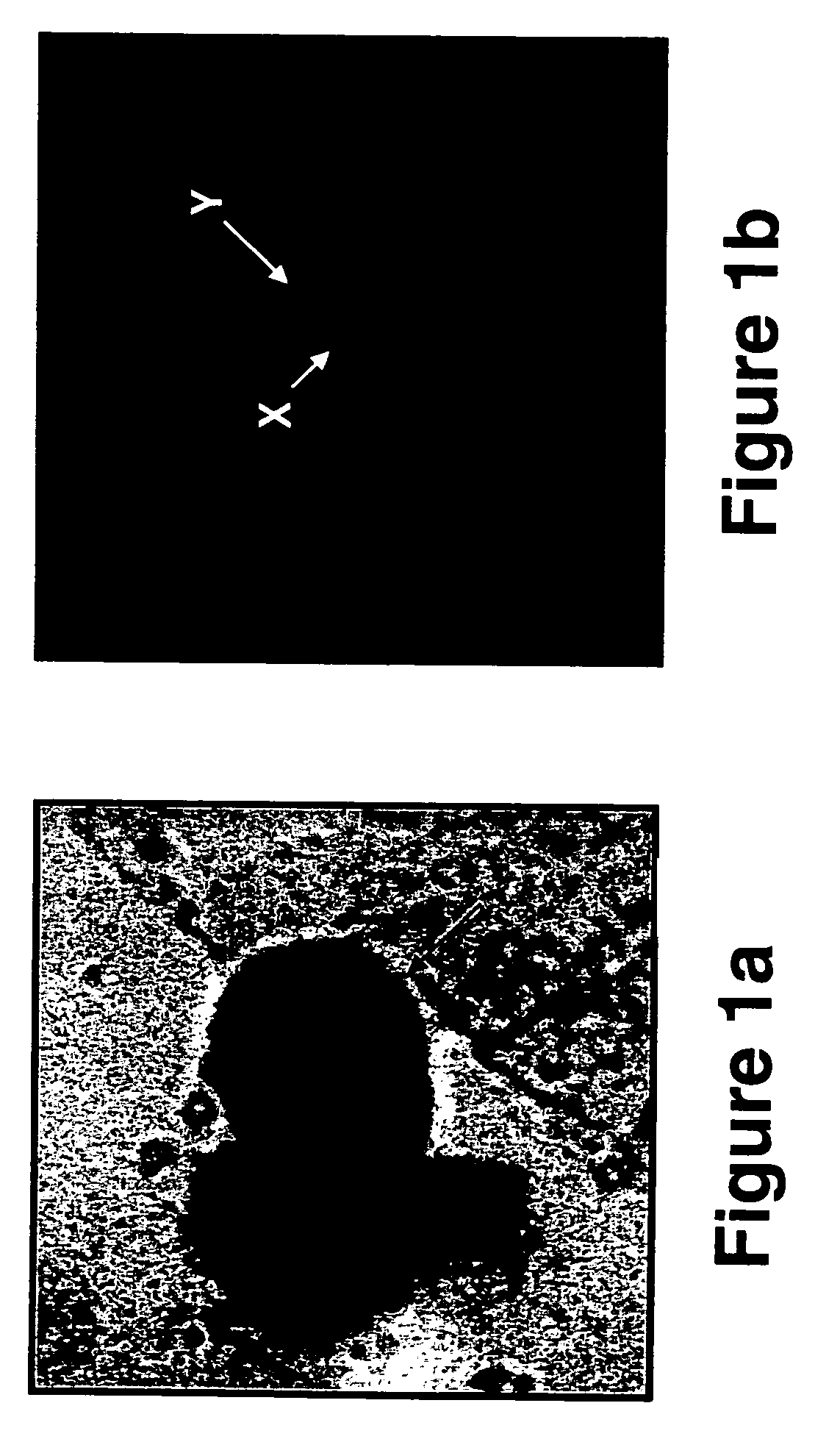
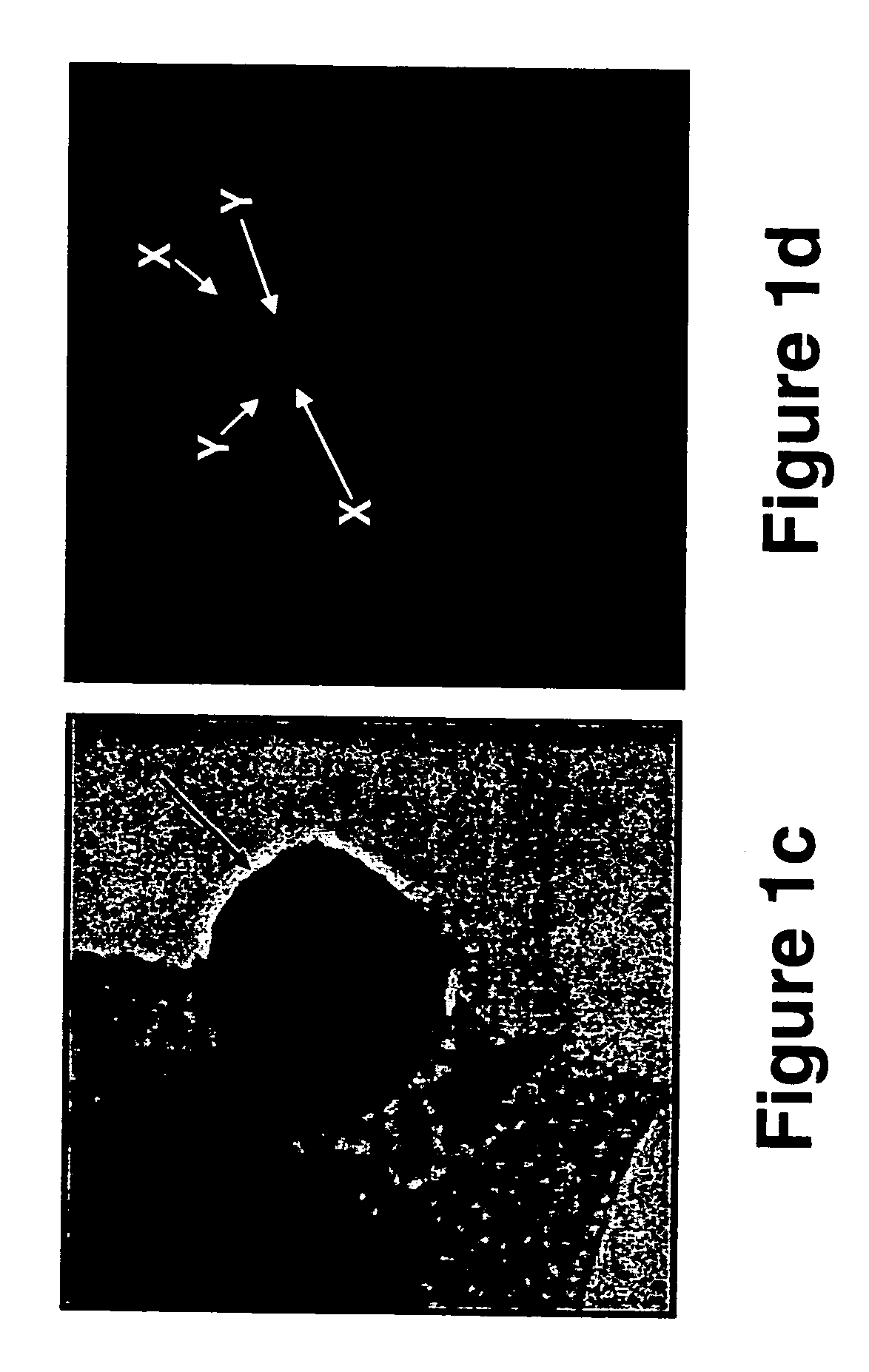
Determination of Fetal Fish Pattern from ExtraVillous Trophoplast Cells Obtained from Transcervical Specimens

[0277] Transcervical cells obtained from pregnant women between 6th and 15th Week of gestation were analyzed using immunohistochemical staining followed by FISH analysis, as follows.

[0278] Materials and Experimental Methods

[0279] Study subjects—Pregnant women between 6th and 15th week of gestation, which were either scheduled to undergo a pregnancy termination or were invited for a routine check-up of an ongoing pregnancy, were enrolled in the study after giving their informed consent.

[0280] Sampling of transcervical cells—A Pap smear cytobrush (MedScand-AB, Malmö, Sweden) was inserted through the external os to a maximum depth of 2 cm (the brush's length), and removed while rotating it a full turn (i.e., 360°). In order to remove the transcervical cells caught on the brush, the brush was shaken into a test tube containing 2-3 ml of the RPMI-1640 medium (Beth Haemek, Isra...

example 2

Fetal Fish Pattern Can be Determined on Extravillous Trophoblast Cells Using the HLA-G and the CHL1 Antibodies

[0310] To increase the detection rate of fetal trophoblasts in human transcervical cells, the present inventors have employed the CHL1 antibody, a new extravillous trophoblast-recognizing antibody, raised against the chorion leave from a fetal membrane (Higuchi T, et al., 2003, Mol. Hum. Reprod. 9: 359-366; Fujiwara H, et al., 1993, J. Clin. Endocrinol. Metab. 76: 956-961; Higuchi T, et al., 1999, Mol. Hum. Reprod. 5: 920-926), as follows.

[0311] Materials and Experimental Methods

[0312] CHL1 antibody—The CHL1 antibody which recognizes the melanoma cell adhesion molecule [MCAM, Mel-CAM, S-endo 1 or MUC18 / CD 146, Higuchi, 2003 (Supra)] was obtained from Alexis Biochemicals [Cat. No. 805-031-T100, monoclonal antibody to human CD146 (F4-35H7, S-endo1; anti-MCAM)] and was diluted 1:200 prior to use on transcervical cell samples.

[0313] Immunohistochemistry and FISH analyses wer...

example 3

Identification of Sycitiotrophblasts in Transcervical Specimens Using an NDOG-1 Antibody

[0319] In attempts to improve the sensitivity of trophoblast identification in transcervical specimens, and due to the fact that syncytiotrophoblasts are not stained using common trophoblast antibodies (e.g., HLA-G) the present inventors employed the mouse anti human trophoblast protein NDOG-1 antibody on transcervical specimens obtained from pregnant women.

[0320] Prior to use, the mouse anti human trophoblast protein NDOG-1 antibody (MCA277, Serotec immunological excellence, UK) was diluted 1:50 in the antibody diluent and was incubated on the transcervical specimens according to the immunohistochemistry protocol described in “Materials and Experimental Methods” of Example 1, hereinabove.

[0321] As is shown in FIGS. 6a-b, the NDOG-1 antibody specifically labeled the nuclei of the syncytiotrophoblasts present in transcervical specimens obtained from a pregnant woman at the 7th week of gestation...


PUM

PropertyMeasurementUnit
depthaaaaaaaaaa
pHaaaaaaaaaa
volumeaaaaaaaaaa

Abstract

A non-invasive, risk-free method of prenatal diagnosis is provided. According to the method of the present invention transcervical specimens are subjected to trophoblast-specific immuno-staining followed by FISH, PRINS, Q-FISH and / or MCB analyses and / or other DNA-based genetic analysis in order to determine fetal gender and / or identify chromosomal and / or DNA abnormalities in a fetus. Also provided is a method of in situ chromosomal, DNA and / or RNA analysis of a prestained specimen by incubating the prestained specimen in ammonium hydroxide. Also provided is a method of identifying embryonic cells according to a nucleus / cytoplasm ratio of at least 1:1 and the presence of at least variably condensed chromatin.

Description

[0001] This application is a continuation-in-part of U.S. patent application Ser. No. 10 / 921,899, filed Aug. 20, 2004, which is a continuation-in-part of PCT Patent Application No. PCT / IL2004 / 000304, filed Apr. 1, 2004, which claims the benefit of priority from U.S. patent application Ser. No. 10 / 405,698, filed Apr. 3, 2003, now abandoned. The contents of the above applications are hereby incorporated by reference in their entirety.FIELD AND BACKGROUND OF THE INVENTION [0002] The present invention relates to a method of diagnosing genetic abnormalities using trophoblast cells from transcervical specimens, and, more particularly, to the biochemical and genetic analysis of trophoblast cells for determination of fetal gender and / or chromosomal abnormalities in a fetus. [0003] Prenatal diagnosis involves the identification of major or minor fetal malformations or genetic diseases present in a human fetus. Ultrasound scans can usually detect structural malformations such as those involvi...

Claims


Application Information

Patent Timeline
Patent Type & Authority Applications(United States)
IPC IPC(8): C12Q1/68G01N33/68
CPCC12Q1/6841C12Q1/6879C12Q1/6883C12Q2600/156G01N2800/36G01N2800/368G01N2800/387G01N33/689
Inventor FEJGIN, MOSHEAMIEL, ALIZALIBERMAN, MEYTAL
Owner MONALIZA MEDICAL
Why Eureka
Try Eureka