
Three-dimensional peripheral lymphoid organ cell cultures

Inactive Publication Date: 2005-09-01
UNIVERSITY OF ROCHESTER
View PDF9 Cites 44 Cited by
  • Summary
  • Abstract
  • Description
  • Claims
  • Application Information

AI Technical Summary

Benefits of technology

[0025] The long-term in vitro maintenance of normal peripheral lymphocytes provided by the present invention represents a major advance for a number of basic immunological studies. Furthermore, culture of lymphocytes and other immune accessory cells in the appropriate microenvironment may allow replication of an antigen-specific immune response in an accessible, easily modifiable setting. Potential applications for lymphoid organ cell culture method of the present invention range from the testing of immunogenicity of vaccine candidates in vitro, to the generation of human monoclonal antibodies, to immunotherapy interventions. The present invention provides a culture system highly suited to these studies because of its physiologic relevance, its limited size, easy sampling, and increased access.

Problems solved by technology

The long-term maintenance of resting B lymphocytes in the absence of over-expression of proto-oncogenes is therefore unfeasible using current technology.
Despite its ability to support hematopoietic differentiation, several limitations are obvious in the Dexter culture system.
In addition, the optimal temperature for cell output and duration of Dexter culture is 33° C., a temperature not optimal for growth of all cells.
The essential role these factors play in immune system function has hampered the faithful replication of normal immune mechanisms in vitro.
Indeed, even the long-term culture of peripheral lymphocytes in the absence of activating stimuli or cell transformation has not been achieved so far.

Method used


Image

  • Three-dimensional peripheral lymphoid organ cell cultures
  • Three-dimensional peripheral lymphoid organ cell cultures
  • Three-dimensional peripheral lymphoid organ cell cultures

Examples


example 1

Three-D Culture System BioReactors

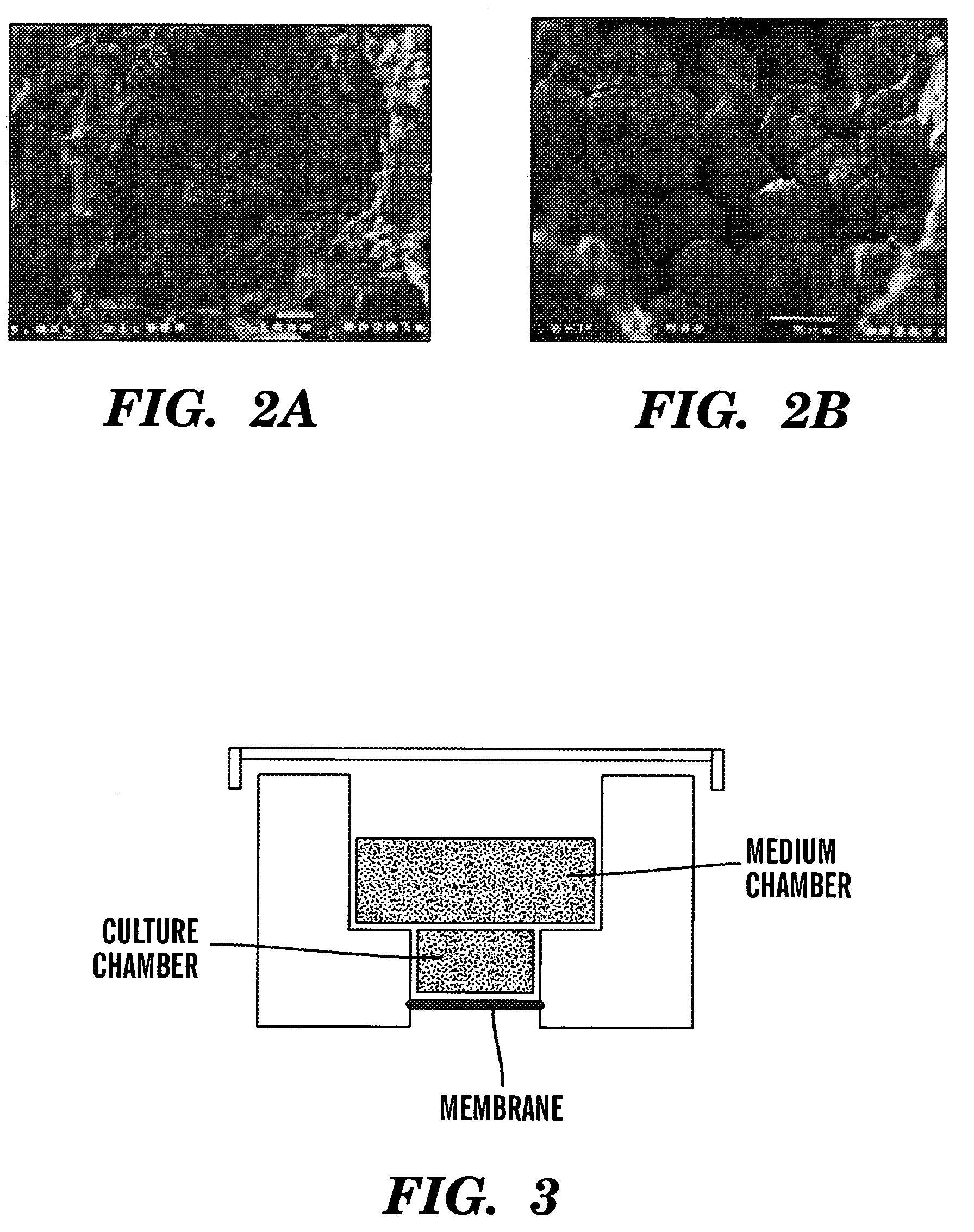
[0107] The 3D bioreactor system of the present invention was based on the hypothesis that lack of 3D structure and the consequent morphological distortion of the physiologic microenvironment could be responsible for the inability of long term in vitro cell cultures to support long-term maintenance of lymphoid cell cultures, as well as for the other alterations observed in the Dexter cultures (Wang et al., “Multilineal Hematopoiesis in a Three-Dimensional Murine Long-Term Bone Marrow Culture,”Exp. Hematol. 23:26-32 (1995); and Mantalaris et al., “Engineering a Human Bone Marrow Model: A Case Study on Ex vivo Erythropoiesis,”Biotech. Progr. 14:126-133 (1998)), which are hereby incorporated by reference in their entirety).

[0108] To test the feasibility of recapitulating the 3D structure and function of lymphoid organs in vitro, the scaffold-based, packed-bed bioreactor was adapted for culturing murine spleen cells using the principle previously demon...

example 2

Long-Term Culture of Unstimulated Splenic Lymphocytes

[0112] Total splenocyte preparations were generated by gently grinding fresh spleens from C57B1 / 6 mice in PBS, 5% fetal calf serum using frosted glass slides. About 20×106 live leukocytes were seeded onto each bioreactor, and cultured in complete RPMI medium, 10% FCS. A small amount of cells was harvested weekly by gentle pipetting from the matrix bed, and analyzed for expression of B and T-cell specific surface markers.

[0113] In striking contrast to flask cultures, significant fractions of B220 / IgM positive B cells were routinely detected as far as 8 weeks into culture (when the cultures were terminated). As shown in FIGS. 4A-F, these B cells expressed somewhat lower B220 and IgM levels than the majority of B2 cells in the spleen. They also shared some features with peritoneal B 1 cells, most notably expression of low levels of the CD5 antigen, shown in FIGS. 5A-H. However, unlike B1 cells, 3D B cells expressed CD23, a defining...

example 3

Long-Term 3D Culture B Cells Capable of Responding to in vitro Stimulation

[0117] To test whether 3D culture B cells are functionally competent, B cells were stimulated from a 2-week culture with the polyclonal activator LPS, and their response to this stimulus was compared with that of normal, ex vivo-derived splenic B cells. As shown in FIG. 9, 3D B lymphocytes proliferated to an extent comparable to primary cells, and upregulated the co-stimulatory activation markers CD80 and CD86, as well as the marker Syndecan-1, characteristic of Ig-secreting plasma cells, to levels similar to controls. In addition, they were able to undergo immunoglobulin class switching to IgG, a highly specific gene rearrangement process unique to activated B cells (Bottaro et al., “Local and General Regulatory Elements of Immunoglobulin Class Switch Recombination,” In Molecular Mechanisms of IgE Regulation, Vercelli, ed., Chichester, England: J. Wiley and Sons, pp. 155-177 (1997), which is hereby incorpora...


PUM

PropertyMeasurementUnit
Gene expression profileaaaaaaaaaa
Immunogenicityaaaaaaaaaa
Cytotoxicityaaaaaaaaaa

Abstract

The present invention relates to a method of culturing peripheral lymphoid organ cells on a three-dimensional scaffolding which is covered or surrounded with culture medium, under conditions effective to generate and maintain mature and functional lymphoid cells, where the three-dimensional scaffolding allows cells in the culture medium to have cell to cell contact in three dimensions. The present invention also provides methods of screening for vaccine candidates for efficacy in eliciting an immune response, identifying genes or proteins which are related to peripheral lymphoid organ cell formation or function, screening for drugs effecting peripheral lymphoid organ cell maturation, treating a patient for a disease condition using antigen-specific lymphoid cells, and effecting gene expression of peripheral lymphoid organ cells.

Description

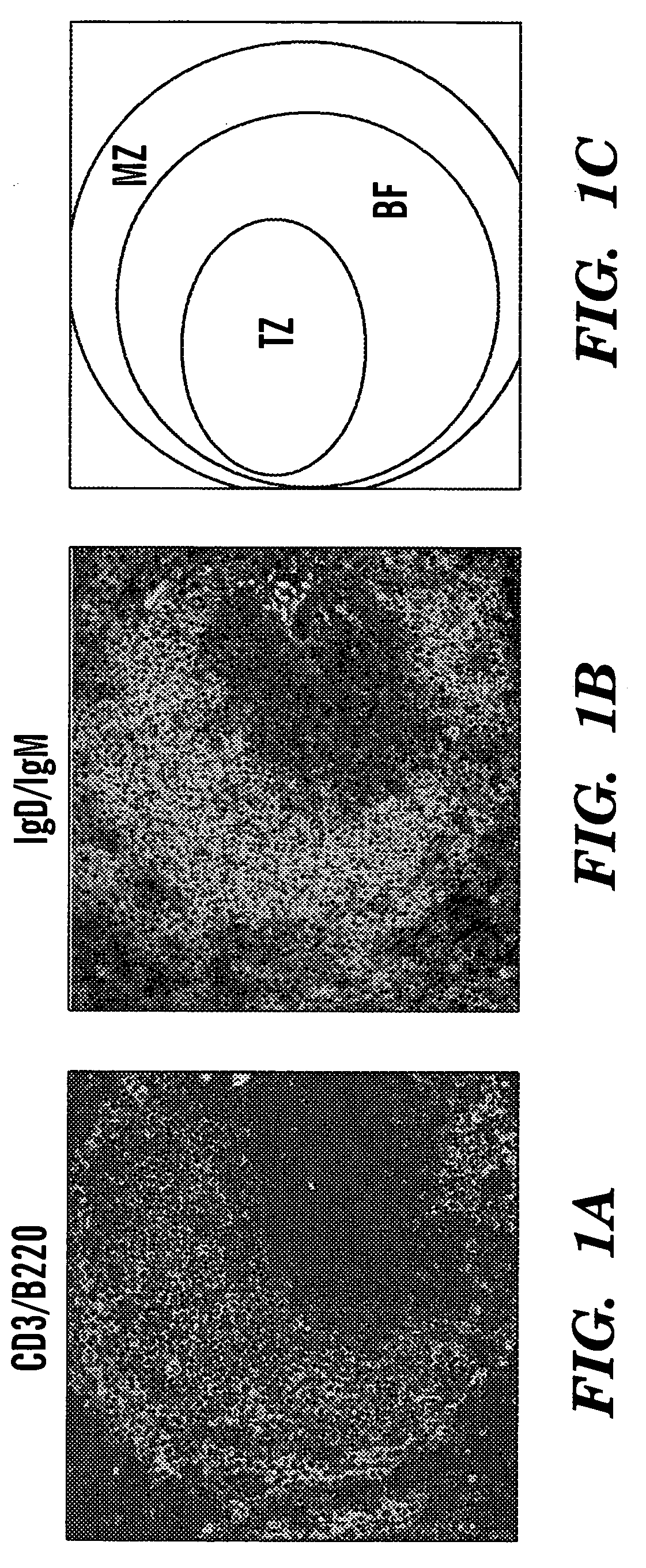
[0001] This application claims the benefit of U.S. Provisional Patent Application Ser. No. 60 / 415,944, filed Oct. 3, 2002.[0002] The subject matter of this application was made with support from the National Aeronautics and Space Administration, NASA Grant No. NAG 9-1360 and NIH-NIAID Grant No. 1 R21 AI47988-01A1. The U.S. Government may have certain rights.FIELD OF THE INVENTION [0003] The present invention relates to the field of cell culture, and, in particular, to methods and compositions related to cultured immune system cells derived from peripheral lymphoid organ cells. BACKGROUND OF THE INVENTION [0004] Peripheral lymphoid organs represent the principal sites of adaptive immune responses and their complex anatomy reflects the presence of well-defined subregions enriched for specific immune system cell subsets. As a model peripheral lymphoid organ, the ultrastructure of the spleen is reviewed here (see also review in Tarlinton D., “Germinal Centers: Form and Function,”Curr. O...

Claims


Application Information

Patent Timeline
IPC IPC(8): A61K39/395C12N5/071C12N5/078
CPCC12N5/0648C12N2533/78C12N2531/00C12N5/0651
Inventor WU, J.H. DAVIDBOTTARO, ANDREA
Owner UNIVERSITY OF ROCHESTER
Learn More