

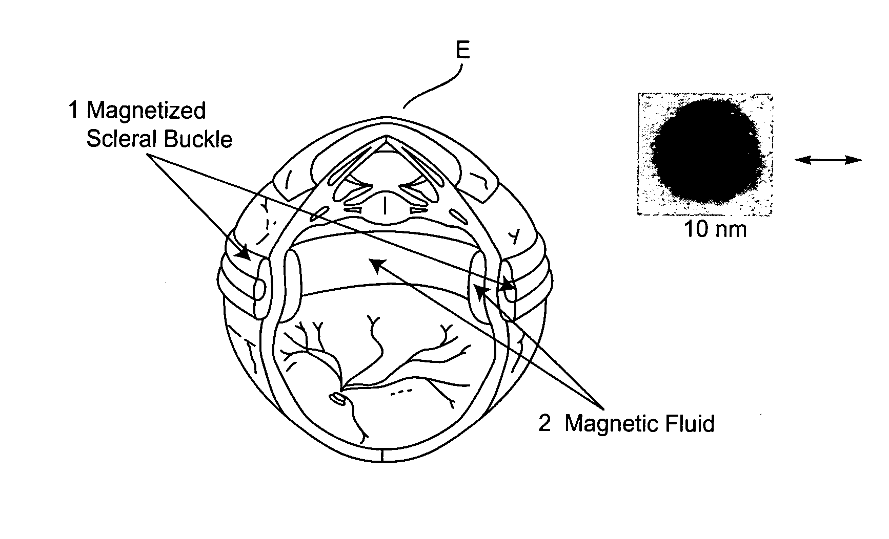
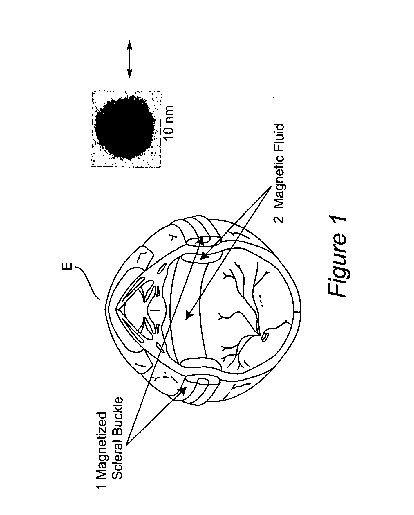
Magnetized scleral buckle, polymerizing magnetic polymers, and other magnetic manipulations in living tissue
a technology of magnetic manipulation and living tissue, applied in the field of biocompatible magnetic systems and medical procedures, can solve the problems of partial or complete loss of vision in several million people worldwide, retinal detachment, and insufficient scleral buckling to close retinal holes in all patients, and achieve the effect of invasiveness of some conventional surgeries
- Summary
- Abstract
- Description
- Claims
- Application Information
AI Technical Summary
Benefits of technology
Problems solved by technology
Method used
Image

Examples
example 1
[0058] Magnetic Nb—Fe—B microparticles were placed into a medium of Silastic A (Dow-Corning) and magnetic properties were measured via SQUID magnetometry.
[0059] Preparation of a Dispersion of Nb—Fe—B Microparticles in Silastic-A:
[0060] A 5-mL vial was charged with 2.9954 g of Silastic A 9280-50 and 0.9200 g of Nb—Fe—B microparticles (MagneQuench, Inc.). The mixture was stirred with a stainless steel spatula and sonicated for 5 minutes at 50% power. The resulting material was a highly viscous dispersed mixture that showed little signs of settling after several days of observation.
[0061] Measurement of Response of Nb—Fe—B Particles to an Applied Magnetic Field
[0062] A 15-mg sample of dry Nb—Fe—B particles was measured at room temperature in response to applied magnetic fields. Measurements were made from 0 to 70 kOe at 25° C. While the magnetic field was insufficient to saturate the particles, FIG. 4 demonstrates that the slope of the magnetic response decreased substantially as t...
example 2
Preparation of Magnetite Nanoparticles
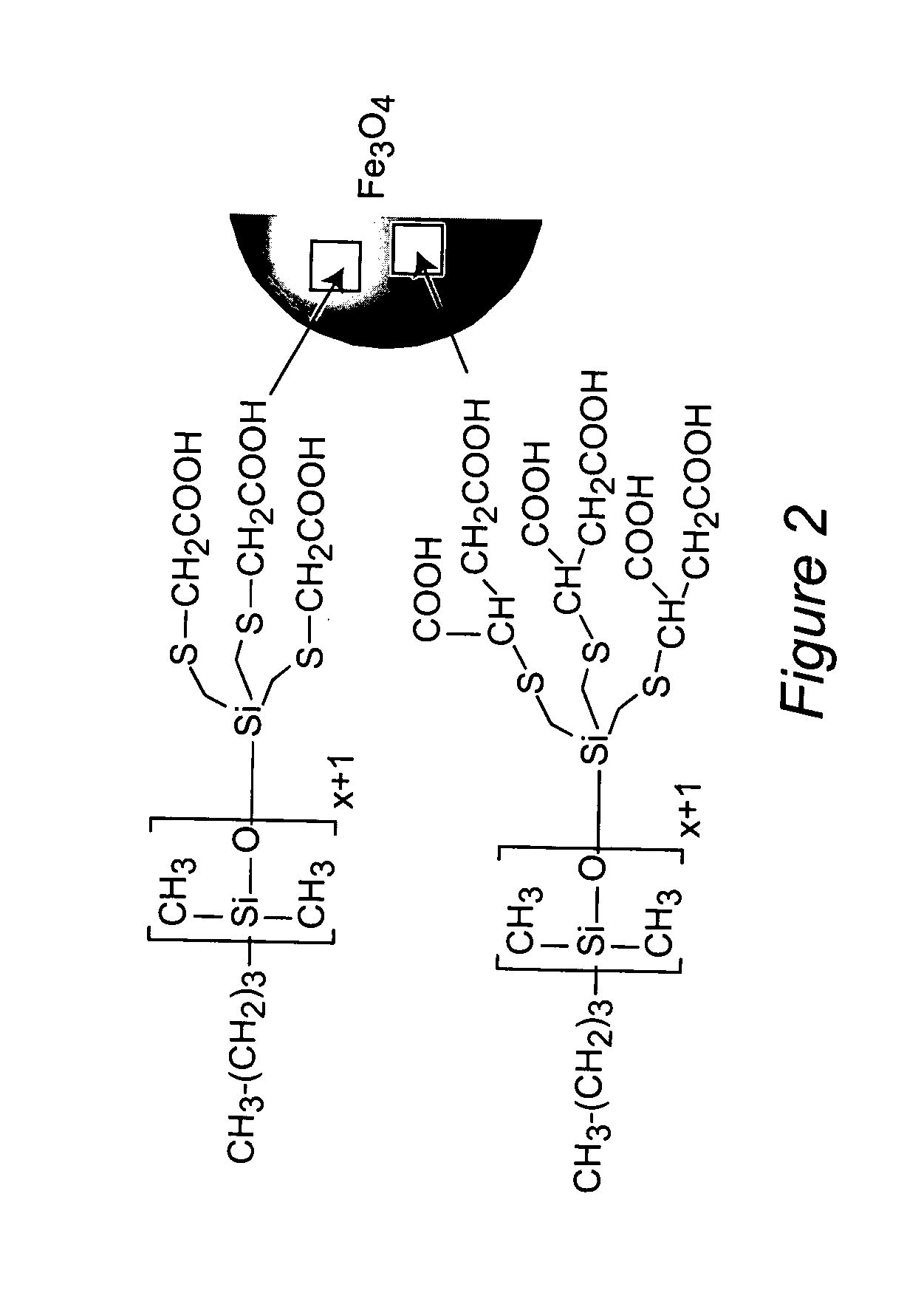
[0066] Carboxylic acid-functionalized PDMS surfactants were synthesized for steric stabilization of magnetite nanoparticle dispersions in biocompatible polysiloxane carrier fluids (FIG. 2). Trivinylsilyl-terminated PDMS was prepared via living polymerization of D3, then reacted with either mercaptoacetic acid or mercaptosuccinic acid using a free radical thiol-ene addition to afford PDMS containing either three or six carboxylic acid groups at one end (FIG. 3). Magnetite nanoparticles were prepared by chemically co-precipitating FeCl2 and FeCl3 at pH 9-10, then the PDMS-magnetite nanoparticle complexes were prepared via interfacial adsorption of the carboxylate groups of the PDMS stabilizer onto aqueous magnetite particles at a slightly acidic pH.
[0067] Repeated centrifugations to remove any aggregates resulted in well-dispersed polymer-magnetite nanoparticle complexes. The complexes were characterized with transmission electron microscopy to ...
example 3
In Vitro Evaluations of Magnetite Silicone Magnetic Fluid
[0070] In-vitro evaluations of the magnetite silicone magnetic fluid of Example 2 have been conducted using the well-established MTT Assay in which the tetrazolium salt MTT (3-(4,5-dimethylthiazol-2-yl)-2,5-diphenyl-tetrazolium bromide) is enzymatically reduced in living, metabolically-active cells but not in dead cells. The reactions were carried out in multi-well plates, and the purple formazan reaction product was disssolved in dimethylsulfoxide, and measured by visible spectroscopy (FIG. 8). Referring to FIG. 8, magnetic fluids or their supernatants were incubated with human retinal pigment epithelial cells (HRPE) or C4-2 prostate cancer cells for 48 to 72 hours. Viability was then measured in a 96-well plate. Healthy cells oxidize the yellow dye MTT into a blue formazan product, which is then quantified at 540 nm in a well plate reader. The assay results (FIG. 9) suggest that the magnetite-polydimethylsiloxane fluids wer...
PUM
| Property | Measurement | Unit |
|---|---|---|
| surface area | aaaaa | aaaaa |
| inner diameter | aaaaa | aaaaa |
| speed | aaaaa | aaaaa |
Abstract
Description
Claims
Application Information