Preparation and application of anti-tumor bifunctional fusion proteins

a technology of fusion proteins and tumors, applied in the field of tumor immunology, can solve the problems of inoperable tumor burden in patients with hepatoma, untreated micrometastases, and inability to detect micrometastases, and achieve the effects of preventing tumor growth recurrence, reducing tumor burden, and infiltrating tumor tissues

Inactive Publication Date: 2005-10-20
SYMBIGENE ACQUISITION CO +1
View PDF30 Cites 36 Cited by
  • Summary
  • Abstract
  • Description
  • Claims
  • Application Information

AI Technical Summary

Problems solved by technology

However, two thirds of hepatoma patients have inoperable tumor burdens at the time of diagnosis.
More importantly, even if the modality of surgical resection is available to such patients, the problem of distant, undetected micrometastases remains untreated by such therapy.
Likewise, the traditional therapies of radiotherapy and chemotherapy also have significant limitations, most prominently the systemic inhibition of the hematopoietic and immune system.
Thus, the toxic effects of radiotherapy and chemotherapy limit efficacy of these therapies in the cases where radical treatment is most desired—in the patient with significant tumor burden at the time of diagnosis.
However, one of the recognized limitations of such antibody therapy is the likelihood that distant metastases may still escape such therapy or that antigen-negative variants will develop, leading to a later relapse with metastatic disease.
While tumor vaccines clearly confer long term protection against tumor metastatic outgrowth and even subsequent tumor challenges, the clinical application of this knowledge has proved to be difficult.
First, it has proven difficult to reliably expand functional DCs in ex vivo expansion protocols.
Second, reproducible activation of DCs in vivo has not yet been achieved.
Third, no clear protocol has been established that permits the activation and antigen loading of the desired DC population, i.e., those capable of eliciting an anti-tumor response.
In sum, the expansion of activated DCs selectively located at tumor site that present immunogenic tumor antigens is a problem that remains unsolved.
Therefore, while it is clear that immune molecules, e.g., tumor-specific antibodies, and vaccines eliciting immune responses can effect tumor growth, a unified approach that permits the simultaneous reduction of tumor growth and the generation of lasting protective immune response is still lacking.

Method used


Image

  • Preparation and application of anti-tumor bifunctional fusion proteins
  • Preparation and application of anti-tumor bifunctional fusion proteins
  • Preparation and application of anti-tumor bifunctional fusion proteins

Examples


example 1

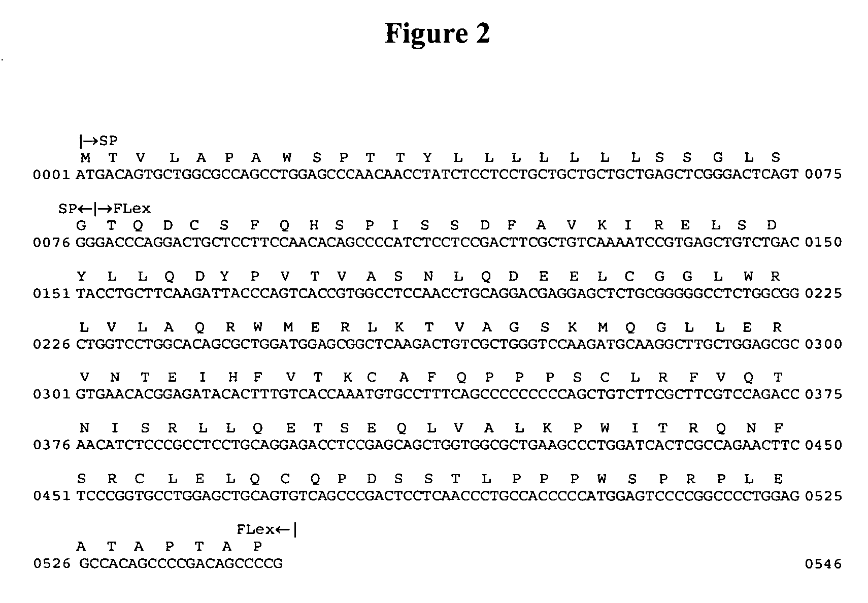
Human Flt3 Ligand Extracellular Region (hFLex) cDNA Synthesis

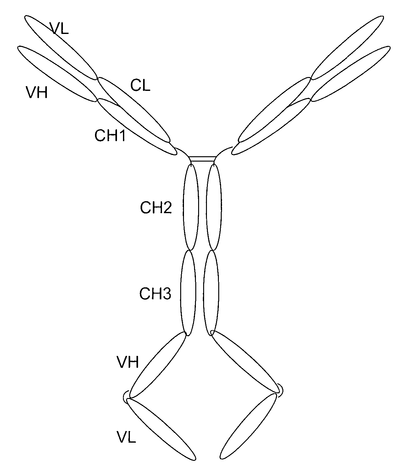
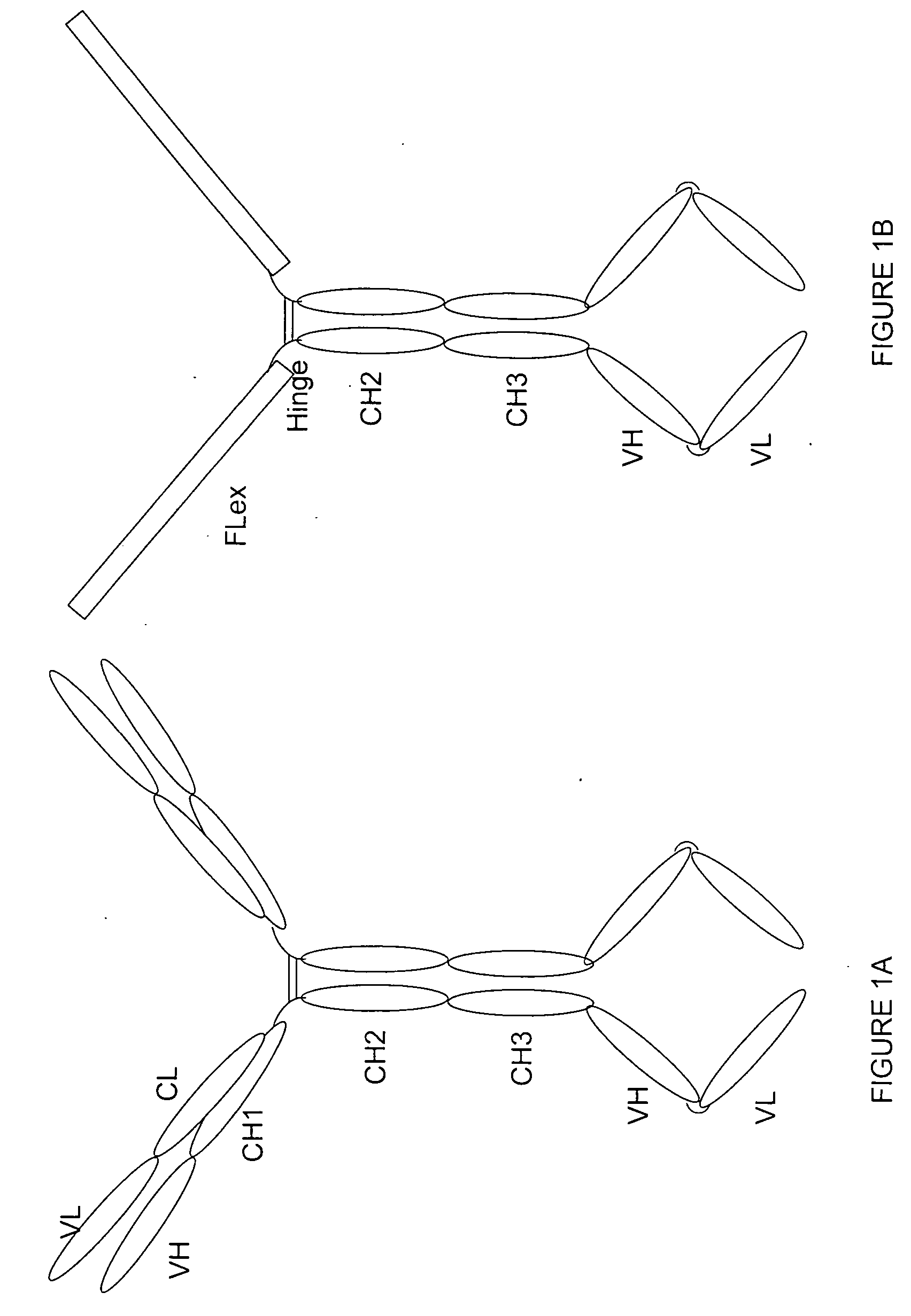
[0164] Purpose: Because the Flt3 ligand is a type I transmembrane protein whose extracellular region is at the N terminus, modification of the N terminus of FL may adversely affect its biological activities. Therefore, we employed a methodology used to construct a tetravalent biospecific antibody (see FIG. 1A). See Column et al., Nat Biotech 15:159-163 (1997). Typically, the tetravalent bispecific antibodies were constructed by fusing the DNA encoding a single chain antibody at the C terminus of an antibody with a different specificity. In order to obtain bifunctional fusion protein with high biological activities, we constructed a fusion protein with FLex at N the terminus and the antibody molecule at the C terminus (see FIG. 1B). First, the FLex gene was fused to the 5′end of a human IgG1 cDNA (hinge plus CH2 plus CH3) to generate the Flex-Ig fusion gene. Then the hFLex-Ig fusion gene was fused to the 5′ end of a single...

example 2

Cloning and Identification of the Constant Region of Human IgG1

[0169] The native human IgG1 cDNA of 1416 bp encodes 471 amino acids and a translation termination codon. The constant region of IgG1 was cloned by RT-PCR using the following protocol: Human peripheral blood mononuclear cells (PBMCs) were isolated from heparinized blood of healthy volunteers by Ficoll-Hypaque density gradient centrifugation. RNA was isolated from PBMCs with TRIzol Reagent (Gibco BRL). The cDNA of IgG1 Fc fragment was obtained by Onestep RT-PCR (Qiagen). The primers for RT-PCR were as follows: Fc sense, 5′-gca ctc gag ttt tac ccg gag aca ggg aga g-3′; Fc antisense, 5′-gag ccc aaa tct tgt gac aaa ac-3′. The RT-PCR products were separated on agarose gel. The correct DNA fragment was gel-purified and cloned into pGEM-T vector (Promega), and its sequence was verified. The clone was denoted pGEM-T / IgFc.

example 3

Construction of SM5-1 Chimeric Antibody and Humanized Antibody

[0170] 1. Cloning of mouse SM5-1 heavy and light chain variable region genes. RNA was isolated from SM5-1 (IgG1, κ) hybridoma cells (deposited at ATCC having ATCC Designation No. HB-12588) with TRIzol Reagent (Gibco BRL, Grand Island, N.Y.). The heavy and light variable region cDNAs of SM5-1 were cloned from hybridoma cells using 5′RACE system (Gibco BRL, Gaithersburg, Md.) according to the manufacture's instructions. The nested PCR products were analyzed by agarose gel electrophoresis (FIG. 5). The specific heavy chain PCR fragments of about 590 bp and light chain fragment of about 530 bp were gel-purified and cloned into pGEM-T vector (Promega, Madison, Wis.) for sequence determination, respectively. The DNA sequences of heavy (SM VH) and light (SM, VL) variable region are SEQ ID NO:7 (FIG. 6) and SEQ ID NO:9 (FIG. 7), respectively.

[0171] 2. Construction of expression vectors for chimeric antibodies. The two vectors p...


PUM

PropertyMeasurementUnit
solubleaaaaaaaaaa
nucleic acidaaaaaaaaaa
β-lactamaseaaaaaaaaaa

Abstract

Provided herein is a chimeric protein, which chimeric protein comprises a Flt3 ligand, or a biologically active fragment thereof, and a proteinaceous or peptidyl tumoricidal agent, or a targeting agent which binds to a receptor expressed on a tumor, and uses thereof, particularly in the treatment of malignancy. Other embodiments and uses are disclosed.

Description

CROSS-REFERENCE TO RELATED APPLICATIONS [0001] This application is a continuation-in-part of U.S. application Ser. No. 10 / 723,003, filed Nov. 26, 2003, which claims the benefit of PRC application serial no. 03129290.9, filed Jun. 13, 2003, and PRC application 200310119930.0, filed Nov. 25, 2003 (title: Preparation and application of anti-tumor bifunctional fusion proteins), all of which are incorporated herein in their entirety including the drawings by reference thereto.BACKGROUND OF THE INVENTION [0002] This invention relates to the field of tumor immunology, mainly about the anti-tumor bifunctional fusion proteins and their nucleic acid sequences, methods of preparation and application of them in preparation of antitumor drugs. [0003] Tumor immunotherapy involves the induction of tumor regression by modulation of natural host defense mechanisms or by manipulation with a immunological agent. Immunotherapy is a recognized therapeutic modality for the treatment of malignancies along...

Claims


Application Information

Patent Timeline
Patent Type & Authority Applications(United States)
IPC IPC(8): A61K39/00C07K14/475C07K14/82C07K16/28C07K16/32
CPCC07K14/4747C07K2317/52C07K16/2809C07K16/2863C07K16/2896C07K16/30C07K16/32C07K2317/24C07K2317/31C07K2317/53C07K2317/622C07K2317/626C07K2317/73C07K2319/00C07K2319/01C07K2319/33C07K2317/34C07K14/475
Inventor MA, JINGGUO, YANJUN
Owner SYMBIGENE ACQUISITION CO
Why Eureka
Try Eureka