Method for cryopreserving microencapsulated living animal cells enclosed in immunoisolation membranes, such microencapsulated living animal cells in immunoisolation membranes, and biohybrid artificial organ modules using such microencapsulated living animal cells in immunoisolation membranes

Active Publication Date: 2005-12-01
SHOWA UNIVERSITY
View PDF1 Cites 20 Cited by
  • Summary
  • Abstract
  • Description
  • Claims
  • Application Information

AI Technical Summary

Benefits of technology

[0022] (3) According to the method for cryopreserving the living animal cells in the immunoisolation membranes of the present invention, since no complicated chemi

Problems solved by technology

However, there may be problems in infection with swine retrovirus, overcoming of immunological reactions, adjustment of the number of hepatocytes in reactors, securement of cell sources, establishment

Method used


Image

  • Method for cryopreserving microencapsulated living animal cells enclosed in immunoisolation membranes, such microencapsulated living animal cells in immunoisolation membranes, and biohybrid artificial organ modules using such microencapsulated living animal cells in immunoisolation membranes
  • Method for cryopreserving microencapsulated living animal cells enclosed in immunoisolation membranes, such microencapsulated living animal cells in immunoisolation membranes, and biohybrid artificial organ modules using such microencapsulated living animal cells in immunoisolation membranes
  • Method for cryopreserving microencapsulated living animal cells enclosed in immunoisolation membranes, such microencapsulated living animal cells in immunoisolation membranes, and biohybrid artificial organ modules using such microencapsulated living animal cells in immunoisolation membranes

Examples


experiment 1

[0060] (Experiment 1)

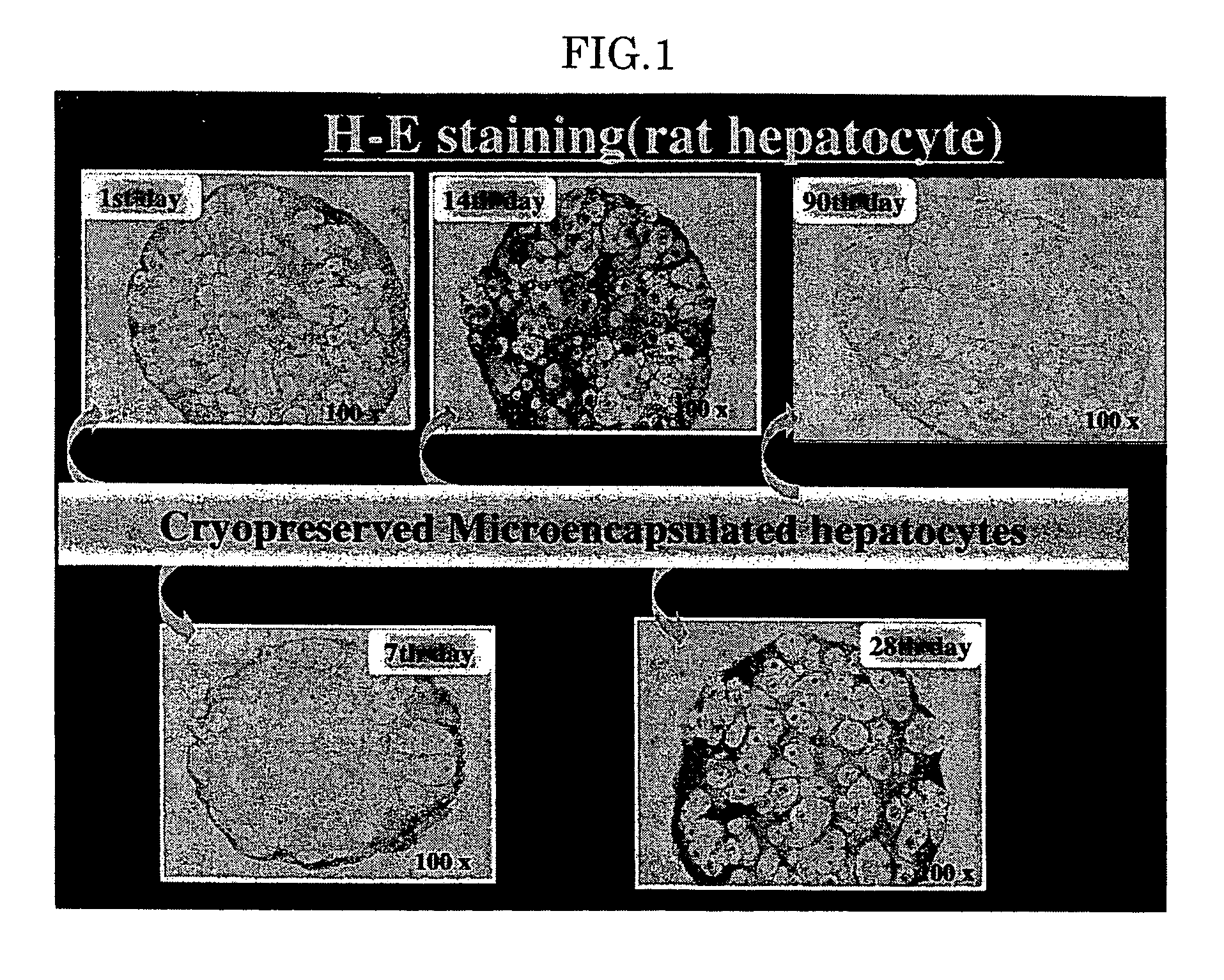
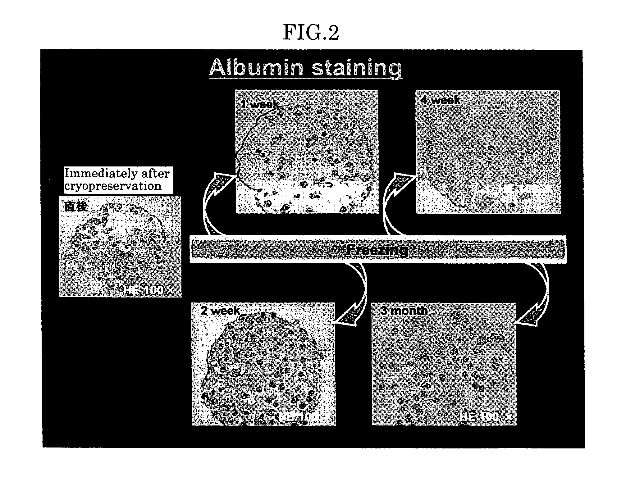
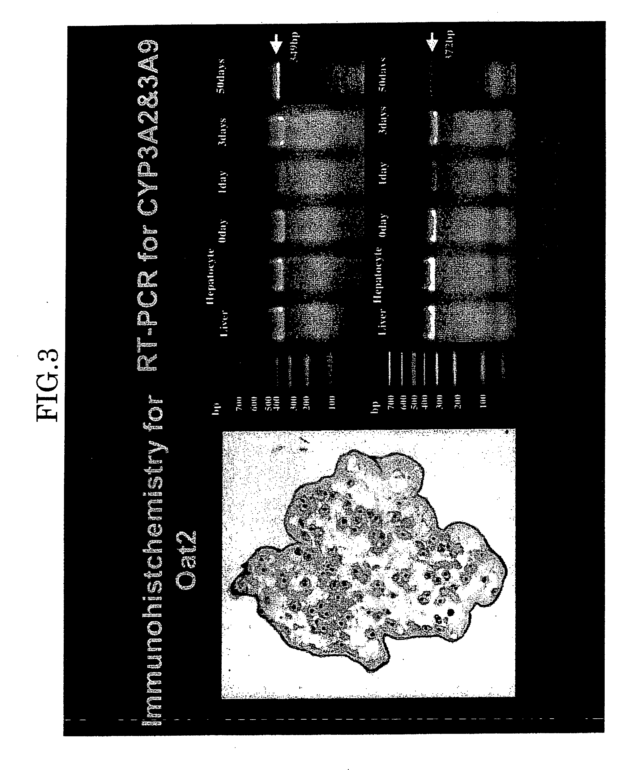
[0061] The hepatocytes were microencapsulated by the alginate / poly-L-lysine method, and then suspended in DMEM containing 10% fatal bovine serum (FBS) and 10% dimethylsulfoxide. The suspension was immediately placed in liquid nitrogen and cryopreserved. The cryopreserved hepatocyte microcapsules in the immunoisolation membrane were unfrozen with the lapse of time in a water bath at 37° C., and histogenetic evaluations (Hematoxylin & Eosin (H-E), PAS (Periodic Acid Schiff Staining) albumin (ALB) and drug-metabolizing capacity (cytochrome P450IIIA2, 9) were examined for them. The microencapsulated rat hepatocytes were unfrozen 1st day, 7th day, 14th day, 28th day and 90th day after beginning the cryopreservation, and their cell activity was examined. They exhibited “positive” for ALB and PAS, and expression of OAT2 and CYP450III1A2 and 9 was observed. It was also revealed that the cryopreserved microencapsulated rat hepatocytes maintained viability.

[0062]FIG. 1 s...

experiment 2

[0063] (Experiment 2)

[0064] The function of the rat hepatocytes in the immunoisolation membranes was histogentically evaluated with respect to specific hepatocyte-metabolizing capacity (albumin synthesis ability and urine nitrogen synthesis ability, H-E, PAS, ALB).

[0065] The immunoisolation membrane-enclosed cells cryopreserved for one week were unfrozen in water bath at 37° C., and cultured for one week. FIG. 4 shows results on this culturing. FIG. 4 shows H-E staining test results in an upper portion and albumin staining test results in an lower portion (1st day, 3rd day and 7th day from the left), revealing that the cryopreserved capsules maintained the metabolizing function peculiar to the liver even after one-week cultivation.

[0066]FIG. 5 shows results in observing synthesized amounts of urea during culturing, which demonstrates that the cryopreserved rat cells in the immunoisolation membranes exhibited higher urea synthesis and cell activity (an upper polygonal line in FIG. ...

experiment 3

[0067] (Experiment 3)

[0068] The cryopreserved hepatocytes in the immunoisolation membranes originated from the primary human hepatocytes obtained in the same manner as in Experiment 1 were unfrozen in water bath at 37° C. The function of the rat hepatocytes in the immunoisolation membranes was histogentically evaluated with respect to specific hepatocyte-metabolizing capacity (albumin synthesis ability and urine nitrogen synthesis ability, H-E, PAS, ALB). It was examined whether the cryopreserved hepatocytes in the immunoisolation membranes functioned normally or not.

[0069]FIG. 6 gives photographs obtained by observing with a phase microscope, preparations in which unfrozen human hepatocytes in an immunoisolation membrane was treated with formalin and fixed with paraffin, which shows that the shape of the human hepatocytes in the immunoisolation membrane did not change by freezing, unfreezing and culturing. FIG. 7 gives photographs in H-E staining results of human hepatocytes in an...


PUM


Abstract

A method is disclosed for cryopreserving living animal cells in immunoisolation membranes, including: (1) cutting out a living organ from an animal, (2) digesting the cutout organ into the discrete living animal cells and separating the discrete cells, (3) suspending the separated cells in a solution of sodium chloride containing sodium alginate and collagen, (4) forming microcapsules of the living animal cells by using the resulting suspension, (5) forming immunoisolation membranes around outer surfaces of the microcapsules of the living animal cells by covering the outer surfaces with alginate-(poly-L-lysine) and thereby obtaining the living animal cells enclosed in the immunoisolation membranes, (6) suspending the resulting living animal cells enclosed in the immunocapsules in a cell damage-preventing solution, and (7) immediately with liquid nitrogen.

Description

[0001] A method for cryopreserving microencapsulated living animal cells enclosed in immunoisolation membranes, such microencapsulated living animal cells in immunoisolation membranes, and biohybrid artificial organ modules using such microencapsulated living animal cells in immunoisolation membranes BACKGROUND OF THE INVENTION [0002] (1) Field of the Invention [0003] The present invention relates to a method for cryopreserving microencapsulated living animal cells enclosed in immunoisolation membranes, such microencapsulated living animal cells enclosed in the immunoisolation membranes, and biohybrid artificial organ modules using such microencapsulated living animal cells enclosed in the immunoisolation membranes. In particular, the present invention relates to a method for cryopreserving microencapsulated living hepatocytes in immunoisolation membranes, such microencapsulated living hepatocytes in the immunoisolation membranes, and biohybrid artificial organ modules using such mi...

Claims


Application Information

Patent Timeline
IPC IPC(8): A01N1/02A61K45/00
CPCA01N1/0221A01N1/02
Inventor AOKI, TAKESHIKUSANO, MITSUOSHIMIZU, YOSHINORIKOIZUMI, TOMOTAKEYASUDA, DAISUKEIZUMIDA, YOSHIHIKOMURAI, NORIYUKIJIN, ZHENGHAOKOBAYASHI, YASUNAKATO, HIROHISAHUA, LUCHUN
Owner SHOWA UNIVERSITY
Why Eureka
Try Eureka