Eureka AIR delivers breakthrough ideas for toughest innovation challenges, trusted by R&D personnel around the world.

Activation of natural killer (NK) cells and methods of use

a technology applied in the field of natural killer cells and methods of use, can solve the problems of unknown natural reservoir of filoviruses, poor immune responses of natural killer cells (nk), t, uncontrolled spread and growth of virus, etc., and achieves enhanced immune response, high-effect adjuvant effect, and enhanced immune system response

Inactive Publication Date: 2006-05-11
BAVARI SINA +1
View PDF1 Cites 3 Cited by
  • Summary
  • Abstract
  • Description
  • Claims
  • Application Information

AI Technical Summary

Benefits of technology

[0008] Thus current methods for activating NK cells in vitro involve culturing such cells in the presence of different cytokines (such as IL-1, IL-2, IL-12, IL-15, IFNα, IFNγ, IL-6, IL-4, IL-18 in certain circumstances), used alone or in combination, which activation can be considerably increased by adhesion factors or co-stimulation factors such as ICAM, LFA or CD70. Similarly, in vivo, the efficacy of NK cells in anti-tumoral immunity is not dissociable from co-administration of cytokines such as IL-2 / IL-15 or IL-12, IL-18, and IL-10. The activation methodologies described in the prior art thus all depend on using cytokines. Such methods have certain disadvantages, however, linked to the cost of preparing the cytokines, to the toxic nature of many cytokines, which cannot be used in in vivo applications, or to the non-specific nature of many cytokines, the in vivo use of which risks being accompanied by undesirable effects. Further, since the natural killing function is often altered in patients with tumors, the possibility of collecting such cells to activate them ex vivo can be considerably reduced.
[0009] There is thus a real need for novel methods for expanding and activating NK cells to enhance both cellular immunity mediated by cytotoxic T lymphocytes and humoral immunity mediated by antibodies. The present application provides a solution to this problem. In particular, the present application demonstrates for the first time the possibility of activating resting NK cells with virus-like particles (VLPs). The present application also describes, for the first time, a method of activating NK cells which is not dependent on the presence of cytokines, and which can thus overcome the disadvantages described in the prior art. The present invention thus describes novel methods for preparing activated natural killer cells and means for carrying out these novel methods.
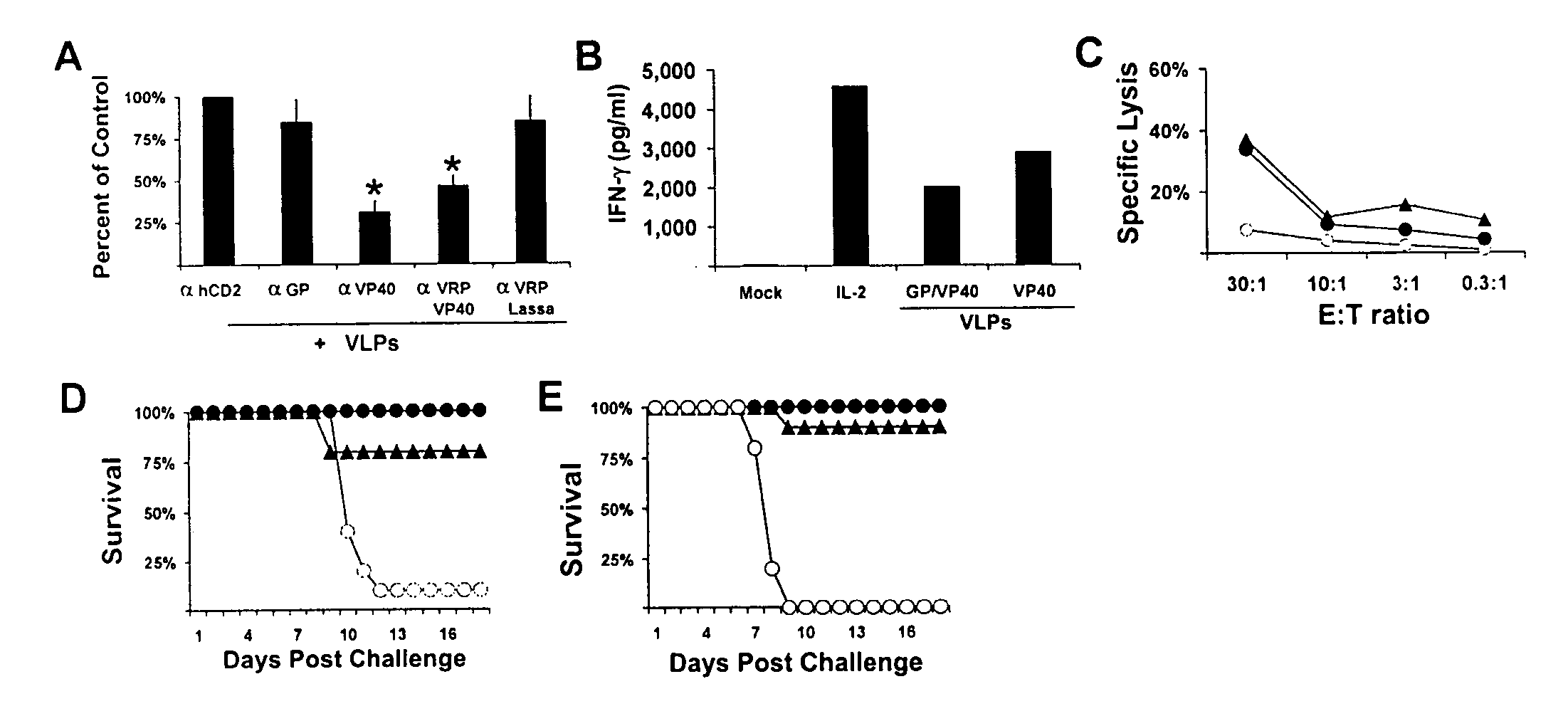
[0011] The present invention satisfies the needs discussed above. The present invention is directed to a composition and method for activating NK cells in order to enhance the immune system response against a foreign cell or organism. When the composition of the invention is administered with an immunogen, the composition enhances the immune response to said immunogen and therefore constitutes a highly effective adjuvant. In addition, we found that Ebola VLPs enhanced the number of natural killer cells in lymphoid tissue. Ebola VLPs containing only the matrix viral protein (VP)40 were sufficient to induce natural killer cells responses and provide protection from infection in the absence of the viral glycoprotein.
[0012] We have previously shown that virus-like particles, comprised of the EBOV glycoprotein (GP) and VP40 efficiently mature and activate murine and human myeloid dendritic cells (Warfield K. L., 2003, Proc. Natl. Acad. Sci. USA., 100, 15889-15894; Bosio C. M., 2004, Virology, 326, 280-287). In addition to their potent activation of DC, which are critical mediators of innate and adaptive immune responses, VLP activate T and B cells in vivo following intraperitoneal administration to mice (Warfield, 2003, supra). Therefore, since VLP are highly immunogenic in mice in the absence of adjuvant, we utilized the genome-free Ebola VLPs to study the contribution of NK cells to innate immune responses to lethal EBOV infection. We found that VLPs enhanced the number of natural killer cells in lymphoid tissue. VLPs containing only VP40 were sufficient to induce natural killer cells responses and provide protection from infection in the absence of the viral glycoprotein.

Problems solved by technology

Unfortunately, the natural reservoir of filoviruses is not known.
This lack of DC activity most likely results in poor immune responses by natural killer (NK), T, and B cells, which in turn contributes to the uncontrolled spread and growth of the virus.
Therefore, the rapid initiation of early immune responses may limit EBOV infection, and is critically linked to host survival.
In this respect, current techniques for activating NK cells are all based on using cytokines, generally in high doses which are not tolerated well by the host.
Such methods have certain disadvantages, however, linked to the cost of preparing the cytokines, to the toxic nature of many cytokines, which cannot be used in in vivo applications, or to the non-specific nature of many cytokines, the in vivo use of which risks being accompanied by undesirable effects.

Method used


Image

  • Activation of natural killer (NK) cells and methods of use
  • Activation of natural killer (NK) cells and methods of use
  • Activation of natural killer (NK) cells and methods of use

Examples


example 1

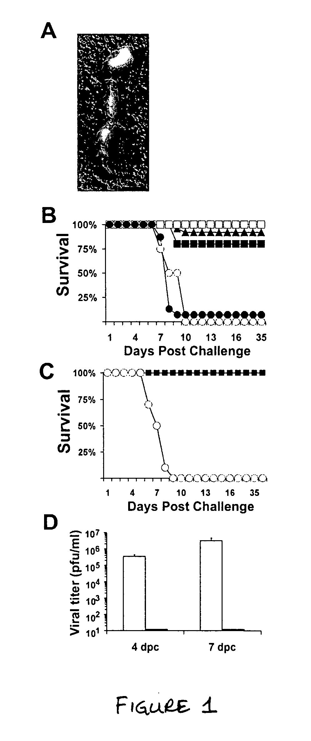
[0104] VLPs rapidly induce protection from lethal EBOV infection. Morphologically, the VLPs are almost indistinguishable from inactivated EBOV by electron microscopy (Warfield et al., 2003, supra; Bavari et al., 2002, J. Exp. Med. 195, 593-602) or by atomic force microscopy (FIG. 1A and (Feldmann et al., 2003, Nat. Rev. Immunol. 3, 677-685). The VLPs induced potent innate immune responses, as mice injected intraperitoneally once with VLPs, 1-3 days before challenge with more than 3,000 LD50 of EBOV (Bray et al., 1999, supra) were 80-100% protected from death (FIG. 1B). However, mice injected 3 days before challenge with either irradiated, inactivated EBOV or the sucrose-purified supernatants from mock-transfected cells succumbed to EBOV challenge (FIG. 1B). Irradiating the VLPs had no effect on the outcome of these experiments (unpublished observations), suggesting that the failure of the inactivated EBOV to protect mice from EBOV infection was not simply due to the irradiation. Int...

example 2

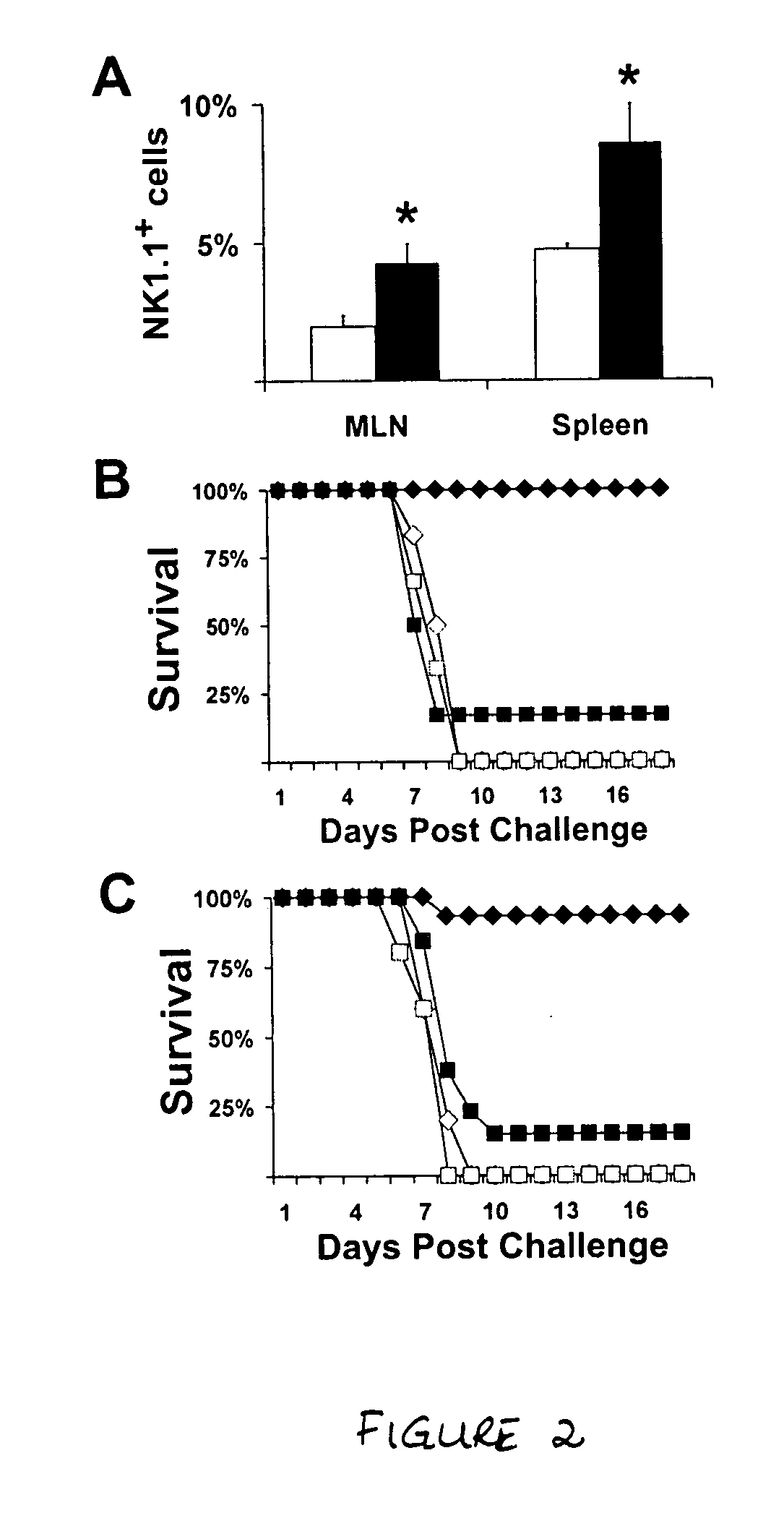
[0105] Innate protection against EBOV requires NK cells. Although many different factors may have contributed to VLP-induced innate protection, we narrowed our search to the role of NK cells. Marked increases in NK cell activity occur early in microbial invasions and results in the recruitment of NK cells to the site of infection (Yokoyama and Scalzo, 2002, supra). VLPs recruited almost twice the number of NK cells in both the mediastinal lymph node and spleen compared to animals receiving PBS alone (FIG. 2A), suggesting VLP administration induces NK cell proliferation and / or trafficking in lymphoid tissues. To directly examine the role of NK cells in EBOV infections, NK cell-deficient mice (Kim et al., 2000, supra) were administered VLPs 3 days prior to lethal EBOV challenge. VLP-pretreatment of mice lacking functional NK cells did not protect from EBOV infection (1 / 6, FIG. 2B), unlike VLP-injected wild-type C57Bl / 6 mice (6 / 6, P=0.0076). Further, mice depleted of NK cells using ant...

example 3

[0107] NK cell responses to Ebola virus. Ebola VLPs are morphologically and antigenically similar to live EBOV [FIG. 1A and Warfield et al., 2003, supra; Bavari et al., 2002, supra; Swenson et al., 2004, FEMS Immunol. Med. Microbiol. 40, 27-31]. However, unlike VLPs, inactivated EBOV did not induce innate protection from EBOV infection or stimulate NK cell responses in vitro (FIG. 1B). Therefore, we set out to determine if murine NK cells possessed the ability to respond to live EBOV. Unlike exposure to IL-2 or VLPs, live EBOV did not induce secretion of IFN-γ, MIP-1α, or TNF-α from NK cells (FIG. 4A-C).

[0108] Several viruses, including human cytolomegalovirus, HIV, and Epstein-Barr virus replicate efficiently in NK cells (Rice et al., 1984, Proc. Natl. Acad. Sci. USA 81, 6134-6138; Chehimi et al., 1991, J. Virol. 65, 1812-1822; Kanegane et al., 2002, Crit. Rev. Oncol. Hematol. 44, 239-249; Valentin and Pavlakis, 2003, Anticancer Res. 23, 2071-2075). To determine whether the lack o...


PUM

PropertyMeasurementUnit
Compositionaaaaaaaaaa
Immunogenicityaaaaaaaaaa

Abstract

The present invention relates to filovirus VLPs and their use in activating innate immunity, specifically natural killer cells, and in enhancing an immune response to an antigen in an animal.

Description

[0001] This application is claims the benefit of priority under 35 U.S.C. 119(e) from U.S. Application Ser. No. 60 / 562,803 filed on Apr. 13, 2004, still pending and herein incorporated by reference in its entirety.INTRODUCTION [0002] Marburg (MARV) and Ebola (EBOV) viruses, members of the family Filoviridae, cause an acute and rapidly progressive hemorrhagic fever with mortality rates up to 90% (Feldmann H., 1996, Arch. Virol. Suppl., 11, 77-100). These viruses are fast-acting, with death often occurring within seven to ten days post infection; however, the incubation period is considered to be two to twenty-one days (Borio L., 2002, JAMA, 287, 2391-2405; Peters C. J., 1999, J. Infect. Dis., 179 Suppl. 1, 9-16). Unfortunately, the natural reservoir of filoviruses is not known. Filoviruses are transmitted through contact with bodily fluids or tissues of humans or nonhuman primates (Brown D. W., 1997, Rev. Med. Virol., 7, 239-247; Pinzon J. E., 2004, Am. J. Trop. Med. Hyg., 71, 664-67...

Claims


Application Information

Patent Timeline
IPC IPC(8): C12Q1/68C12P21/06
CPCA61K39/12A61K2039/5258A61K2039/555A61K2039/55577C07K16/10C12N2760/14123C12N2760/14134C12N2760/14223C12N2760/14234A61P31/14A61P35/00A61P37/04A61P37/08A61K39/464838A61K39/4613A61K2039/5158
Inventor BAVARI, SINAWARFIELD, KELLY L.
Owner BAVARI SINA
Why Eureka
Eureka Blog
Learn More