Biodegradable dual porous scaffold wrapped with semi-permeable membrane and tissue cell culture using thereof

a biodegradable, semi-permeable membrane technology, applied in the direction of prosthesis, biomass after-treatment, bandages, etc., can solve the problems of scaffolds with relatively low porosities, uncontrollable pore size, and non-absorbable cartilage substitutes used up to date that develop various side effects and complications

Inactive Publication Date: 2006-07-06
IND ACADEMIC CORP FOUND YONSEI UNIV
View PDF5 Cites 39 Cited by
  • Summary
  • Abstract
  • Description
  • Claims
  • Application Information

AI Technical Summary

Benefits of technology

[0016] Further, the present invention aims to provide a method of proliferating tissue cells, including sectioning a scaffold into small pieces; seeding tissue cells onto each of the scaffold pieces and loading the scaffold pieces into a mold having a morphology of a tissue to be regenerated; adding a mixture of a semi-permeable

Problems solved by technology

However, the non-absorbable cartilage substitutes used up to date develop various side effects and complications, such as skin necrosis and inflammation.
However, the conventional fabrication techniques generally result in scaffolds with relatively low porosities, uncontrollable pore size and poorly interconnected, open-pore networks.
Also, when tissue cells are seeded onto the scaffold and proliferated thereon, the pores on the surface of the scaffold are often blocked, thereby causing difficulty in preparation of grafts.
Thus, the conventional techniques furthe

Method used


Image

  • Biodegradable dual porous scaffold wrapped with semi-permeable membrane and tissue cell culture using thereof
  • Biodegradable dual porous scaffold wrapped with semi-permeable membrane and tissue cell culture using thereof
  • Biodegradable dual porous scaffold wrapped with semi-permeable membrane and tissue cell culture using thereof

Examples


example 1

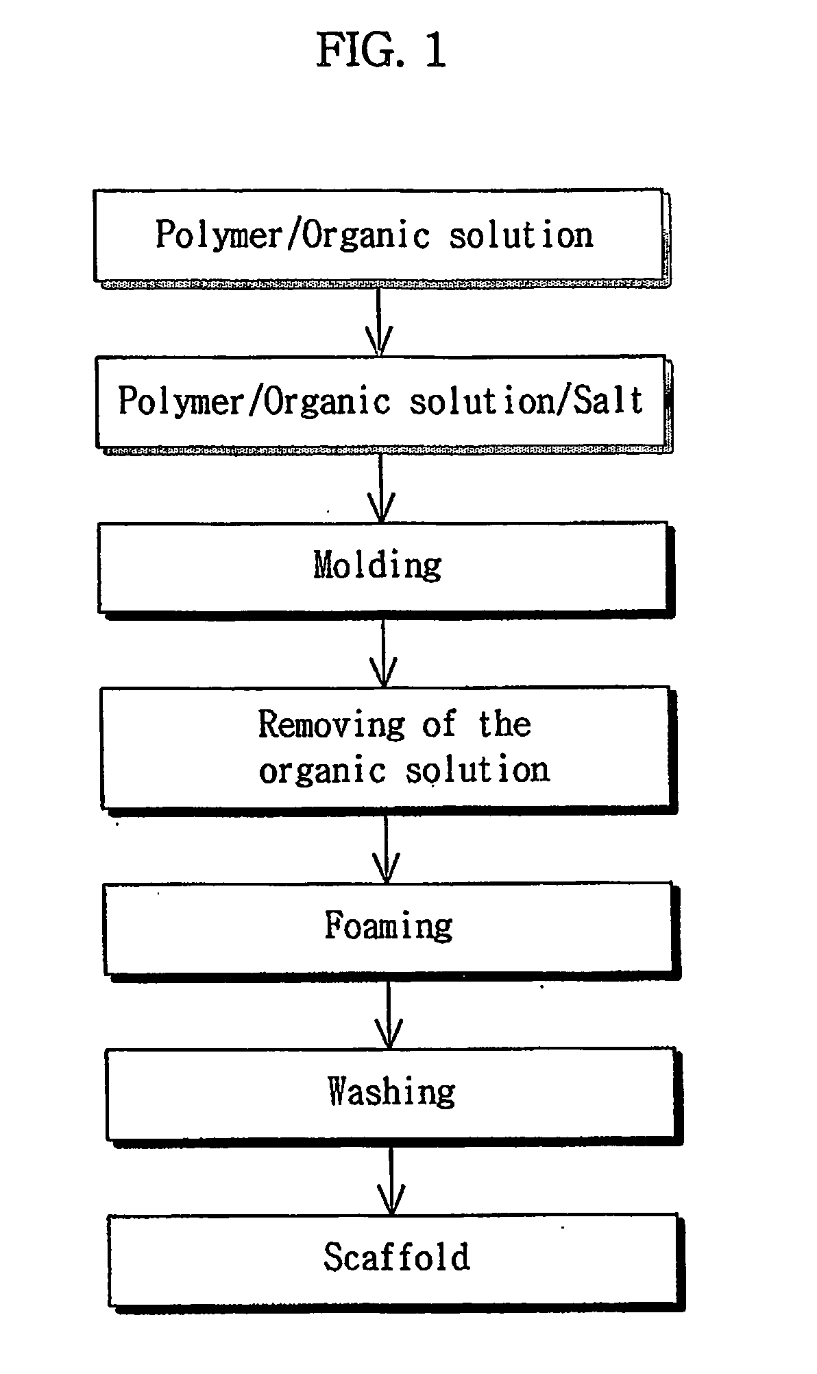
Scaffold Fabrication

[0082] A mixture solvent was prepared by mixing 90% of methylchloride (Ducksan Chemical Co. Ltd., Korea) and 10% of dimethylsulfoxide (Sigma, USA). 10% of PLGA, based on the weight of the mixture solvent, which was composed of lactic acid (Sigma, USA) and glycolic acid (Sigma, USA) at a weight ratio of 75:25, molecular weight of 90,000 to 126,000 Da, was dissolved in the mixture solvent.

[0083] The resulting solution was mixed with ammonium hydrogen carbonate (Junsei Chemical Co. Ltd., Japan) having a particle size of 150-250 μm at a weight ratio of 9:1. The resulting mixture was poured into a cylinder-type schale in size of 100 mm in diameter (Dongsung science Co. Ltd, Korea).

[0084] The methylchloride (Ducksan Chemical Co. Ltd., Korea) contained in the mixture filled into the schale (Dongsung Science Co. Ltd, Korea) was partially evaporated, thus generating a semi-solidified sample.

[0085] Thereafter, the semi-solidified sample was immersed in 30% acetic acid ...

example 2

Regeneration of a Biological Tissue

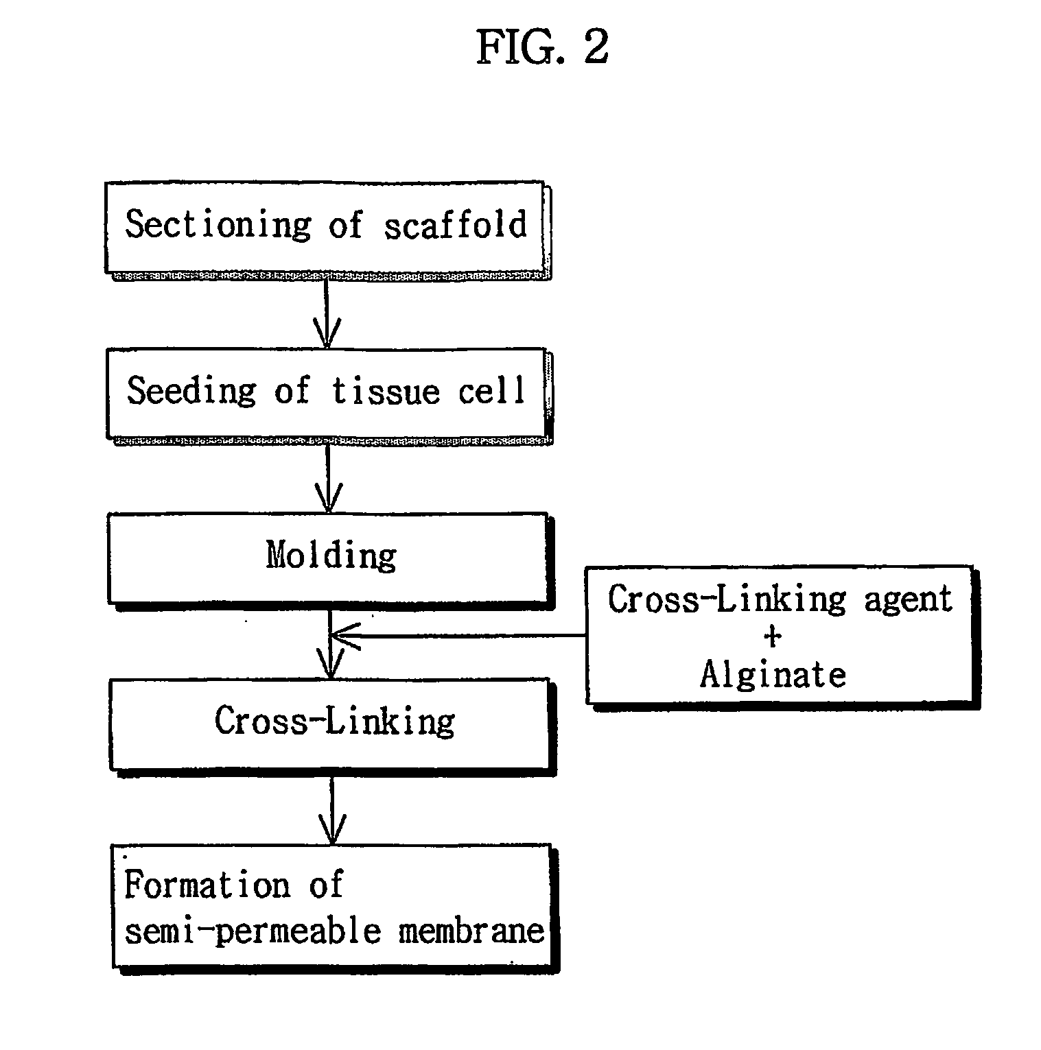
(i) Seeding of Tissue Cells Onto the Scaffold

[0090] The scaffold prepared in Example 1 was sectioned into pieces at the size of 2 mm in diameter and 1 mm thick. Chondrocytes isolated from rabbits were seeded onto the scaffold pieces at a density of 1.0×10⁶ cells / ml.

[0091] The scaffold pieces were incubated at 37° C. in high humidity containing 5% concentration of CO2 for about three hours to allow for cross-linking of the scaffold pieces. Then, they were incubated for three days in DMEM (JBI, Korea) containing 4 ml of an antibiotic / antifungal solution and FBS (Sigma, USA).

(ii) Formation of a Semi-permeable Membrane on an Outer Surface of the Scaffold and Proliferation of the Tissue Cells.

[0092] A 3% sodium alginic acid solution (Sigma, USA) was mixed with an equal volume of DMEM (JBI, Korea) containing 4 ml of an antibiotic / antifungal solution and FBS (Sigma, USA).

[0093] The resulting mixed solution was put into a 50-ml tube (Nunc, USA) con...

experimental examples

Evaluation of Cell Proliferation

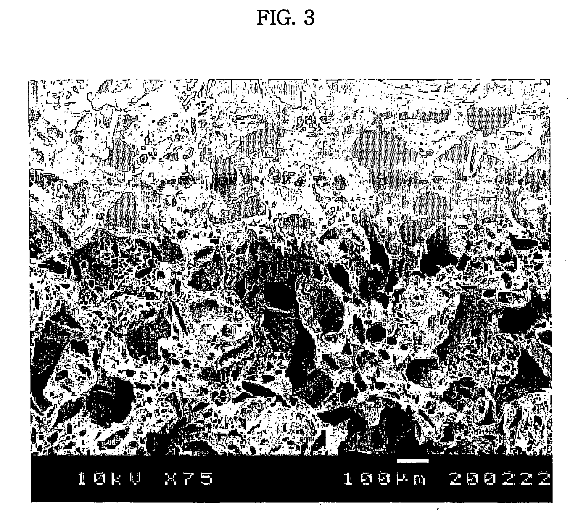
[0096] The gel beads prepared in Example 3 were incubated at 37° C. in high humidity containing 5% CO2 with 90 rpm of shaker's agitation. The culture medium was replaced by a fresh one every three or four days. On Days 7, 14, 21 and 31, to evaluate chondrocyte proliferation, the gel beads were observed under a scanning electron microscope (S-800, Hitachi, Japan), and while a quantitative DNA assay was measured by fluorescence using a luminescence spectrometer (Luminescence Spectrometer LS50B, PERKIN ELMER, Great British).

[0097] The results are given in FIGS. 6, 7 and 8.

[0098]FIG. 6 shows SEM images showing chondrocytes that have been proliferated on the scaffold having a semi-permeable membrane on an outer surface thereof according to the present invention. FIG. 7 is a graph showing DNA synthesis of chondrocytes grown on the scaffold according to the present invention. FIG. 8 is a graph showing glycosaminoglycan synthesis of chondrocytes grown on ...


PUM

PropertyMeasurementUnit
Sizeaaaaaaaaaa
Sizeaaaaaaaaaa
Biological propertiesaaaaaaaaaa

Abstract

Disclosed is a scaffold including a semi-permeable membrane on an outer surface thereof. The present invention also discloses a method of preparing a scaffold covered with a semi-permeable membrane, including loading one or more scaffolds into a mold with a predetermined form and size; and adding a semi-permeable agent and a cross-linking agent to the mold and cross-linking the semi-permeable agent to form the semi-permeable membrane on the outer surface of each of the scaffolds. The scaffold covered with the semi-permeable membrane selectively introduces nutrients into the scaffold by allowing penetration of only external nutrients into the scaffold and excreting metabolic wastes generated by tissue cells to the outside of the scaffold. In addition, the scaffold has the morphology of a biological tissue of interest by cross-linking the small-sized scaffolds, thereby allowing uniform proliferation of tissue cells throughout the whole scaffold.

Description

TECHNICAL FIELD [0001] The present invention relates, in general, to a scaffold and a method of preparing biological tissues using the scaffold. More particularly, the present invention relates to regeneration of biological tissues by preparing a porous scaffold by gas foaming of an effervescent salt using a biodegradable polymer, sectioning the scaffold into small pieces, seeding tissue cells onto the scaffold pieces, forming a semi-permeable membrane on an outer surface of each of the scaffold pieces, and cross-linking the semi-permeable membrane-covered scaffold pieces into a predetermined form. [0002] The term “scaffold”, as used herein, refers to a porous biodegradable polymer construct to support cell growth and migration. BACKGROUND ART [0003] Typically, bone cartilage is a tissue that is not naturally regenerated once damaged. To repair damaged cartilage tissues, cartilage substitutes such as non-absorbable biological substances have been used. However, the non-absorbable ca...

Claims


Application Information

Patent Timeline
IPC IPC(8): A61F13/00C12N5/08A61L27/20A61L27/56C12N5/00
CPCA61F2002/30762A61F2002/30766A61L27/20A61L27/56C12N5/0068C12N2533/40C12N2533/74C08L5/04C12N11/02C12N5/0602
Inventor KIM, JUNG-HYUNLEE, HYE-WONCHOI, SUNG-WOOK
Owner IND ACADEMIC CORP FOUND YONSEI UNIV
Why Eureka
Try Eureka