
Wound care polymer compositions and methods for use thereof

a polymer composition and wound care technology, applied in the field of wound care polymer compositions, can solve the problems of exacerbated injury, increased thrombosis of damaged arterial surfaces within the vascular system, and precipitated new smooth muscle cell proliferation and neointimal growth

Inactive Publication Date: 2006-08-24
MEDIVAS LLC
View PDF17 Cites 104 Cited by
  • Summary
  • Abstract
  • Description
  • Claims
  • Application Information

AI Technical Summary

Benefits of technology

[0032] In still another embodiment, the invention provides a multilayer bioactive wound dressing that includes a non-stick layer comprising a biodegradable hydrogel; a supporting layer of a biodegradable polymer having a

Problems solved by technology

However, damaged arterial surfaces within the vascular system are highly susceptible to thrombus formation.
However, these techniques also serve to exacerbate the injury, precipitating new smooth muscle cell proliferation and neointimal growth.
The effectiveness of this procedure is limited in some patients because the treatment itself damages the vessel, thereby inducing proliferation of smooth muscle cells and reocclusion or restenosis of the vessel.
However, tissue surrounding a porous stent tends to infiltrate the pores.
In certain applications, pores that promote tissue ingrowth are considered to be counterproductive because the growth of neointima can occlude the artery, or other body lumen, into which the stent is being placed.
However, the effect of nitric oxide in the regulation of apoptosis is complex.
This situation results in restenosis following vessel injury, for example following angioplasty.
Damage to the endothelial and medial layers of a blood vessel, such as often occurs in the course of balloon angioplasty and stent procedures, has been found to stimulate neointimal proliferation, leading to restenosis of atherosclerotic vessels.
An unfortunate consequence of this procedure is the nearly total destruction of the endothelial layer by expansion of the angioplasty balloon and precipitation of foreign body inflammatory response to the stent.
Since mechanical intervention has destroyed the natural blood / artery barrier, all too often the result is a local uncontrolled proliferative response by smooth muscle cells leading to restenosis.
Initial wound hypoxia is important for fibroblast proliferation and angiogenesis; however, continued hypoxia at the wound site delays wound healing.
As a result, if an occlusive dressing is continuously applied to an ischemic wound, healing is severely impaired.
These wounds can sometimes have large amounts of exudates that require frequent debridement.

Method used


Image

  • Wound care polymer compositions and methods for use thereof
  • Wound care polymer compositions and methods for use thereof
  • Wound care polymer compositions and methods for use thereof

Examples


example 1

[0266] Amide Bond Formation This example illustrates the coupling of a carboxyl group of a polymer with an amino functional group of the bioactive agent, or equally, the coupling of a carboxyl group of the bioactive agent with an amino functional group of a polymer.

[0267] Coupling Through Pre-Formed Active Esters; Carbodiimide Mediated Couplings—Conjugation of 4-Amino-Tempo to Polymer The free carboxylic acid form of the PEA polymer is converted first to its active succinimidyl ester (PEA-OSu) or benzotriazolyl ester (PEA-OBt). This conversion can be achieved by reacting dried PEA-H polymer (i.e. PEA with free pendant carboxylic acids) with N-Hydroxysuccinimide (NHS) or 1-Hydroxybenzotriazole (HOBt) and a suitable coupling agent, such as dicyclohexylcarbodiimide (DCC), in anhydrous CH2Cl2 at room temperature for 16 hrs. After filtering away the precipitated dicyclohexylurea (DCU), the PEA-OSu product may be isolated by precipitation, or used without further purification, in which c...

example 2

[0269] Ester Bond Formation This example illustrates coupling of a carboxyl group of a polymer with a hydroxyl functional group of the bioactive agent, or equally, coupling of a carboxyl group of the bioactive agent with a hydroxyl functional group of a polymer.

[0270] Carbodiimide Mediated Esterification For the conjugation, a sample of the carboxyl-group-containing polymer was dissolved in DCM. To this slightly viscous solution was added a solution of the hydroxyl-containing-drug / biologic and DMAP in DCM. The flask was then placed in an ice bath and cooled to 0° C. Next, a solution of 1,3-diisopropylcarbodiimide (DIPC) in DCM was added, the ice bath removed, and the reaction warmed to room temperature. The conjugation reaction was stirred at room temperature for 16 hours during which time TLC was periodically performed to monitor consumption of the hydroxyl functional group of the bioactive agent. After the allotted time, the reaction mixture was precipitated, and the Polymer-bioa...

example 3

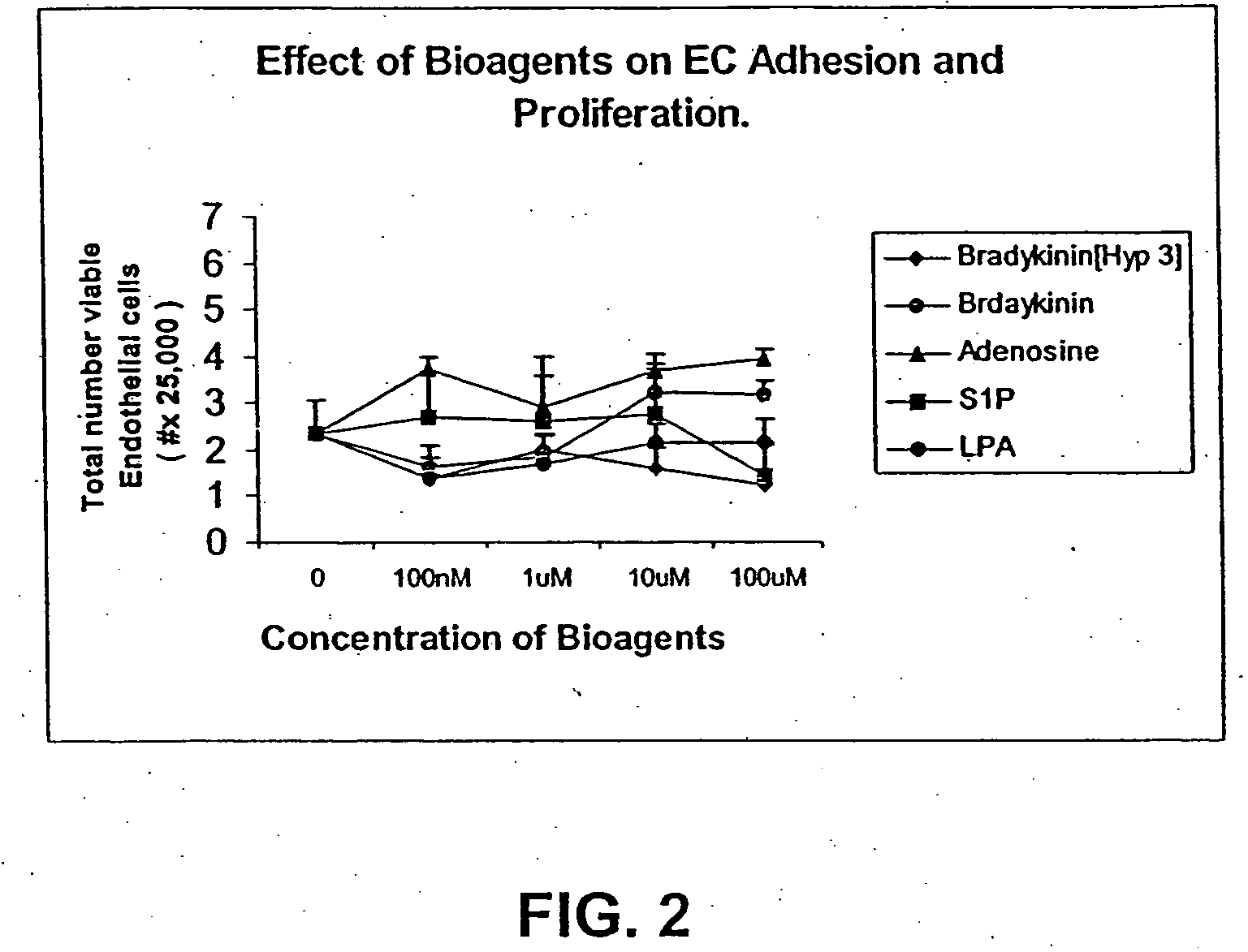
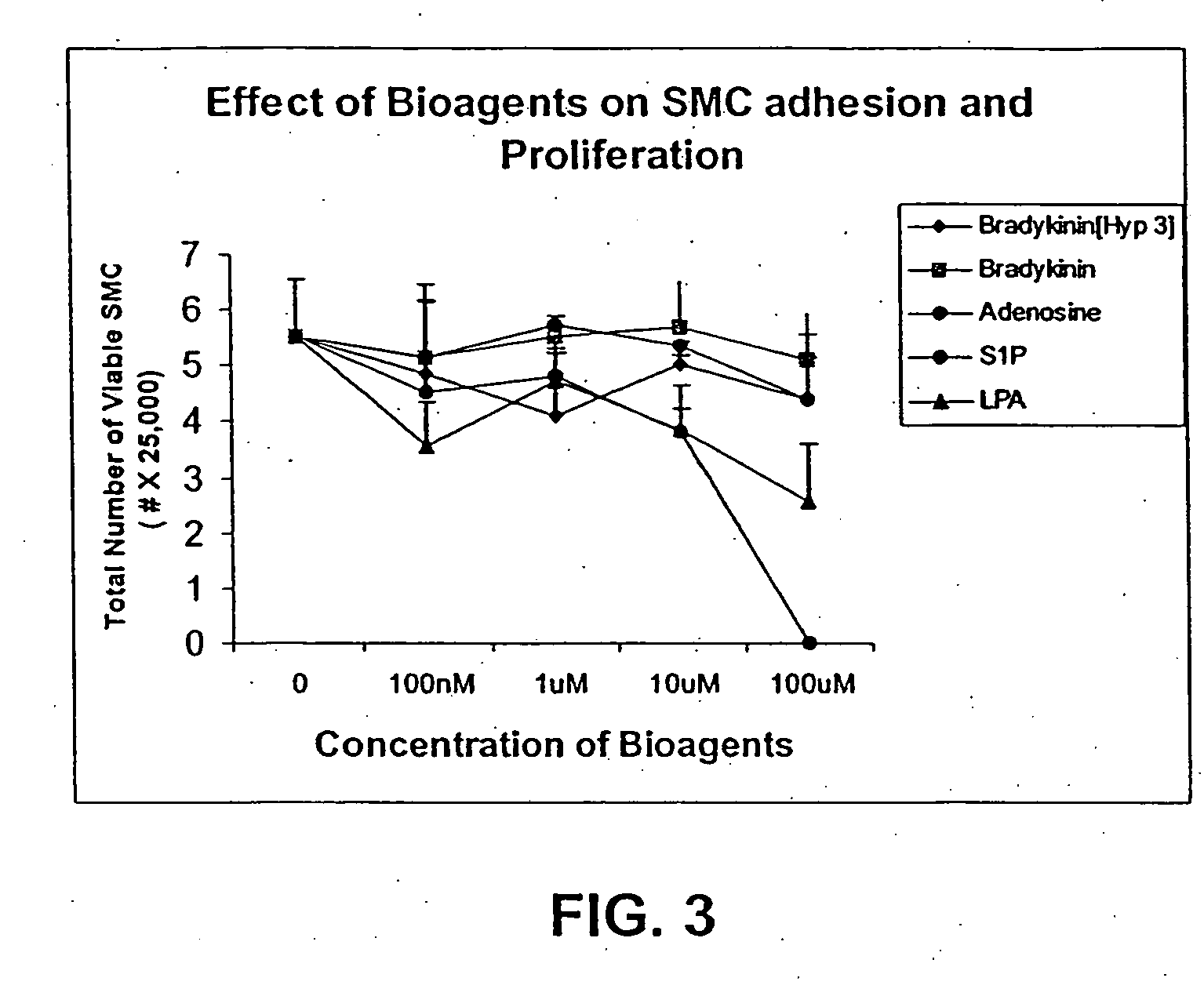
[0271] This Example illustrates the effect of different concentrations of bioactive agents on adhesion and proliferation of epithelial cells (EC) and smooth muscle cells (SMC) on gelatin coated surfaces.

[0272] Human Coronary artery endothelial cells (EC) plated on gelatin coated culture plates were co-cultured with EC special media containing one of the bioactive agents shown in Table 1 below in the various concentrations shown.

TABLE 1Bioagents100 μM10 μM1 μM100 nmABradykinin[Hyp 3]37237.233.720.372BBradykinin322.832.283.2280.3228CAdenosine80.168.0160.8160.0816DSphingosine 1-113.8511.3851.13850.11385Phosphate (S1P)ELysophosphatidic137.5513.7551.3750.1376Acid (LPA)FControlNoadditives

Cells cultured under similar conditions without adding bioagents are considered as ‘Control.’

[0273] Twenty-four hours later the cells were observed microscopically, stained with trypan blue and counted. The results of the microscopic observation of cell morphology and confluency of culturing the EC in...


PUM

PropertyMeasurementUnit
Timeaaaaaaaaaa
Timeaaaaaaaaaa
Timeaaaaaaaaaa

Abstract

The present invention provides wound healing or wound care polymer compositions that can be formulated to release a wound healing agent at a controlled rate by adjusting the various components of the composition. The compositiona can be used in an external wound dressing, as a polymer implant for delivery of the wound healing agent to an internal body site, or as a coating on the surface of an implantable surgical device to deliver wound healing agents that are dispersed in a biodegradable polymer or hydrogel, or both. Methods of using the invention bioactive polymer compositions to deliver wound healing agents that promote natural healing of wounds, especially chronic wounds, are also provided.

Description

RELATED APPLICATIONS [0001] This application is a continuation-in-part and relies for priority under 35 U.S.C. § 120 of U.S. Ser. No. 11 / 128,903, filed May 12, 2005, which relies for priority under 35 U.S.C. § 119(e) on U.S. Provisional Application Nos. 60 / 570,668, filed May 12, 2004, and 60 / 605,381, filed Aug. 27, 2004 and is also a continuation-in-part and relies for priority under 35 U.S.C. § 120 of U.S. Ser. No. 10 / 362,848, filed Oct. 14, 2003 and the content of each of which is incorporated herein by reference in its entirety.FIELD OF THE INVENTION [0002] The invention relates generally to compositions used in wound care and healing, and in particular to biodegradable polymer compositions that promote healing at wound sites. BACKGROUND INFORMATION [0003] The normal endothelium, which lines blood vessels, is uniquely and completely compatible with blood. Endothelial cells initiate metabolic processes, like the secretion of prostacylin and endothelium-derived relaxing factor (EDR...

Claims


Application Information

Patent Timeline
IPC IPC(8): A61K35/12A61F2/00A61L15/00A61K35/44
CPCA61L15/26A61L15/44A61L31/10A61L31/16A61L2300/80C08L79/02A61K38/18A61K38/39A61K45/06A61M35/00A61K35/44A61K2300/00
Inventor CARPENTER, KENNETH W.ZHANG, HUASHIMCCARTHY, BRENDAN J.SZINAI, ISTVANTURNELL, WILLIAM G.GOPALAN, SINDHU M.KATSARAVA, RAMAZ
Owner MEDIVAS LLC
Learn More