Since the APC
gene helps to regulate the growth of new cells (i.e., it is a
tumor suppressor gene), the inactivation of it disrupts the natural balance found in
healthy tissue and allows the over production of potentially malignant tissue.
Villous adenomas are flat and found in the
rectum more often than tubular; they also are at
increased risk of becoming malignant.
When this mutated
gene does not reconstruct the
DNA in the correct sequence, errors occur that can lead to cancerous growth.
However, these criteria do not work well in a
population-based examination for colon
cancer because up to 2% of non-hereditary colon
cancer patients meet them.
There have not been enough conclusive epidemiological studies to determine a percent intake of
dietary fat that causes
cancer or percent intake of
fiber that might be protective.
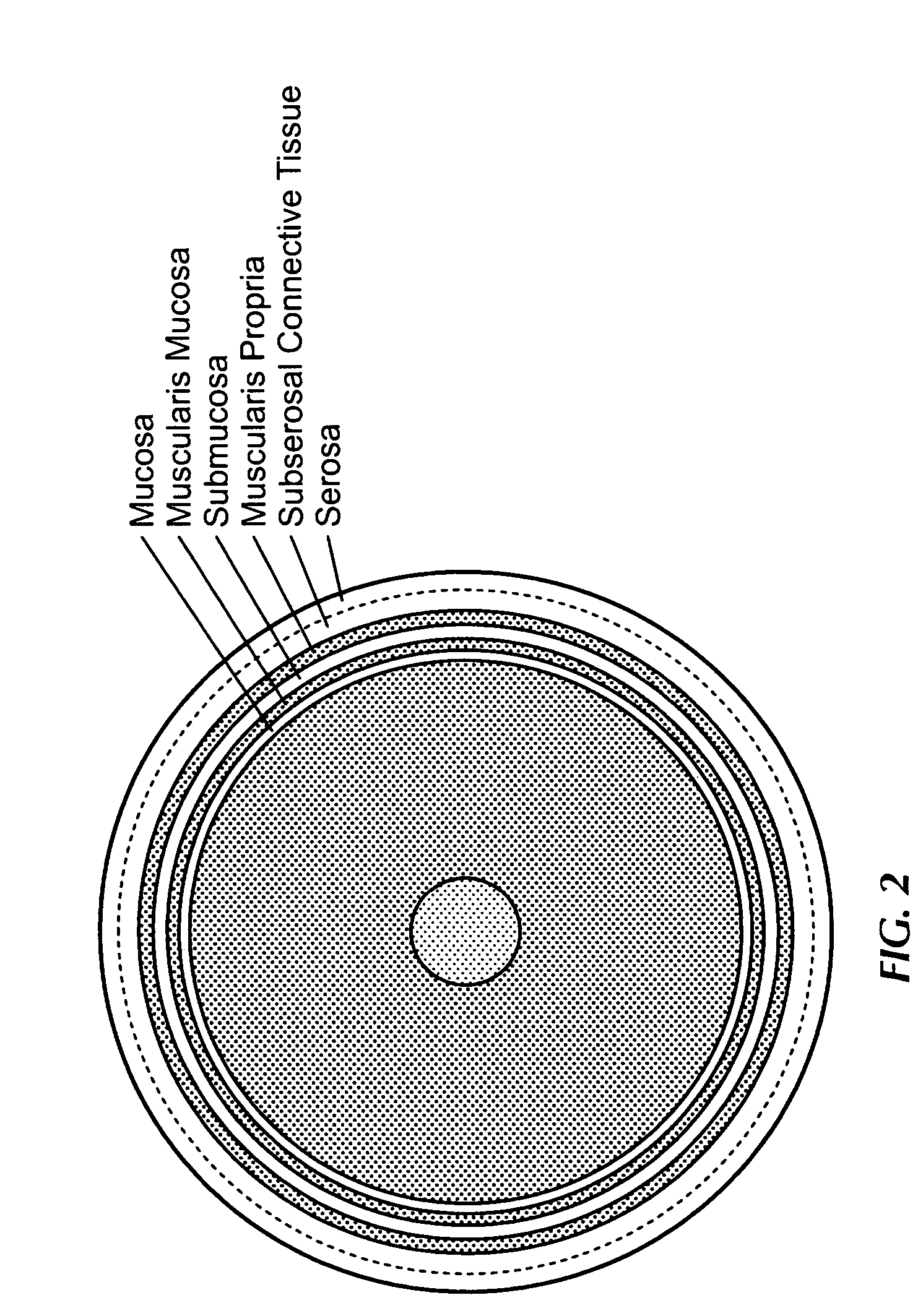
In this region, the composition of the rectal wall changes making tumor staging difficult.
Because of the
bony pelvis, the principle of wide local removal of the cancer-bearing bowel segment is subject to severe limitations by the
anatomy of the pelvic
rectum.
Currently, there is not an accurate manor to stage
lymph nodes associated with rectal cancer.
Therefore, improved
lymph node staging does not assure an improvement in the outcome of rectal cancer.
Attempts have been made to use PET for T staging of rectal cancer, but have not been successful.
Therefore, it is thought that it is useful in looking at the recurrence of the cancer, or at the spread of metastatic cells to other organs, but is currently not useful in the anatomic, primary staging of rectal cancer.
MRI has seen limited clinical use for various reasons, one of which being that there has not been an overwhelming
consensus of its utility by the surgeons treating rectal cancer.
However, with only a few exceptions, the
consensus of the majority of the experts in the field show that there are major limitations for a widespread use of MR in T staging of rectal cancer using body coils.
One major
disadvantage is that it is likely to overstage T2 tumors.
However, this same study reported some problems with this
system that limits its usefulness as a T staging technique.
This procedure requires a long examination time, the costs are relatively high, and movement-related artifacts in the images sometimes decreases the resolution so that the individual
layers of the
rectum cannot be seen.
Another study used an endorectal coil MRI and found it to be a reliable local staging technique for rectal cancer, but even though its accuracy is very high, ERUS is still the preferred method because of ease of application and cost.
However, for T staging, its many limitations were quickly discovered.
CT has a wide spread of inter- and intra-observer accuracies, and is inaccurate in the identification of
lymph node metastases.
The ability to detect local tumor extension is stage dependent, being accurate in the identification of
late stage (T4) tumors, and very poor at the identification and differentiation of T2 and T3 tumors.
However, using CT to look for recurrent tumors has problems.
But, a major difficulty with CT in detecting recurrent cancer is its insensitivity to local tumor at the anastomatic site, inability to detect tumor within a fibrotic surgical scar, and inability to differentiate between hyperplastic and tumorous lymph nodes.
Some of the problems using ERUS arise from 1) inter-user variability (including T2 vs T3 discrepancies), 2) user to user portability, 3)
adjuvant therapy, and 4)
stenosis.
Another problem encountered with the use of ERUS is that
adjuvant, preoperative radiotherapy can increase the
echogenicity of the rectal wall thereby reducing its accuracy in T staging.
ERUS is also less accurate in the lower rectum because changes in
anatomy cause the examination to be more difficult; although, the position of the tumor with respect to the circumference of the rectum does not decrease the accuracy in T staging.
There have been problems with overstaging and understaging of mid-stage tumors for two reasons: 1) the overstaging is caused by the lack of differentiation between tumor and
inflammation and is a problem associated with T2 tumors, and 2) the understaging is thought to be caused by a lack of specific, cellular information at the
microscopic level.
However, there is a
trade off for increased frequency, which is decreased imaging depth.
ERUS is limited in the
visualization of stenotic tumors of the rectum.
However, this problem will not be solved in men unless improvements are made in the ERUS systems, or another imaging modality is shown to image stenotic tumors well.
However, ERUS in its current form is not readily portable among examiners.
False
instrumentation may include problems caused by inadequate contact of
balloon to irregular tumor surface and a gap formed by air, fluid or
feces.
Any images in this category would not give the examiner a clear and accurate view of the tumor, thereby reducing the likelihood of a correct diagnosis.
The second preventable category, interpretive errors may be due to examiner
confusion and lead mostly to the overstaging of a tumor even though the exam provided clear ultrasonic images.
Imaging error due to anatomic defects are not naturally occurring defects but those caused by previous biopsies, polypectomies, or operations resulting in
edema, abscesses, or
fibrosis.
Although ERUS is presently the
standard of care for the accurate staging of rectal cancer, in its current format, it is limited by its 2D nature, lack of portability, and difficulty in obtaining accurate intra and inter examiner comparisons.
The accurate staging of rectal tumors is necessary because understaging may lead to undertreatment and overstaging results in potentially more invasive operations and subsequent increased morbidity and mortality.
Limitations of two dimensional (2D)
ultrasound that are addressed by 3D imaging include: mentally transforming multiple 2D images to form a 3D impression of the
anatomy and
pathology is not only time-consuming and inefficient, but is also more importantly, variable and subjective, which can lead to incorrect decisions in diagnosis, and in the planning and delivery of therapy; diagnostic (e.g. obstetric) and therapeutic (e.g. staging and planning) decisions often require
accurate estimation of organ or tumor volume; conventional 2D
ultrasound techniques calculate volume from simple measurements of height, width and length in two orthogonal views, by assuming an idealized (e.g. ellipsoidal) shape, which can potentially lead to low accuracy,
high variability and operator dependency; conventional 2D ultrasound is suboptimal for monitoring therapeutic procedures, or for performing quantitative prospective or follow-up studies, due to the difficulty in adjusting the
transducer position so that the 2D
image plane is at the same anatomical site and in the same orientation as in the previous examination; the validity of diagnostics with 3D as compared to 2D ultrasound is being tested, and the reported conclusions to date show 3D to be superior.
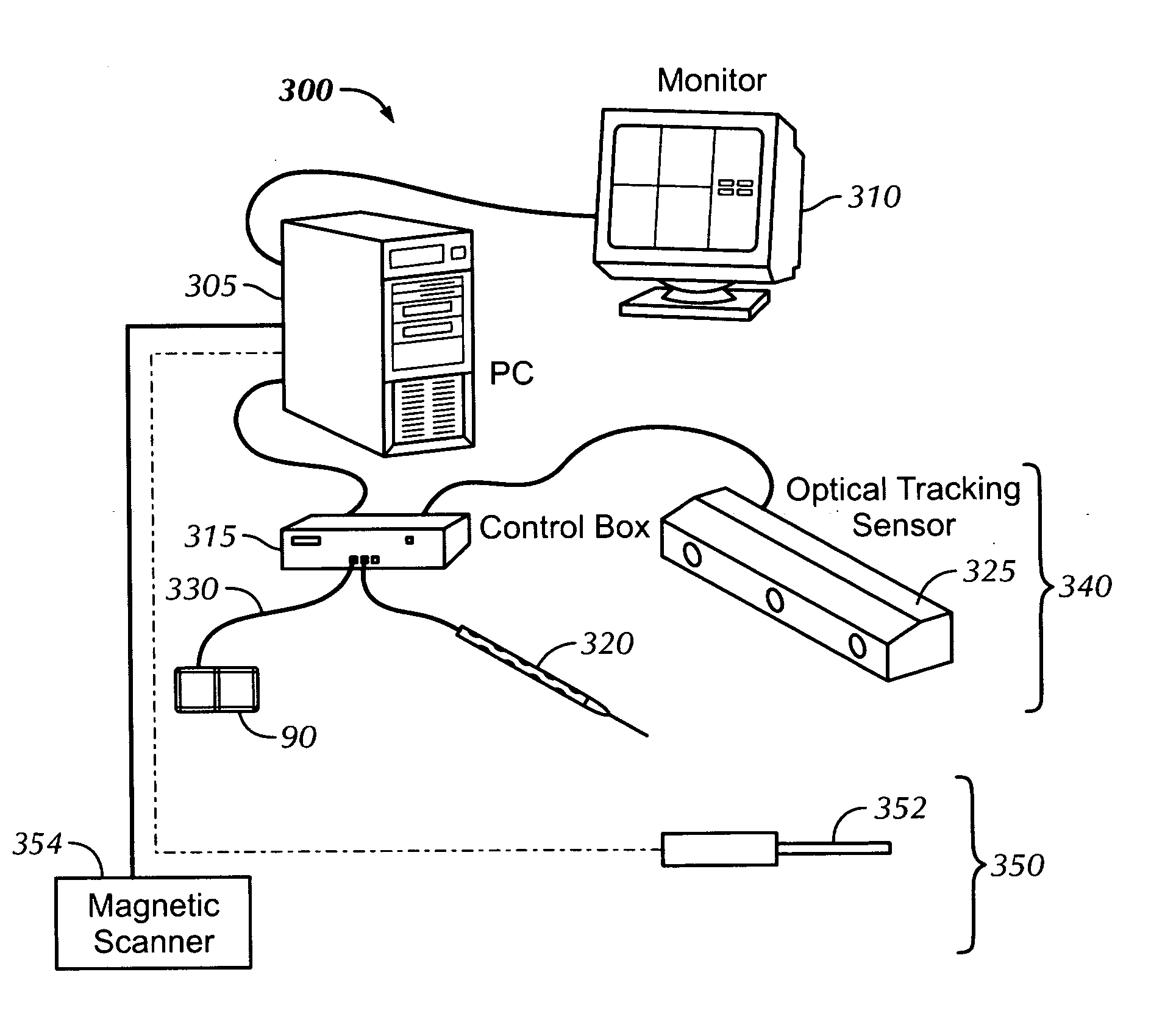
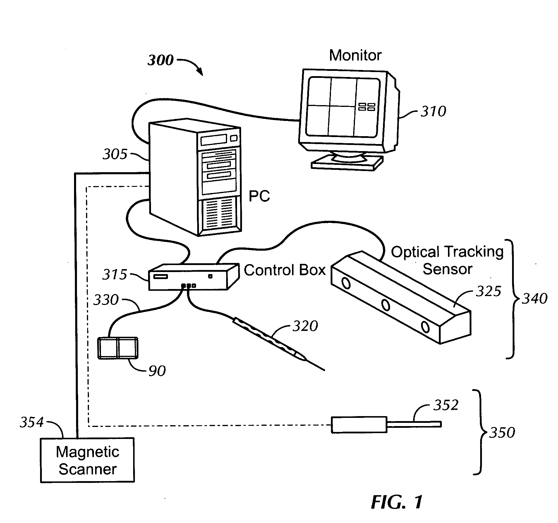
Magnetic localization systems are currently not as accurate as optical localization systems.

