Self-assembling nanoparticle drug delivery system

a drug delivery system and nanoparticle technology, applied in the field of self-assembling drug delivery systems, can solve problems such as system non-replicability, and achieve the effect of facilitating membrane transduction

Inactive Publication Date: 2006-12-28
CHIMEROS
View PDF58 Cites 67 Cited by
  • Summary
  • Abstract
  • Description
  • Claims
  • Application Information

AI Technical Summary

Benefits of technology

[0010] The present invention provides for a novel nanoparticle drug delivery system that can be administered across mucosal barriers and is able to transport a wide range of molecules including therapeutic proteins into the circulatory system. The nanoparticles of the present invention comprise building blocks re-engineered from natural proteins self-assemble to form nanocages. During the assembly process, drugs of choice will be captured by the specific chemistries of the inward facing surfaces of the cage-forming blocks by simple diffusion/concentration mechanics. The assembled cage has special functionalities to guide the assembly of a surrounding envelope, which is an encapsulating self-assembling double layer of neutral, a

Problems solved by technology
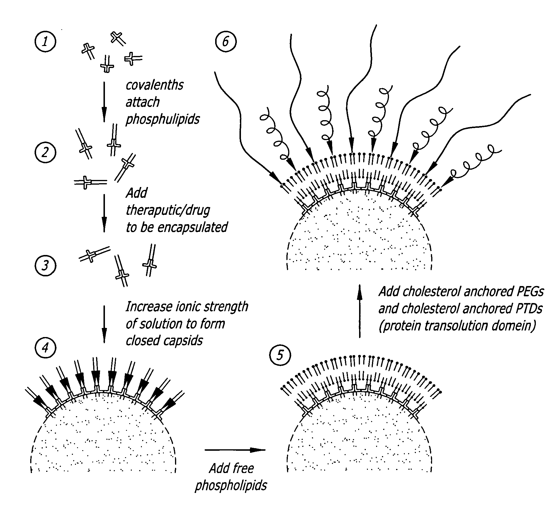
Although this design is inspired by the natural be

Method used


Image

  • Self-assembling nanoparticle drug delivery system
  • Self-assembling nanoparticle drug delivery system
  • Self-assembling nanoparticle drug delivery system

Examples


example 1

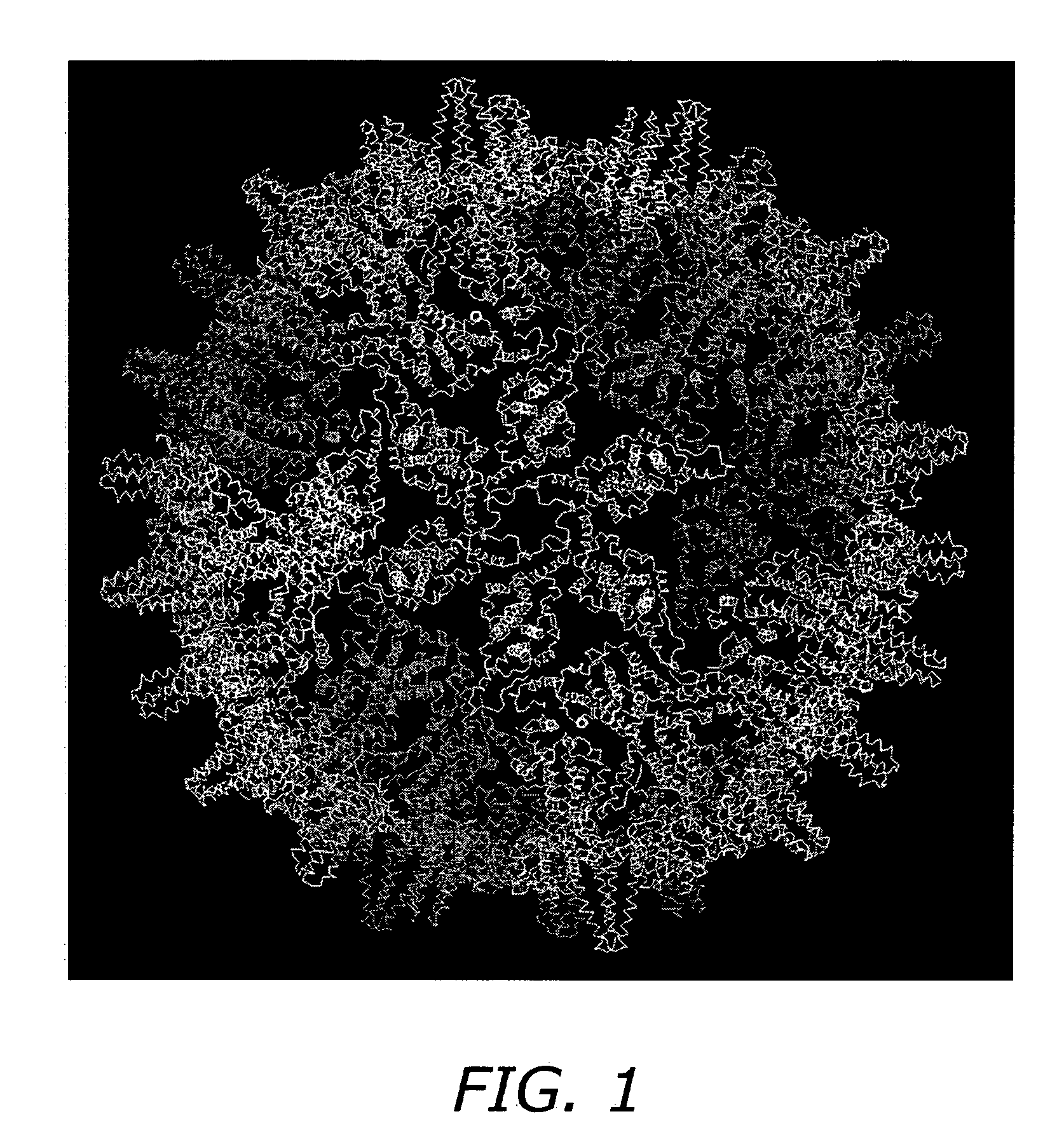
Core Protein Expression and Purification

[0084] A pET-11a vector containing the full-length HBV C-protein gene, is transformed into E. coli DE3 cells and grown at 37° C. in LB media, fortified with 2-4% glucose, trace elements and 200 ug / mL carbenicillin. Protein expression is induced by the addition of 2 mM IPTG (isopropyl-beta-D-thiogalactopyranoside). Cells are harvested by pelleting after three hours of induction. SDS-PAGE is used to assess expression of C-protein.

[0085] Core protein is purified from E. coli by resuspending in a solution of 50 mM Tris-HCl, pH 7.4, 1 mM EDTA, 5 mM DTT, 1 mM AEBSF, 0.1 mg / mL DNase1 and 0.1 mg / mL RNase. Cells are then lysed by passage through a French pressure cell. The suspension is centrifuged at 26000×G for one hour. The pellet is discarded and solid sucrose added to the supernatant to a final concentration of 0.15 M and centrifuged at 10000×G for one hour. The pellet is discarded and solid (NH4)2SO4 is then added to a final concentration of 40...

example 2

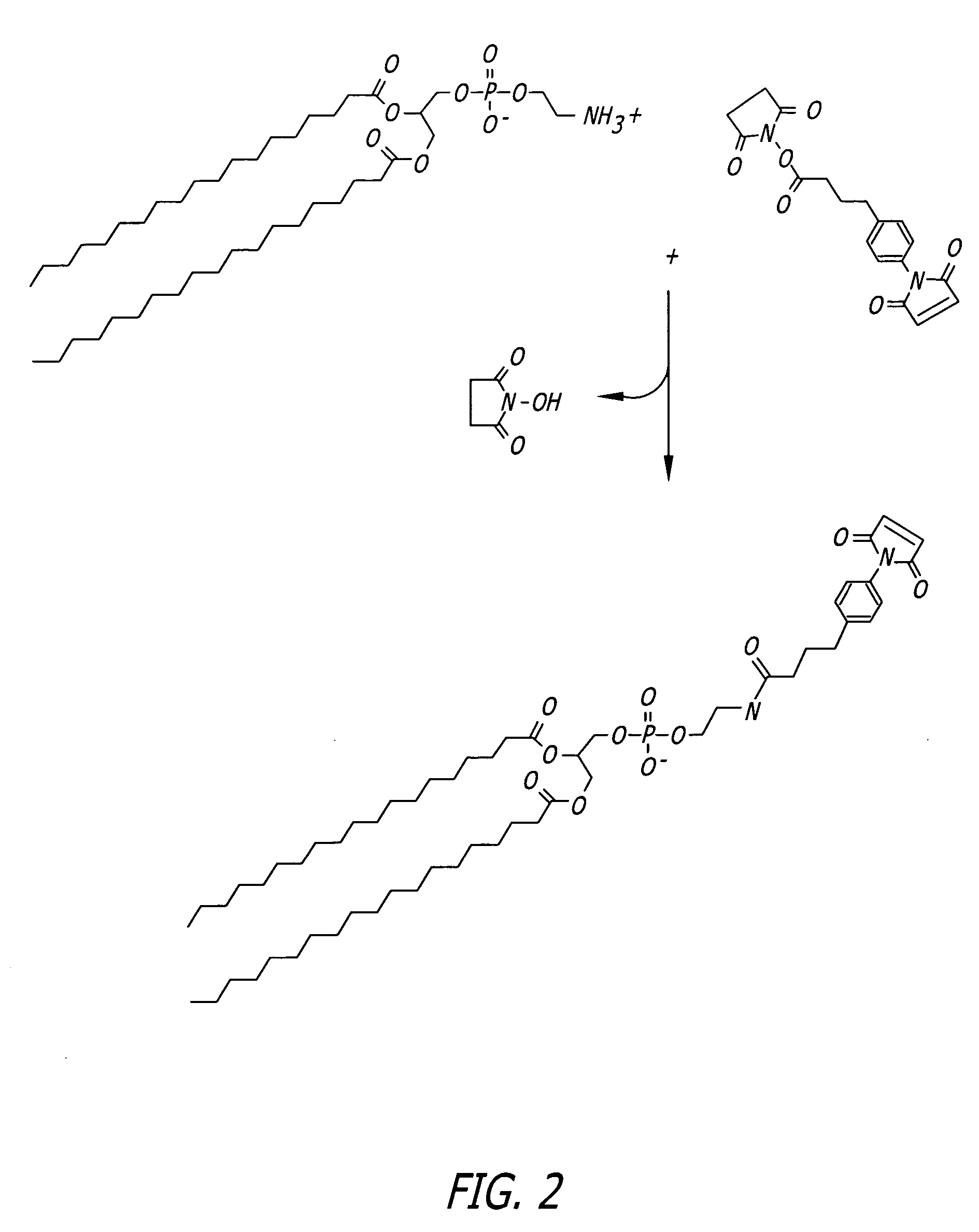
Protocol for Phospholipid Conjugation via SMPB Intermediate (FIG. 2)

[0086] 1. Dissolve 100 micromoles of phosphatidyl ethanolamine (PE) in 5 mL of argon-purged, anhydrous methanol containing 100 micromoles of triethylamine (TEA). Maintain the solution under an argon or nitrogen atmosphere. The reaction may also be done in dry chloroform.

[0087] 2. Add 50 mg of SMPB (succinimidyl-4-(p-maleimidophenyl)butyrate, Pierce) to the PE solution. Mix well to dissolve.

[0088] 3. React for 2 hours at room temperature, while maintaining the solution under an argon or nitrogen atmosphere.

[0089] 4. Remove the methanol from the reaction solution by rotary evaporation and redissolve the solids in chloroform (5 mL).

[0090] 5. Extract the water-soluble reaction by-products from the chloroform with an equal volume of 1% NaCl. Extract twice.

[0091] 6. Purify the MPB-PE derivative by chromatography on a column of silicic acid (Martin F J et al., Immunospecific targeting of liposomes to cells: A novel a...

example 3

Protocol for Phospholipid Conjugation via MBS Intermediate (FIG. 3)

[0093] 1. Dissolve 40 mg of PE in a mixture of 16 mL dry chloroform and 2 mL dry methanol containing 20 mg triethylamine, maintain under nitrogen.

[0094] 2. Add 20 mg of m-maleimidobenzoyl-N-hydroxysuccinimide ester (MBS) to the lipid solution and mix to dissolve.

[0095] 3. React for 24 hours at room temperature under nitrogen.

[0096] 4. Wash the organic phase three times with PBS, pH 7.3, to extract excess cross-linker and reaction by-products.

[0097] 5. Remove the organic solvents by rotary evaporation under vacuum.


PUM

PropertyMeasurementUnit
Ionic strengthaaaaaaaaaa

Abstract

A self-assembling nanoparticle drug delivery system for the delivery of drugs including peptides, proteins, nucleic acids or synthetic chemical drugs is provided. The self-assembling nanoparticle drug delivery system described herein includes viral capsid proteins, such as Hepatitis B Virus core protein, encapsulating the drug, a lipid bi-layer envelope and targeting or facilitating molecules anchored in the lipid bilayer. A method for construction of the self-assembling nanocparticle drug delivery system is also provided.

Description

RELATED APPLICATIONS [0001] This application claims the benefit of priority under 35 U.S.C. §119(e) of U.S. Provisional Patent Application No. 60 / 574,409 filed May 25, 2004.FIELD OF THE INVENTION [0002] The present invention relates to methods for drug delivery. Specifically, the present invention relates to a self-assembling drug delivery system comprised of pharmacologic drugs captured within viral capsid proteins and encapsulated in a lipid envelope. BACKGROUND OF THE INVENTION [0003] Nanotechnology, the term derived from the Greek word nano, meaning dwarf, applies the principals of both physical and biological sciences at a molecular or submicron level. The materials at nanoscale can be a device or system, or supramolecular structures, complexes or composites. Nanotechnology is making significant advances in biomedical applications, including drug delivery techniques. [0004] The development of drug delivery systems for small molecules, proteins and DNA have been greatly influenc...

Claims


Application Information

Patent Timeline
IPC IPC(8): C12Q1/68G01N33/53A61K39/12
CPCA61K9/5184A61K47/48776B82Y5/00C12N2730/10123C07K2319/00C12N7/00C12N2730/10122C07K14/005Y10S977/795Y10S977/801Y10S977/802Y10S977/797Y10S977/798Y10S977/80A61K47/6901A61K47/6913A61P1/04A61P1/08A61P11/00A61P11/06A61P11/08A61P17/04A61P19/02A61P19/06A61P25/00A61P25/04A61P25/06A61P25/08A61P25/16A61P25/18A61P25/20A61P25/22A61P25/24A61P29/00A61P3/02A61P31/00A61P31/04A61P31/10A61P31/12A61P35/00A61P3/06A61P3/08A61P37/02A61P37/06A61P7/00A61P7/02A61P7/04A61P9/00A61P9/06A61P9/10A61P9/12A61P3/10A61K9/1277A61K9/5192
Inventor DE LOS RIOS, MIGUELOH, KENNETH
Owner CHIMEROS
Why Eureka
Try Eureka