Pharmaceutical compositions
- Summary
- Abstract
- Description
- Claims
- Application Information
AI Technical Summary
Problems solved by technology
Method used
Image
Examples
example 1
[0268] This example describes the production, purification, chemical coupling of the recombinant Sip (rSip) polypeptide to DPPE-MCC phospholipid and a procedure to generate liposome.
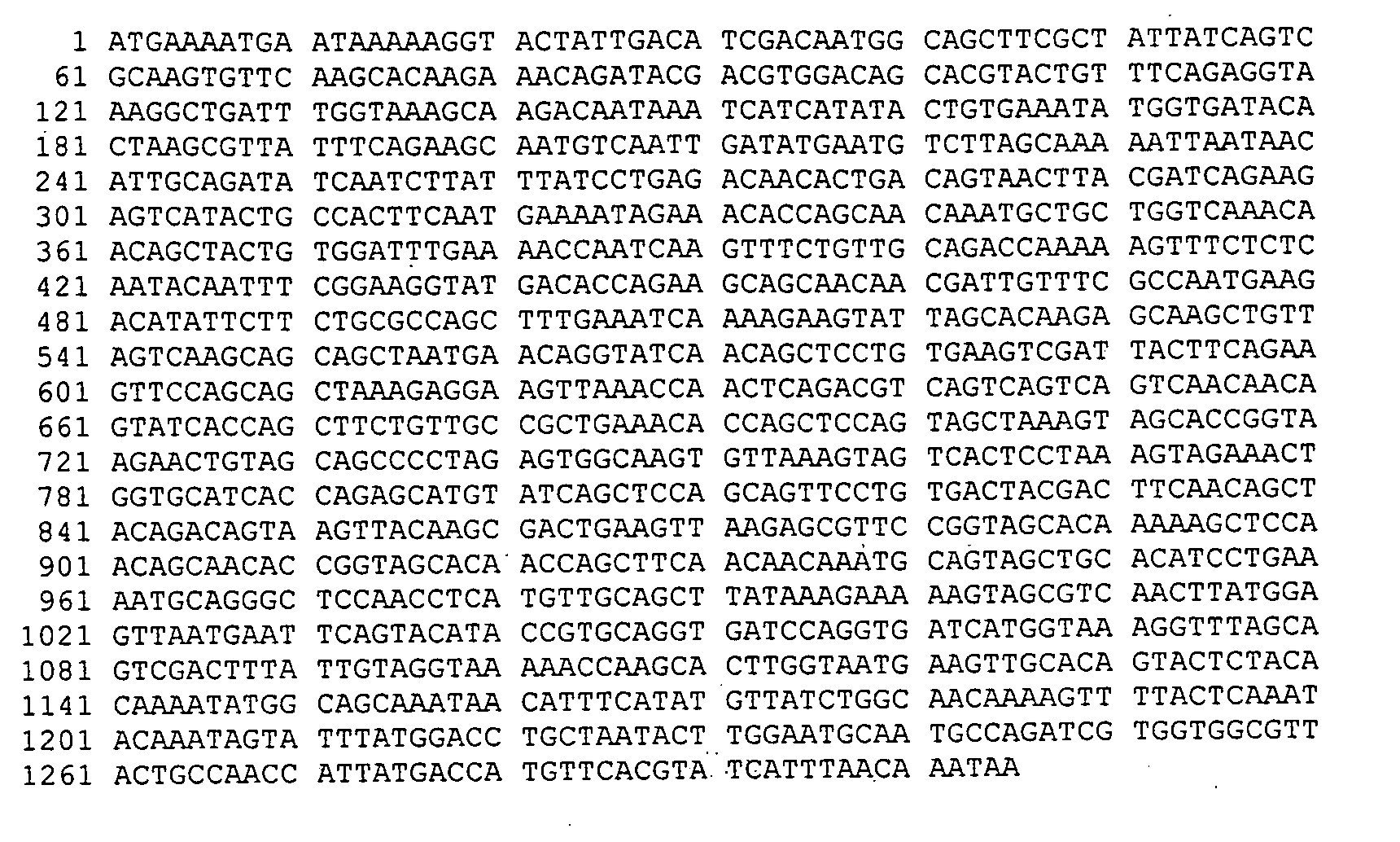
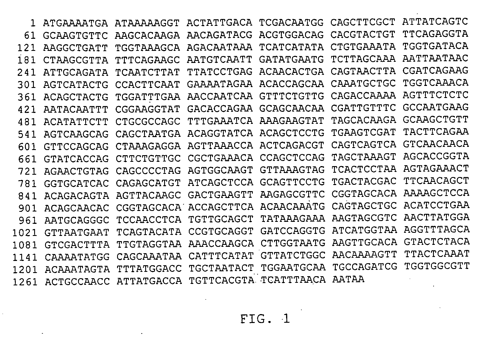
[0269] The sip gene from strain C388 / 90 (serotype Ia / c) was amplified from purified chromosomal DNA by PCR with recombinant Taq DNA Polymerase (Amersham Pharmacia Biotech) as described by the manufacturer. The following two primers OCRR-259 (5′-CCGGCAGATCTATGAAAATGAATAAAAAGGTACTATTG-31) (SEQ ID No:13) and OCRR-260 (5′-GGCCGTCTAGATTATTTGTTAAATGATACGTGAACA-3′) (SEQ ID No:14) which respectively contained the BglII and XbaI restriction sites were used to perform the amplifications. PCR was performed with 10 cycles of 30 sec at 94° C., 20 sec at 56° C. and 30 sec at 72° C., followed by 25 cycles of 30 sec at 94° C., 20 sec at 70° C. and 30 sec at 72° C. and a final elongation period of 5 min at 72° C. The amplification product was ligated into plasmid pURV22 and after sequencing, the recombinant plasmid name...
example 2
[0274] This example describes the cloning strategy used to generate C1rSip and C2rSip, the production of these recombinant polypeptides in E. coli, and their purification.
[0275] In order to increase the efficiency of the coupling reaction and to generate Sip-liposomes that are more stable, modified rSip polypeptides where at least one cysteine residue was added to the amino acid sequence have been designed. Preferably, these cysteine residues are added at positions where it does not remove the antigenic properties of the original Sip polypeptide.
[0276] Sip polypeptides of the invention include Sip polypeptides to which at least one cysteine has been added in order to be coupled to the liposome.
[0277] The modifications to the original sip gene sequence are presented in Table 1. To generate the polypeptides C1rSip and C2rSip, mutagenesis experiments using the Quickchange Site-Directed Mutagenesis kit from Stratagene, the oligonucleotides described in Table 2, and the sip genes clon...
example 3
[0280] This example illustrates the coupling of C1rSip and C2rSip to DPPE-MCC phospholipid.
[0281] In order to couple the C1rSip and C2rSip to DPPE-MCC phospholipid, liposomes containing DPPE-MCC lipid were prepared as described in Example 1. Before coupling, the C1rSip and C2rSip polypeptides were treated with 25 mM DTT during 30 min at room temperature. After the incubation, DTT was removed by gel filtration using a P-6 desalting column. The treated-C1rSip and -C2rSip polypeptides were incubated under nitrogen atmosphere at a final concentration of 2 mg / ml with the liposome suspension containing 0.01% Triton X-100. The resulting C1rSip-, C2rSip-liposomes were separated from free protein by ultracentrifugation (200000×g for 1 h at room temperature) and resuspended in an equal volume of PBS buffer containing 0.3M sucrose. Liposome preparations were sterilized by filtration through a 0.22 μm membrane and stored at −80° C. until used. The amount of C1rSip- and C2rSip-bound to the lipo...
PUM
| Property | Measurement | Unit |
|---|---|---|
| Fraction | aaaaa | aaaaa |
| Fraction | aaaaa | aaaaa |
| Fraction | aaaaa | aaaaa |
Abstract
Description
Claims
Application Information
 - R&D
- Intellectual Property
- Life Sciences
- Materials
- Tech Scout
- Unparalleled Data Quality
- Higher Quality Content
- 60% Fewer Hallucinations
Browse by: Latest US Patents, China's latest patents, Technical Efficacy Thesaurus, Application Domain, Technology Topic, Popular Technical Reports.
© 2025 PatSnap. All rights reserved.Legal|Privacy policy|Modern Slavery Act Transparency Statement|Sitemap|About US| Contact US: help@patsnap.com


