Fiber constructs and process of fiber fabrication

Inactive Publication Date: 2007-01-25
THE JOHN HOPKINS UNIV SCHOOL OF MEDICINE
View PDF3 Cites 10 Cited by
  • Summary
  • Abstract
  • Description
  • Claims
  • Application Information

AI Technical Summary

Benefits of technology

[0023] In another aspect, an embodiment provides kits for producing the fiber compositions herein. In one embodiment, the kit includes an effective amount of polyanionic polymer, polycationic polymer, a bioactive material, and instructions for forming a fiber from an interface of solutions of the two polymers.
[0024] Another aspect is a method of making a fiber composition of any of the formulae herein, comprising taking a precursor compound (or intermediate) and reacting it with one or more chemical reagents to provide the compound of the formulae herein. The method can include one or more of the synthetic steps specifically delineated herein. Accordingly, another aspect is a compound made by a process delineated herein. The process can include one or more steps, reagents and starting materials as delineated herein using chemical reactions, techniques and protocols as delineated herein.

Problems solved by technology

However, optimal tissue engineering requires more than an inert scaffold that serves merely as a substrate for cell attachment and growth.

Method used


Image

  • Fiber constructs and process of fiber fabrication
  • Fiber constructs and process of fiber fabrication
  • Fiber constructs and process of fiber fabrication

Examples


example 1

[0098] Fiber Fabrication.

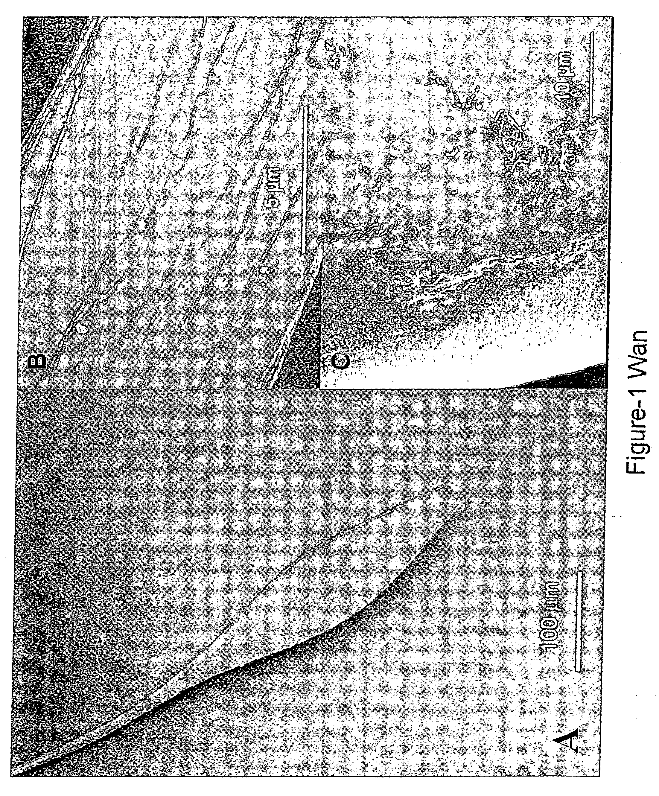
[0099] Fiber was fabricated in the following way: two polyelectrolyte solutions were placed in close proximity on a level surface, then brought into contact by means of forceps or a needle. The mixture in the region of the interface was then scooped up in a continuous upward motion to create a fiber, which could be drawn continuously until one of the polyelectrolyte solutions was depleted. A scanning electron micrograph of a typical fiber is shown in FIG. 1. The fiber surface exhibits a pattern of parallel ridges, as if it were composed of a conglomerate of finer fibers. Interestingly, beads are present at regular intervals along its axis. In contrast to its polyelectrolyte precursors, the fiber is water insoluble.

[0100] Fiber was fabricated by drawing up the interface between two oppositely charged polyelectrolyte solutions using a bent syringe needle (25G3 / 8) attached to the slider of a linear motor (LinMot, Switzerland) with a stroke length of 30 cm. A ...

example 2

[0102] Encapsulation and Release of Protein.

[0103] Bovine serum albumin (BSA) was encapsulated in water soluble chitin-alginate (WSC-A) fibers at different polyelectrolyte concentration ratios. The protein was uniformly distributed, as evident from the light microscope image of encapsulated FITC-BSA. (FIG. 2A) Even after washing, a protein loading level of at least 40% (mass protein / mass polymer) could be achieved. This loading level is considerably higher than that typically obtained by conventional solvent evaporation techniques for formulation of controlled release microspheres. Since the solution goes completely into fiber formation, the efficiency of protein encapsulation is close to 100%.

[0104] The cumulative release profiles of BSA from the fiber are shown in FIG. 2B, typical of a diffusion-controlled release mechanism. As the chitin to alginate concentration ratio increased, a more sustained release of protein was observed. This can be explained by the net negative charge ...

example 3

[0109] Encapsulation of Cells.

[0110] Cells encapsulated in the fiber retained good viability. Two primary cell lines of human dermal fibroblasts (HDF) and bovine pulmonary artery endothelial cells (BPAEC) were encapsulated in WSC-A fibers. Cell viability was established by means of a Live / Dead viability / cytotoxicity kit (Molecular Probes) and a WST-1 assay (Roche Diagnostics GmbH). The WST-1 assay for BPAEC gave relative absorbances of 100%, 95%, 96% and 98% at days 2, 5, 7 and 10 respectively, reflecting the activity of viable cells. The relative absorbances for HDF were 100%, 91%, 74% and 82% for the same time periods.

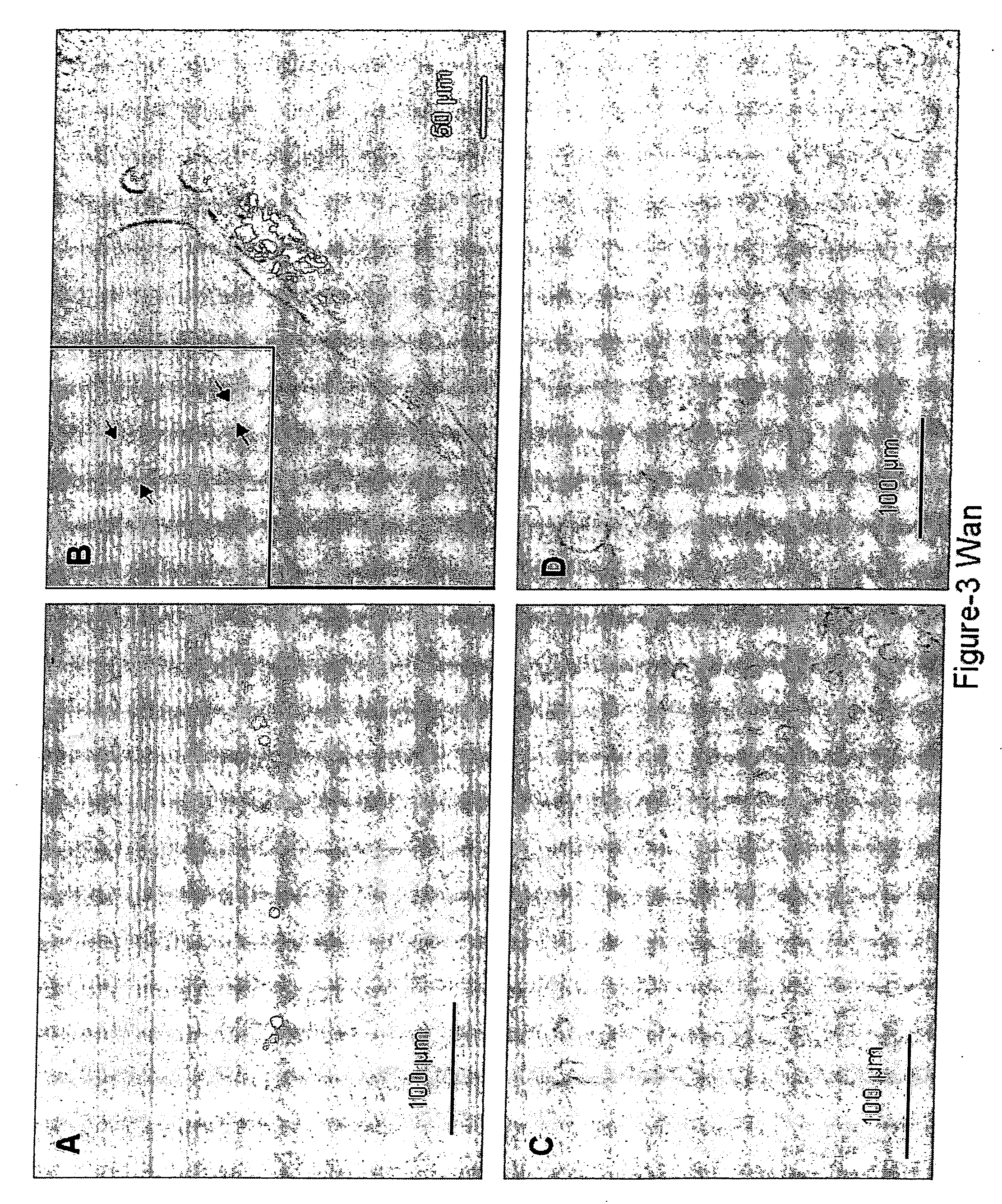
[0111] In contrast to their morphology in monolayer culture, encapsulated HDF and BPAEC were more spherical in shape (FIG. 3A,B). Cells could be entrapped within both bead and fiber regions, and remain viable. Cell clumps in the fiber region appeared as if they were prying open thinner fibers that composed the main fiber, enlarging its diameter significantly.

[0112...


PUM

PropertyMeasurementUnit
diameteraaaaaaaaaa
diameteraaaaaaaaaa
diameteraaaaaaaaaa

Abstract

Described herein are fiber compositions, methods of generating the fiber compositions, and methods of using the fiber compositions in various applications utilizing fiber constructs, including for example, tissue engineering.

Description

[0001] The present application claims the benefit of U.S. provisional application 60 / 667107, filed Mar. 30, 2005, which is incorporated herein by reference in its entirety.BACKGROUND OF THE INVENTION [0002] In the emerging field of tissue engineering, biodegradable polymeric scaffolds are used as templates for tissue regeneration. Science 260, 920-926 (1993). Cells attach to these scaffolds, proliferate, differentiate if necessary, and develop into tissue-like materials to replace or augment the damaged tissue functions. So far, most of these scaffolds play a mainly structural role of supporting cell adhesion and defining the framework of tissue growth. However, optimal tissue engineering requires more than an inert scaffold that serves merely as a substrate for cell attachment and growth. Optimal tissue engineering demands that cues or signal molecules in the form of adhesion molecules, growth and differentiation factors, or even plasmid DNA be incorporated into these scaffolds in ...

Claims


Application Information

Patent Timeline
Patent Type & Authority Applications(United States)
IPC IPC(8): A61K39/12A61K48/00A61K38/16A61K35/12A61K9/70
CPCA61L27/20A61L27/54A61L27/58A61L2300/252A61L2300/258A61L2300/64A61L2300/44A61L2300/45A61L2300/604A61L2300/622A61L2300/624A61L2300/414D01D5/30D01F1/10D01F8/00
Inventor AUN WAN, ANDREW C.LEONG, KAM W.
Owner THE JOHN HOPKINS UNIV SCHOOL OF MEDICINE
Why Eureka
Try Eureka