Ocular gene therapy using avalanche-mediated transfection

a technology of ocular gene therapy and avalanche, applied in the field of medicine, can solve the problems of short half-lives of proteins and aptamers, need intravitreal administration, and potential long-term gene therapy approaches, and achieve the effect of prolonging the therapy time, and prolonging the treatment tim

Active Publication Date: 2007-03-15
THE BOARD OF TRUSTEES OF THE LELAND STANFORD JUNIOR UNIV
View PDF34 Cites 23 Cited by
  • Summary
  • Abstract
  • Description
  • Claims
  • Application Information

AI Technical Summary

Benefits of technology

[0006] The present invention provides a method of treating an ocular disease in a subject. In a first step, a nucleic acid is introduced into cells or a tissue. The nucleic acid is introduced by electron avalanche-mediated transfection. With this technique, a high electric field induces a vapor bubble and plasma discharge between an electrode and the surrounding medium. The formation of a vapor bubble generates mechanical stress. Plasma discharge through the ionized vapor in the bubble enables connectivity between the electrode and the surrounding medium, so that the mechanical stress and electric field are applied simultaneously, which results in permeabilization of the cells or tissue. This permeabilization in turn allows the nucleic acid to enter the cell or tissue. Cells or tissue containing the nucleic acid are then transplanted into an ocular region of the subject.

Problems solved by technology

Therapies involving delivery of proteins or aptamers are examples of such approaches, with the drawback that proteins and aptamers have short half-lives and require intravitreal administration every 4-6 weeks for life for maximal efficacy.
Gene therapy approaches are potentially more long-term, with the possibility of lasting many months or years.
Viral vectors, such as retroviruses and adenoviruses, enable high expression of the introduced DNA but have safety concerns.
Non-viral methods, such as liposomes, have low host immunogenicity but tend to suffer from inefficient DNA delivery to cells.

Method used


Image

  • Ocular gene therapy using avalanche-mediated transfection
  • Ocular gene therapy using avalanche-mediated transfection
  • Ocular gene therapy using avalanche-mediated transfection

Examples


example 1

Construction of a Plasmid for Transfection

[0053] The plasmid shown in FIG. 7 contains the sequence SEQ ID NO: 1. SEQ ID NO: 1 includes a cytomegalovirus (CMV) promoter (1-589 bp), a nucleotide sequence encoding for pigment epithelium-derived factor (PEDF; 590-2131 bp), an internal ribosome entry site (IRES) coding sequence (b2151-2735 bp), and a nucleotide sequence encoding for enhanced green fluorescent protein (eGFP; bp 2739-3455), an SV40 polyA sequence (3612-3662 bp), a phi C31 attB site (3952-4245 bp), a bacterial kan promoter (4541-4576 bp), SV40 origin and promoter enhancer (4653-4955 bp), neo for G418 selection (5004-5798 bp), and an pUC origin (6383-7026 bp).

[0054] To make this plasmid, begin with vector pIRES-EGFP, commercially available from Clontech. Cut the vector with the restriction enzyme BsaI (New England Biolabs) to linearize the vector, make blunt ends (e.g., using DNA Polymerase I, Large (Klenow) Fragment, New England Biolabs), and treat with a phosphatase to r...

example 2

Transfection of Conjunctival Tissue with Luciferase Gene

[0056] A study was conducted in support of the method described herein, where a luciferase marker gene was transfected into conjunctiva tissue. Conjunctival tissue was explanted from adult New Zealand White rabbits and placed in tissue culture dishes. All samples were placed in 1 mL phosphate buffered saline solution with 100 micrograms of plasmid DNA encoding the luciferase gene under a CMV promoter. All samples were cultured in Dulbecco's Modified Eagle Medium (DMEM) plus 10% serum and antibiotic / antimicotic for 24 hours after transfection. Samples were then treated with luciferin substrate (150 micrograms luciferin per ml medium) and imaged using the IVIS-200 system (Xenogen Corp.).

[0057] The conjunctival tissue, which contained conjunctival fibroblasts, was transfected using electron-avalanche mediated transfection with a luciferase marker gene. A control sample of tissue was contacted with the luciferase gene in the abse...

example 3

Comparison of Electron Avalanche Versus Traditional Electroporation in DNA Transfer

[0058] Because electroporation protocols vary for different tissues, experiments were first conducted to determine the optimal protocol for transfecting CAM from a developing chicken egg using traditional electroporation. CAM is a live, readily available, and inexpensive tissue. Its epithelial layer is uniform and has high resistance, making it a good model for epithelial cell layers, such as retinal pigment epithelium. In this model system, 100 μg of pNBL2 plasmid DNA encoding the luciferase gene was pipetted onto the CAM, and pulses were applied. Specifically, a 250-μs, 150-V phase, followed by a 5-ms, 5-V phase in the same polarity was applied. Optimal results were achieved with 50 cycles applied at 1 Hz. The tissue was then cultured and assayed for luciferase bioluminescence. Luciferase expression using this method was about 104 photons / s.

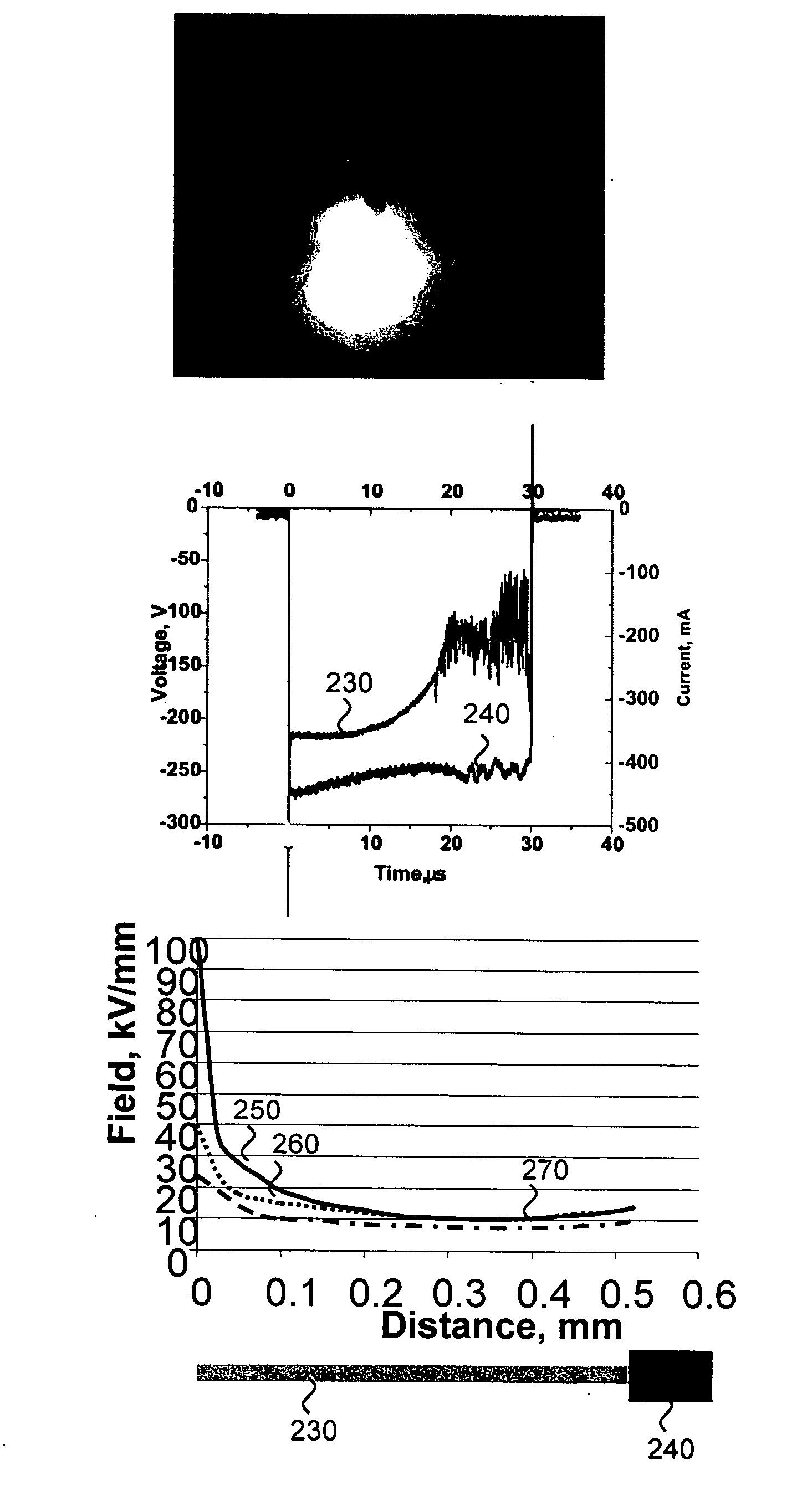
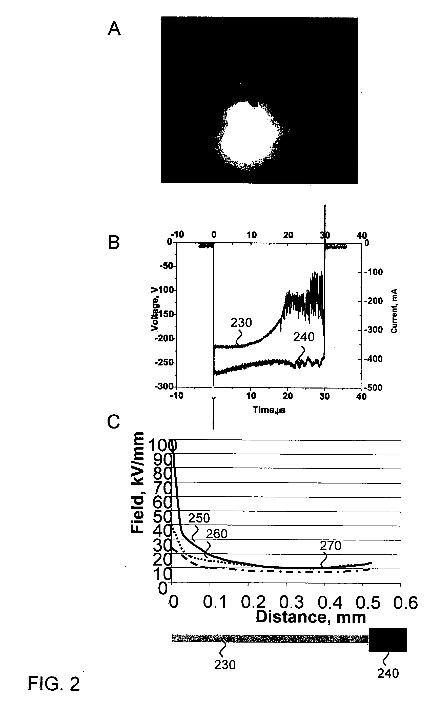
[0059] For electron-avalanche transfection, a 50-μm wire ...


PUM

PropertyMeasurementUnit
electric fieldaaaaaaaaaa
diameteraaaaaaaaaa
diameteraaaaaaaaaa

Abstract

The present invention provides a method of treating an ocular disease in a subject. In a first step, a nucleic acid is introduced into cells or a tissue. The nucleic acid is introduced by electron avalanche transfection. With this technique, a high electric field induces a vapor bubble and plasma discharge between an electrode and the surrounding medium. The formation of a vapor bubble generates mechanical stress. Plasma discharge through the ionized vapor in the bubble enables connectivity between the electrode and the surrounding medium, so that mechanical stress and electric field are applied simultaneously, which results in permeabilization of the cells or tissue. This permeabilization in turn allows the nucleic acid to enter the cell or tissue. Cells or tissue containing the nucleic acid are then transplanted into an ocular region of the subject.

Description

CROSS-REFERENCE TO RELATED APPLICATIONS [0001] This application claims priority from U.S. Provisional Patent Application No. 60 / 708,486, filed Aug. 15, 2005 and from U.S. patent application Ser. No. 11 / 360,984, filed Feb. 22, 2006, which claims priority from U.S. Provisional Patent Application No. 60 / 655,559, filed Feb. 23, 2005, all of which are incorporated herein by reference. FIELD OF THE INVENTION [0002] The present invention relates generally to medicine. More particularly, the present invention relates to a method of treating ocular diseases with gene therapy using avalanche-mediated transfection to genetically modify cells or tissue. BACKGROUND [0003] There are many ocular diseases that affect vision. Diseases of the conjunctiva and cornea, cataracts, uveal diseases, retinal diseases, loss of central acuity and visual field abnormalities and diseases of Bruch's membrane are a few examples. Age-related macular degeneration is a “wet” form of age-related macular degeneration, ...

Claims


Application Information

Patent Timeline
Patent Type & Authority Applications(United States)
IPC IPC(8): C12N15/87
CPCA61K48/00C12N15/87C12N13/00C12M35/00
Inventor CHALBERG, THOMAS W. JR.BLUMENKRANZ, MARKPALANKER, DANIEL V.VANKOV, ALEXANDERHULE, PHILIP JR.MARMOR, MICHAEL F.CALOS, MICHELE P.
Owner THE BOARD OF TRUSTEES OF THE LELAND STANFORD JUNIOR UNIV
Why Eureka
Try Eureka