
Alpha enolase-directed diagnostics and therapeutics for cancer and chemotherapeutic drug resistance

a technology of alpha enolase and cancer, applied in the field of cancer, can solve the problems of increasing the risk of cancer to the patient's life and wellbeing, and the treatment options are significantly limited, and achieve the effect of increasing the sensitivity of neoplastic cells

Inactive Publication Date: 2007-04-05
AURELIUM BIOPHARMA
View PDF0 Cites 11 Cited by
  • Summary
  • Abstract
  • Description
  • Claims
  • Application Information

AI Technical Summary

Benefits of technology

[0011] The present invention is based, in part, upon the discovery that α-enolase, a normal protein involved in carbohydrate metabolism, is expressed at high levels in neoplastic cells as compared to normal cells of the same tissue type, and at yet much higher levels in neoplastic cells that have developed chemotherapeutic drug resistance or metastatic potential. α-enolase expression levels are therefore diagnostic of neoplastic and chemotherapeutic drug resistant cancer cells. α-enolase expression is also diagnostic of metastatic potential. The invention provides a method that uses targeting agents specific for α-enolase to detect and diagnose neoplastic cells and cells with chemotherapeutic drug resistance and metastatic potential in neoplastic cells in a subject. Moreover, the invention provides therapeutic methods for treating neoplastic cells by increasing the sensitivity of the neoplastic cells to chemotherapeutic drugs. The invention also provides therapeutic methods for treating cells that have developed chemotherapeutic drug resistance or developed metastatic potential through the use of targeting agents specific for α-enolase.
[0029] In still another aspect, the invention provides a method of treatment for a neoplasm in a patient. The method comprises administering an effective amount of an α-enolase targeting agent to the patient in which the targeting agent binds to α-enolase expressed in the neoplasm. The patient is administered an effective amount of a chemotherapeutic drug, whereby the α-enolase targeting agent, when bound to the neoplasm, increases the sensitivity of the neoplasm to the chemotherapeutic drug.

Problems solved by technology

The variety of cancer types and mechanisms of tumorigenesis add to the difficulty associated with treating a tumor, increasing the risk posed by the cancer to the patient's life and wellbeing.
The chemotherapeutic drug resistance phenotype can arise to a broad spectrum of functionally distinct drugs, whereby treatment options are significantly limited by chemotherapeutic drug resistance development.
However, because various normal tissues express different amounts of P-glycoprotein, there are significant problems with side effects as any therapy that targets P-glycoprotein on the cell surface of MDR cancer cells would also affect those normal tissues that also have a relatively high level of P-glycoprotein expression, such as liver, kidney, stem cells, and blood-brain barrier epithelium, the latter being a major contributor to the clinical side effects.

Method used


Image

  • Alpha enolase-directed diagnostics and therapeutics for cancer and chemotherapeutic drug resistance
  • Alpha enolase-directed diagnostics and therapeutics for cancer and chemotherapeutic drug resistance
  • Alpha enolase-directed diagnostics and therapeutics for cancer and chemotherapeutic drug resistance

Examples


example 1

[0274] Overexpression of a 44 kD Protein in Cancer Cell Lines

[0275] Studies were performed to determine what proteins, if any, were differentially expressed in chemotherapeutic drug-resistant tumor cell lines as compared to their drug-sensitive counterparts. Drug-sensitive cell lines were obtained from were obtained from ATCC (Manassas, Va., USA). MCF7 / AR, human lung carcinoma small cell H69, H69 / AR, and HL60 / AR cells were obtained from McGill University, Montreal, Qc, Canada. MDA-MB-231 / AR, MOLT4 / AR 250 nM and MOLT4 / AR 500 nM cells were derived at Aurelium BioPharma Inc. (Montreal, QC, Canada). Chemotherapeutic drug-resistant cell lines were derived from a drug-sensitive clone of the “parent” cancer cell line representing a particular tissue.

[0276] The different cell lines used in the Examples below are listed in Table 1.

TABLE 1Drug-Sensitive Cell LinesDrug-Resistant Cell LinesMCF-7MCF-7 / ARMDAMDA-MB231H69H69 / ARH460H460 / ARK562K562 / ARHSB-2HSB-2 / ARRPMI-8226RPMI-8226 / ARMOLT4MOLT4 / A...

example 2

Identification of a 44 kD Protein in MCF-7 / AR Breast Cancer Cells as α-Enolase

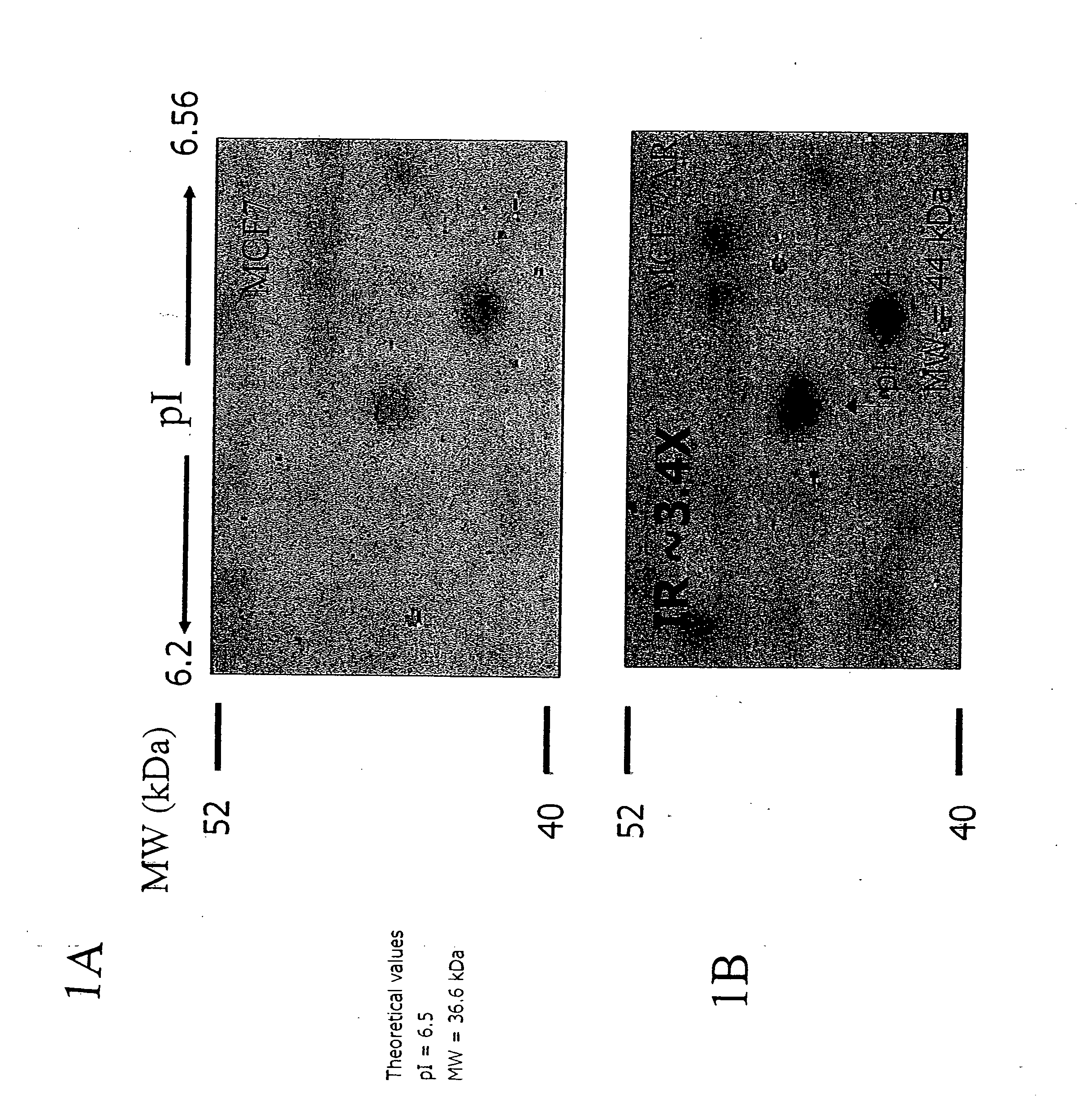
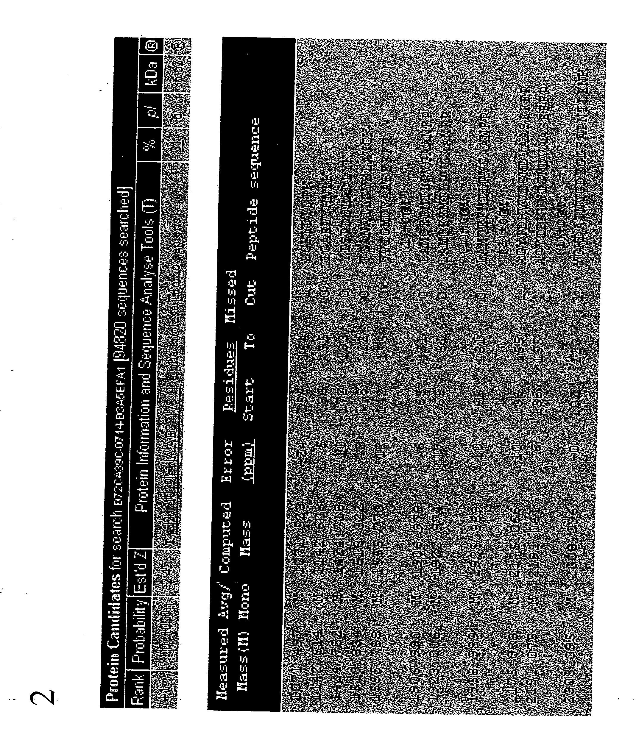
[0282] To discover the identity of the 44 kD protein that was overexpressed in drug-resistant cell lines, the spot located on the 2D gel was subjected to mass spectrometry. FIGS. 1A and 1B show a 44 kD spot to be up-regulated by 3.4 times in the drug-resistant cell samples. The 44 kD spot was isolated and subjected to tryptic digestion in preparation for mass spectrometry. The spot of interest was excised with a clean (clean; acid washed) razor blade and cut into small pieces on a clean glass plate and transfer into a 200 μl PCR tube (MeOH treated). The gel pieces were mixed with 50 μl destainer A and 50 μl destainer B (provided with SilverQuest kit, Life Technologies) (or 100 μl of the destainers premix prepared fresh) and incubated for 15 min. at room temperature (RT) without agitation. The destaining solution was removed using a capillary tip. Water was added to the gel pieces, mix and incubate 10 min ...

example 3

Identification of α-Enolase Overexpression in MOLT4 / AR Cell Lines

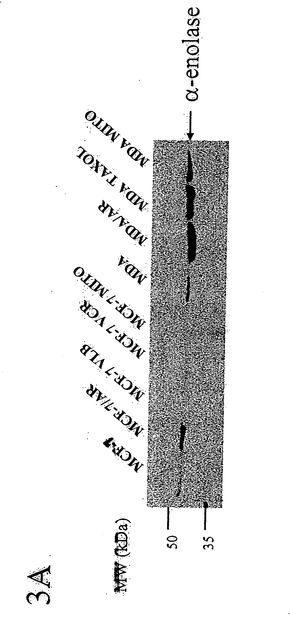
[0284] Western blot analysis utilizing anti-α-enolase antibodies was performed on MOLT4 cell extracts fractionated on SDS-PAGE (FIG. 3). Cells extract were prepared according to the protocol detailed in Example 1. Total cell lysates were thawed, then 100 mg of protein (completed to 50 ml with lysis buffer) were mixed with 10 ml of 5× electrophoresis buffer (60 mM Tris-HCl, pH 6.8, 25% glycerol, 2% SDS, 14.4 mM β-mercaptoethanol, 0.1% bromophenol blue) and these samples were heated at 100° C. for 5 min. and loaded on 10% SDS-PAGE gels. Resolved proteins were electrophoretically transferred onto nitrocellulose membranes (Hybond, Amercham Pharmacia Biotech) for 1 hour. FIG. 3D shows the overexpression of α-enolase in Molt-4 cells selected with adriamycin.

[0285] After blocking the membranes with 5% non-fat milk in 1×PBS overnight at 4° C., all antibody-binding reactions were performed in 5% non-fat milk in 1×PBS for 2 ho...


PUM

PropertyMeasurementUnit
Fractionaaaaaaaaaa
Electrical resistanceaaaaaaaaaa
Cell angleaaaaaaaaaa

Abstract

Disclosed are methods for detecting a neoplasm and / or chemotherapeutic drug resistance or angiogenic potential in neoplastic cells by detecting an increase in the expression of α-enolase in such cells, or in the case of metastatic potential on the surface of such cells, as compared to the level of expression of α-enolase protein in a normal or non-MDR neoplastic cell or on the surface of a non-metastatic neoplastic cell. In addition, methods and a composition are disclosed for increasing the sensitivity of a neoplasm to a chemotherapeutic drug treatment regime, for inhibiting angiogenesis and metastatic potential in chemotherapeutic drug resistant or neoplastic cells, and for inducing apoptosis in chemotherapeutic drug resistant or neoplastic cells.

Description

FIELD OF THE INVENTION [0001] This invention relates to the field of cancer. In particular, this invention relates to the detection, diagnosis, and treatment of neoplastic cells, and to the detection and treatment of chemotherapeutic drug-resistant neoplastic cells and neoplastic cells that show metastatic potential. Furthermore, this invention relates to increasing apoptosis, inhibiting angiogenesis and metastatic potential in neoplastic and chemotherapeutic drug resistant cells. BACKGROUND OF THE INVENTION [0002] Cancer is one of the deadliest illnesses in the United States, accounting for nearly 600,000 deaths annually. This “disease” is in fact a diverse group of diseases, which can originate in almost any tissue of the body. In addition, cancers may be generated by multiple mechanisms including pathogenic infections, mutations, and environmental insults (see, e.g., Pratt et al. (2005) Hum. Pathol. 36(8): 861-70). The variety of cancer types and mechanisms of tumorigenesis add t...

Claims


Application Information

Patent Timeline
IPC IPC(8): C12Q1/68G01N33/574A61K38/16A61K38/14A61K31/7048A61K31/704A61K31/7076A61K31/7072
CPCA61K38/1816A61K38/50C12Q1/527G01N33/57415G01N33/57423G01N33/57426G01N33/57434G01N33/57449G01N33/57484G01N33/57496G01N2500/00G01N2800/44A61K2300/00
Inventor GEORGES, ELIASPRINOS, PANAGIOTIS
Owner AURELIUM BIOPHARMA
Learn More