
Materials and methods for cell-based assays

a cell-based assay and material technology, applied in the field of material and method for cell-based assays, can solve the problems of inherently prone to error, lack of dynamic long-term monitoring, and inability to judge the growth inhibition of cultured cells, and achieve the effect of facilitating three-dimensional cell growth

Inactive Publication Date: 2007-05-03
THE OHIO STATE UNIV RES FOUND
View PDF28 Cites 36 Cited by
  • Summary
  • Abstract
  • Description
  • Claims
  • Application Information

AI Technical Summary

Benefits of technology

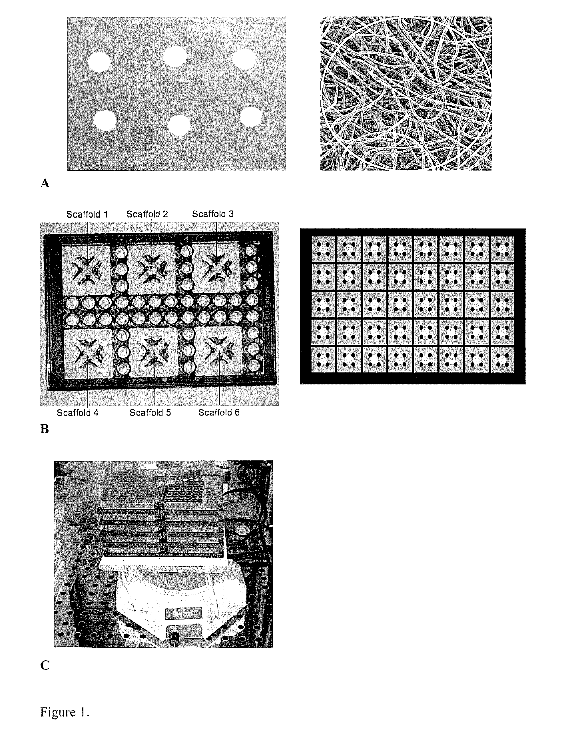
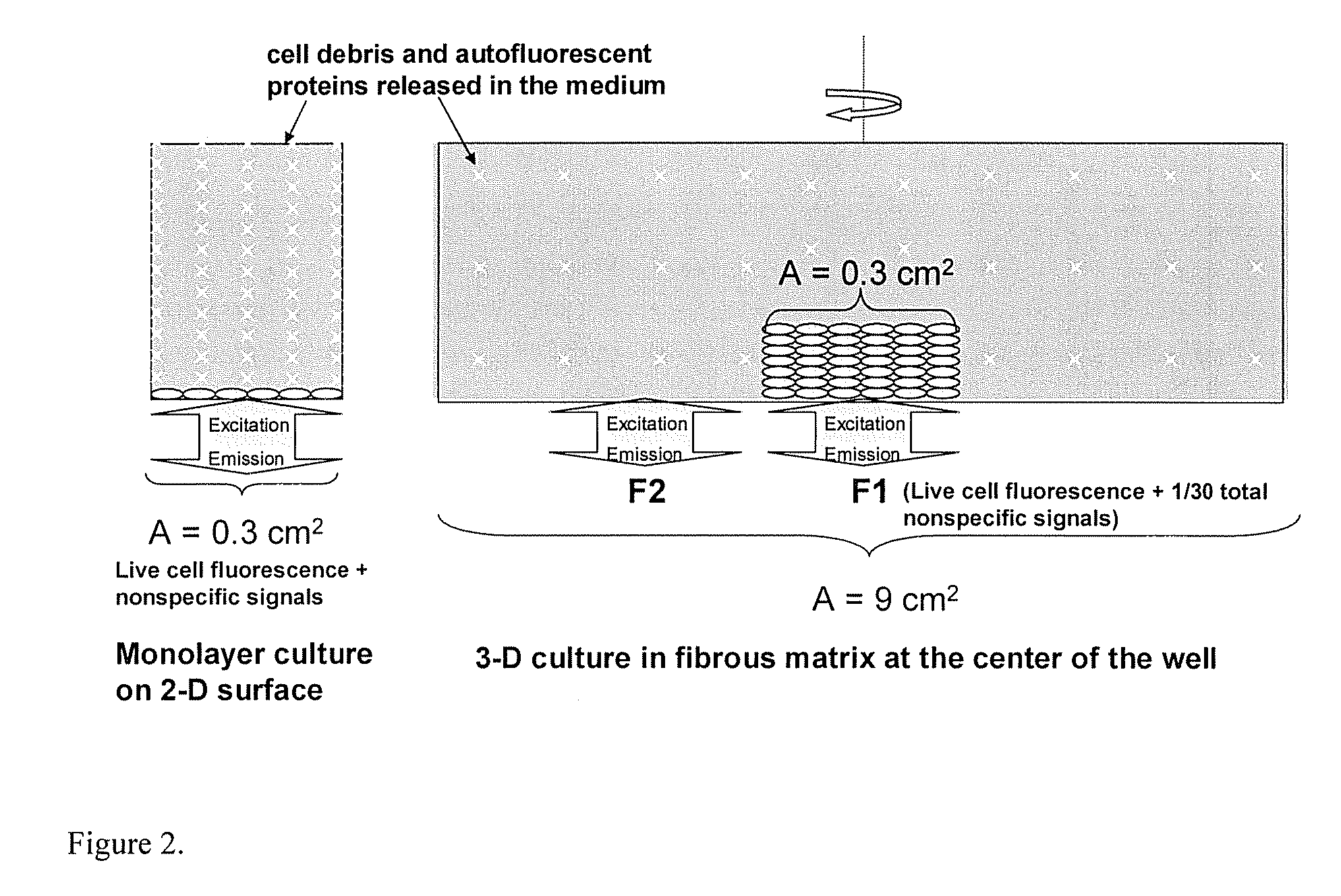
[0006] According to various embodiments, provided herein are methods and apparatuses for analyzing cellular function of cells in three-dimensional culture. The methods and apparatuses provide for culturing cells by seeding for culture a cell of interest within a cell support structure that includes at least one scaffold that is of a suitable porosity to permit the transmission of at least one form of radia

Problems solved by technology

However, there are several major problems associated with conventional in vitro cytotoxicity assays that use static multi-well plate cultures.
Another disadvantage to both systems is that growth inhibition of the cultured cells is usually judged at an arbitrary point of time, lacking a dynamic long-term monitoring.
Cell culture methods that utilize 2-D cultures for cytotoxicity analyses are inherently prone to error because of the lack of a 3-D scaffold to support cell growth and proper tissue function.
So far, almost all cell based assays have been developed in 2-D culture systems, although conventional 2-D cultures usually suffer from contact inhibition and a loss of native cell morphology and functionality.
Currently, pharmaceutical firms spend large amounts of money on compound efficacy and cytotoxicity tests.
However, there is still a 78% failure rate for all drugs, which may be devastating to developing companies.
Effective compounds in vitro may be non-effective in vivo for many reasons, including differences between in vitro and in vivo target biology, interrelated biochemical mechanism, metabolism, poor penetration into solid tissues, etc.

Method used


Image

  • Materials and methods for cell-based assays
  • Materials and methods for cell-based assays
  • Materials and methods for cell-based assays

Examples


example 1

Effects of Serum and Fibronectin on Cell Cultures

[0063] Embryonic stem (ES) cells transfected with constitutive CMV promoter, which is cell cycle sensitive, and a gene encoding a light-emitting protein (e.g., EGFP) can be used for studying proliferation and dynamic responses to growth factor or stimuli. Fetal bovine serum (FBS) is an essential ES growth medium component that is required for the ES cell culture, but its optimal concentration has not been well studied. Fluorescent ES cells can be used to study the effect of FBS on ES cell growth and the result can be used to aid the medium design. One day after seeding, the 3-D cell matrices were cultured in ES media with different FBS contents ranging from 0% to 10% (v / v) and the culture fluorescence signals were monitored. As can be seen in FIG. 7, no cell growth was observed when there was no FBS in the medium and cell growth increased with increasing the FBS content from 0% to 3%. However, further increasing the FBS content to 1...

example 2

Acute Cytotoxicity

[0065] For the live-cell kinetic assays, acute cellular events can be easily characterized with online continuous fluorescence measurements without human intervention. The 3-D cell-based assay system was thus applied to test the response of ES-GFP cells to a surfactant, Triton X-100 (Sigma Chemical Company, St. Louis, Mo.). ES-GFP cells in the 3-D system were cultured to reach a high density with 2000 RFU fluorescence intensity. After applying different doses of Triton X-100, the culture plate was immediately put into the plate reader (Cytofluor Series 4000) at 37° C., with the cycle number set at 80 and cycle time at 3 min. The mixing time before each reading was 10 seconds. As can be seen in FIG. 10, the surfactant caused an acute cell death as indicated by the loss in the culture fluorescence. In general, increasing the surfactant concentration in the medium increased cell death, which followed a first order kinetics as indicated by the linear semilogarithmic ...

example 3

Drug Effects on ES Cell Growth

[0066] One application of the fluorescent cell-based assay is for high-throughput drug screening for cytotoxicity effects. Two weak embryotoxic chemicals, dexamethasone (DM) and diphenylhydantoin (DPH), one non-embryotoxic chemical, penicillin G, and one strong embryotoxic chemical, 5-FU were applied at various concentrations to ES cells grown in 2-D and 3-D at the early proliferation stage (drug exposure one day after seeding the matrices).

[0067] The 2-D cytotoxicity assay was performed in 96-well plates by inoculating 5000 cells into each well containing 150 μl of ES medium. The drug was added one day after inoculation. Fluorescent signals were measured twice per day in the culture plates. The fluorescence given by live cells was calculated as the signals obtained after replacing culture medium with the same volume of PBS Iminius the blanks. As expected, the fluorescence time course data showed the drug effects on ES cell growth: decreasing cell gr...


PUM


Abstract

The methods generally relate to three-dimensional culturing of cells with the steps of seeding a cell support structure in a device with at least one vessel for cell culture having a bottom, at least one wall and a cell support structure for three-dimensional cell culture. The cell support structure is inside the at least one vessel and the device is configured such that assays can be performed. Devices for performing cell culture and assays are also provided. It will be understood that the devices can have many different suitable configurations. In general, the devices are selected such that they can be used with suitable radiation based assays.

Description

CROSS REFERENCE TO RELATED APPLICATIONS [0001] This application claims priority to and any other benefit of U.S. Provisional Application Ser. No. 60 / 732,820, filed on Nov. 2, 2005, and U.S. Provisional Application Ser. No. 60 / 732,821, filed on Nov. 2, 2005, the entire contents of which are incorporated by reference herein.BACKGROUND [0002] In vitro experiments, especially cell culture systems, have been extensively used for cytotoxicity studies. However, there are several major problems associated with conventional in vitro cytotoxicity assays that use static multi-well plate cultures. According to some such conventional methods, cells are cultured on two-dimensional surfaces, which may camouflage authentic cellular responses that would occur in vivo, where cells exhibit morphology and interact with each other in a three-dimensional milieu. According to other conventional methods, cells are cultured in suspension. A relatively long-term response to antigens or foreign materials (e.g...

Claims


Application Information

Patent Timeline
IPC IPC(8): C12M3/00
CPCC12M23/12C12M25/14
Inventor YANG, SHANG-TIANZHANG, XUDONGWEN, YUAN
Owner THE OHIO STATE UNIV RES FOUND
Learn More