Methods and apparatus for atrioventricular valve repair

a technology for atrioventricular valves and surgical repair, applied in the field of medical methods and equipment, can solve the problems of affecting cardiac function, enabling leakage through, and deteriorating the function of the mitral valve, so as to facilitate the reduction of leakage, facilitate the operation of the heart valve, and facilitate the effect of reducing leakag

Active Publication Date: 2007-05-24
CRABTREE TRAVES DEAN
View PDF15 Cites 267 Cited by
  • Summary
  • Abstract
  • Description
  • Claims
  • Application Information

AI Technical Summary

Benefits of technology

[0007] In one aspect, a method of repairing an atrioventricular valve in a patient is provided. The method comprises accessing the patient's atrioventricular valve percutaneously, securing a fastening mechanism to a valve leaflet, and coupling the valve leaflet, while the patient's heart remains beating, to at least one of a ventricular wall adjacent the atrioventricular valve, a papillary muscle, at least one valve chordae, and a valve annulus to facilitate reducing leakage through the valve.
[0008] In another aspect, a method of repairing a mitral valve in the heart of a patient is provided. The method comprises accessing the patient's mitral valve percutaneously, securing a first end of a fastening mechanism to a valve leaflet of the mitral valve, and coupling a second end of the fastening mechanism to a cardiac structure...

Problems solved by technology

More often, however, the mitral valve degenerates with age.
Among the problems that can develop is mitral valve regurgitation in which the mitral valve leaflets become unable to close properly during systole, thus enabling leakage to flow through the mitral valve during systole.
Over time, regurgitation of the mitral valve can adversely affect cardiac function and may compromise a patient's quality of life and/or life-span.
Mitral valve regurgitation can result from a number of different mechanical defects in the mitral valve.
For example, the valve leaflets, the valve chordae which connect the leaflets to the papillary muscles, or the papillary muscles themselves may become damaged or otherwise dysfunctional.
Moreover, the valve annulus may become damaged or weakened an...

Method used


Image

  • Methods and apparatus for atrioventricular valve repair
  • Methods and apparatus for atrioventricular valve repair
  • Methods and apparatus for atrioventricular valve repair

Examples


Embodiment Construction

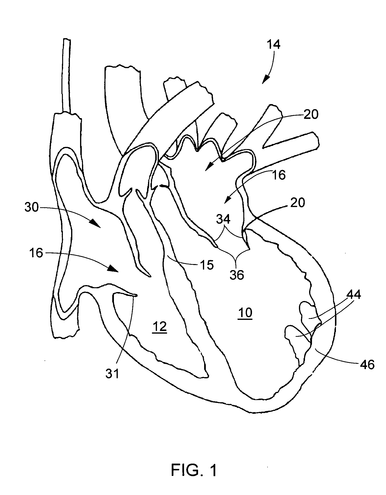
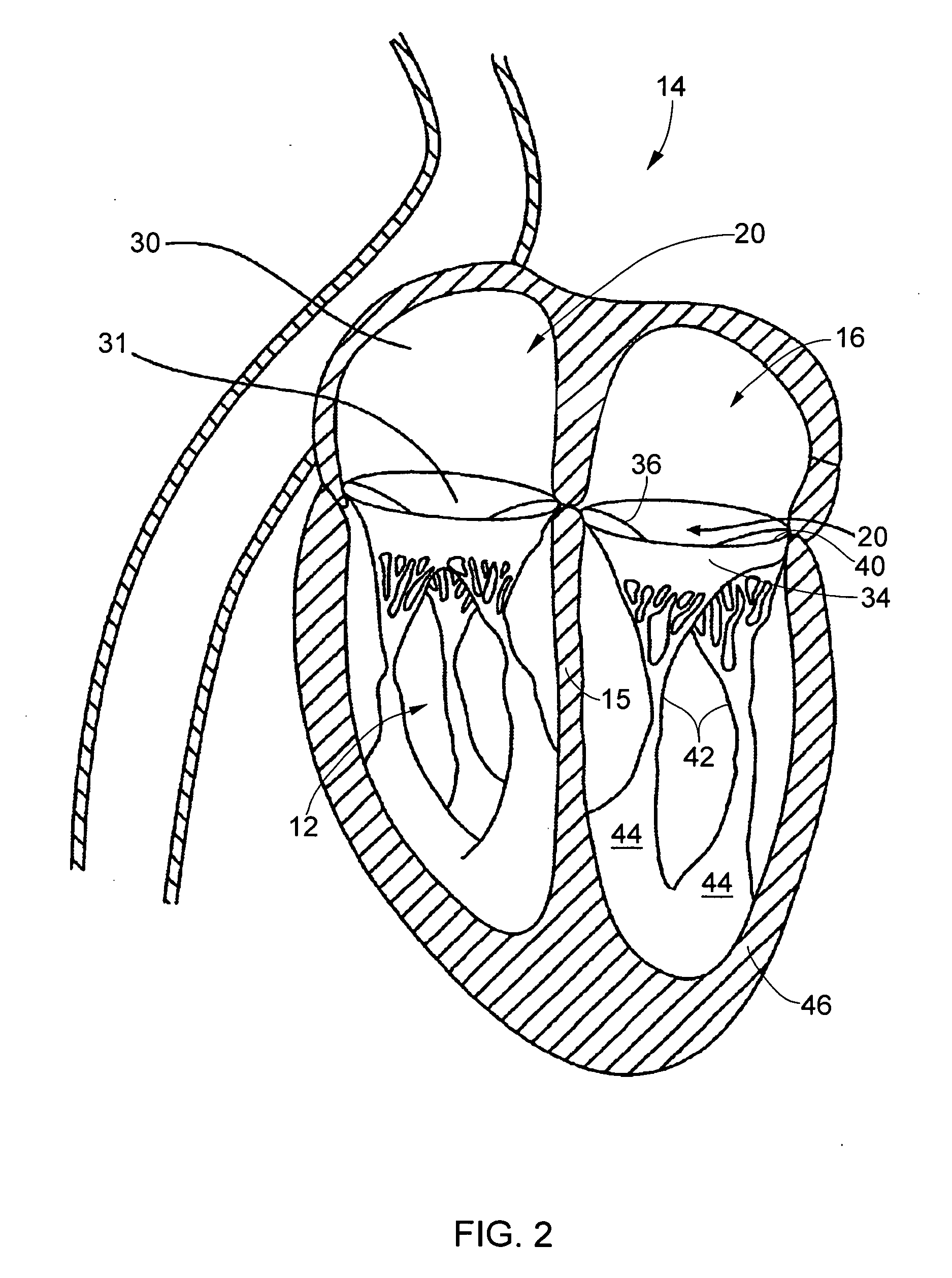
[0019]FIG. 1 is a cross-sectional view of the left and right ventricles 10 and 12, respectively, of a human heart 14 during diastole. Ventricles 10 and 12 are separated by an interatrial septum 15. FIG. 2 is a cross-sectional view of heart 14 during systole. The present invention provides methods and apparatus for the endovascular repair of cardiac valves, particularly atrioventricular valves 16, which inhibit back-flow of blood from a heart ventricle during contraction (systole). In particular, the present invention may be used in repairing, but is not limited to repairing, mitral valves 20.

[0020] As used herein, the term “endovascular,” refers to procedure(s) of the present invention that are performed with interventional tools and supporting catheters and other equipment introduced to the heart chambers from the patient's arterial or venous vasculature remote from the heart. The interventional tools and other equipment may be introduced percutaneously, i.e., through an access sh...


PUM


Abstract

Methods and apparatus for use in repairing an atrioventricular valve in a patient are provided. The methods comprise accessing the patient's atrioventricular valve percutaneously, securing a fastening mechanism to a valve leaflet, and coupling the valve leaflet, while the patient's heart remains beating, to at least one of a ventricular wall adjacent the atrioventricular valve, a papillary muscle, at least one valve chordae, and a valve annulus to facilitate reducing leakage through the valve.

Description

BACKGROUND OF THE INVENTION [0001] This invention relates generally to medical methods and apparatus, and more particularly, to methods and apparatus for the endovascular or minimally invasive surgical repair of atrioventricular valves of the heart, including the mitral valve and the tricuspid valve. [0002] The heart includes four valves that direct blood through the two sides of the heart. The mitral valve lies between the left atrium and the left ventricle and controls the flow of blood into the left side of the heart. The valve includes two leaflets, an anterior leaflet and a posterior leaflet, that close during systole. The leaflets are passive in that they open and close in response to pressure induced to the leaflets by the pumping of the heart. More specifically, during a normal cycle of heart contraction (systole), the mitral valve functions as a check valve to prevent the flow of oxygenated blood back into the left atrium. In this manner, oxygenated blood is pumped into the...

Claims


Application Information

Patent Timeline
IPC IPC(8): A61F2/24
CPCA61B17/0401A61B17/085A61B2017/00783A61B2017/00867A61B2017/0412A61B2017/0437A61B2017/0443A61B2017/0464A61F2/2457
Inventor CRABTREE, TRAVES DEAN
Owner CRABTREE TRAVES DEAN
Why Eureka
Try Eureka