

Cell growth inhibitors containing anti-glypican 3 antibody
a cell growth inhibitor and anti-glypican technology, which is applied in the field of cell growth inhibitors containing antiglypican 3 antibodies, can solve the problems of no clear relationship between
- Summary
- Abstract
- Description
- Claims
- Application Information
AI Technical Summary
Benefits of technology
Problems solved by technology
Method used
Image

Examples
example 1
Preparation of Monoclonal Antibody Against Glypican-3 Synthetic Peptide
[0103] A peptide having an amino acid sequence (the 355th to the 371st amino acids) (RQYRSAYYPEDLFIDKK) of human glypican-3 protein was synthesized. The synthetic peptide was bound to keyhole limpet hemocyanin (KLH) using a maleinimide benzoyloxy succinimide (MBS) type crosslinking agent, thereby preparing an immunogen. Mice (BALB / c, female, 6-week-old) were immunized 3 times with the immunogen at 100 μg / mouse. The antibody titers in serum were assayed. A method employed as an antibody titer assay method involves causing the diluted sera to react with the peptides (0.5 μg) immobilized on a plate, performing reaction with HRP-labeled anti-mouse antibodies, adding a substrate, and then measuring absorbance at 450 nm of the thus developed color (a peptide solid-phase ELISA method). After antibody titers were confirmed, splenocytes were collected, and then fused (Köhler, G, Milstein, C: Nature, 256: 495 (1975)) with...
example 2
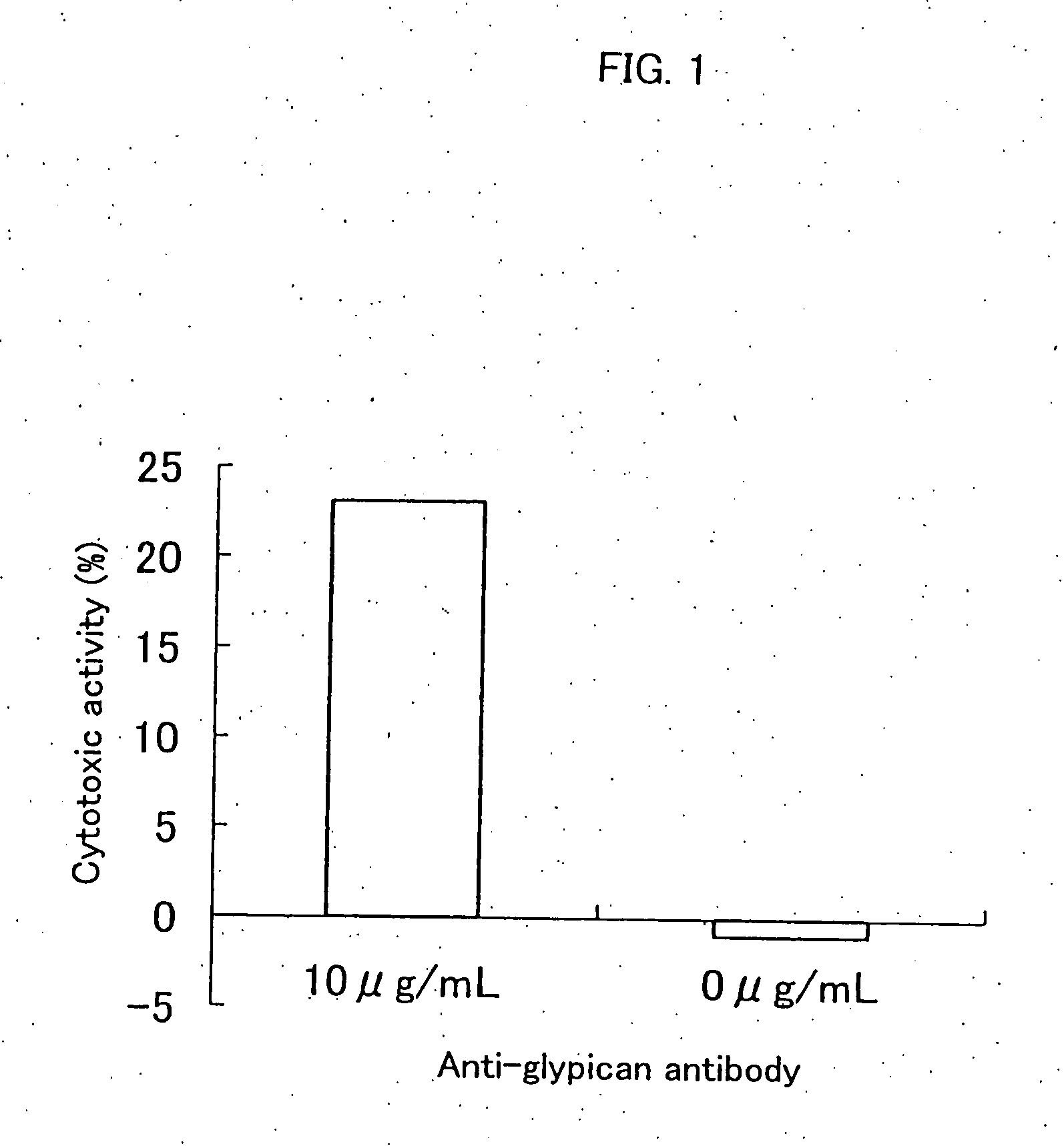
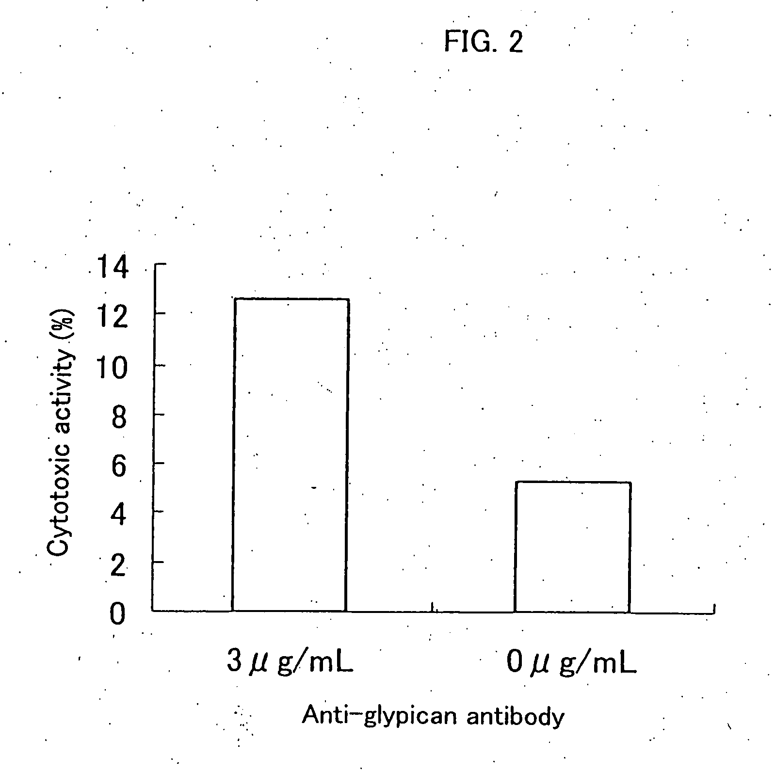
Inhibition of Cell Proliferation Using Anti-Glypican 3 Antibody
[0104] The ADCC (antibody-dependent cell-mediated cytotoxicity) activity and the CDC (complement-dependent cytotoxicity) activity were measured according to the method of Current Protocols in Immunology, Chapter 7. Immunologic studies in humans, Editor, John E, Cologan et al., John Wiley & Sons, Inc., 1993.
1. Preparation of Effector Cell
[0105] The spleen was extracted from a CBA / N mouse (8-week-old, male), and then splenocytes were isolated in RPMI1640 media (GIBCO). The cells were washed in the same media containing 10% fetal bovine serum (FBS, HyClone), and then the cell concentration was prepared at 5×10⁶ / mL, thereby preparing effector cells.
2. Preparation of Complement Solution
[0106] Baby Rabbit Complement (CEDARLANE) was diluted 10 times in 10% FBS-containing DMEM media (GIBCO), thereby preparing a complement solution.
3. Preparation of Target Cell
[0107] An HuH-7 human hepatic cancer cell line (Japanese Col...
example 3
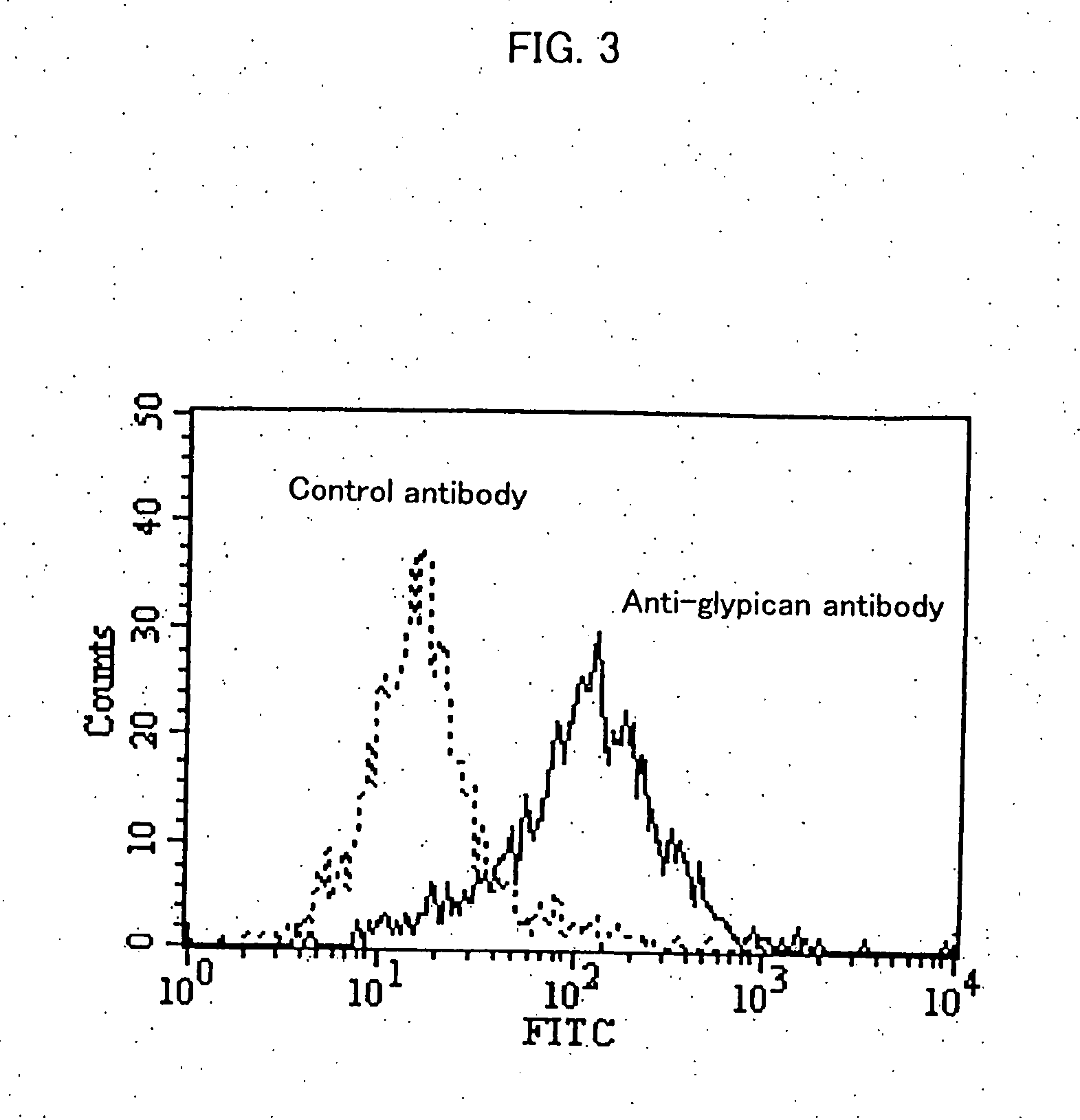
Measurement of the Expression Level of Glypican on HuH-7 Cells
[0111] Approximately 5×10⁵ HuH-7 cells were suspended in 100 μL of FACS / PBS (prepared by dissolving 1 g of bovine serum albumin (SIGMA) in 1 L of CellWASH (Beckton Dickinson)). Then, anti-glypican 3 antibodies (K6511) or mouse IgG2a (Biogenesis) as a control antibody were added at 25 μg / mL, and then the solution was allowed to stand on ice for 30 minutes. After washing with FACS / PBS, the product was suspended in 100 μL of FACS / PBS. 4 μL of Goat Anti-Mouse Ig FITC (Becton Dickinson) was added, and then the solution was allowed to stand on ice for 30 minutes.
[0112] After washing twice with FACS / PBS, the product was suspended in 1 mL of FACS / PBS. Fluorescence intensity of the cells was measured using a flow cytometer (EPICS XL, BECKMAN COULTER).
[0113]FIG. 3 shows the result of flow cytometry. Glypican 3 was expressed on HuH-7 cells, suggesting that anti-glypican 3 antibodies bind to glypican 3 expressed on the cell so as ...
PUM
| Property | Measurement | Unit |
|---|---|---|
| Cytotoxicity | aaaaa | aaaaa |
Abstract
Description
Claims
Application Information