System and method for cytological analysis by raman spectroscopic imaging

a raman spectroscopic imaging and cytological analysis technology, applied in the field of cytological analysis, can solve the problems of inability to detect other disease states (including many physiological states) easily by ordinary cytological methods, and the vivo cytological methods are often impractical, and achieve the effect of stable raman spectral respons

Inactive Publication Date: 2007-08-02
CHEMIMAGE
View PDF45 Cites 84 Cited by
  • Summary
  • Abstract
  • Description
  • Claims
  • Application Information

AI Technical Summary

Benefits of technology

[0055]FIG. 16 is a graph of averaged Raman scattering intensity over a range of Raman shift values for normal RBCs that had been illuminated for analysis of Raman scattering for not more than 100 milliseconds (solid line). The Raman scattering intensity is also shown (dashed line) for the same RBCs that had been illuminated for a sufficient period (about 2-5 seconds) that their Raman spectral response remained stable over time. Spectra obtained from 16 fields of view, each including 3-5 RBCs, were averaged to produce these data.
[0056]FIG. 17 is a graph of averaged Raman

Problems solved by technology
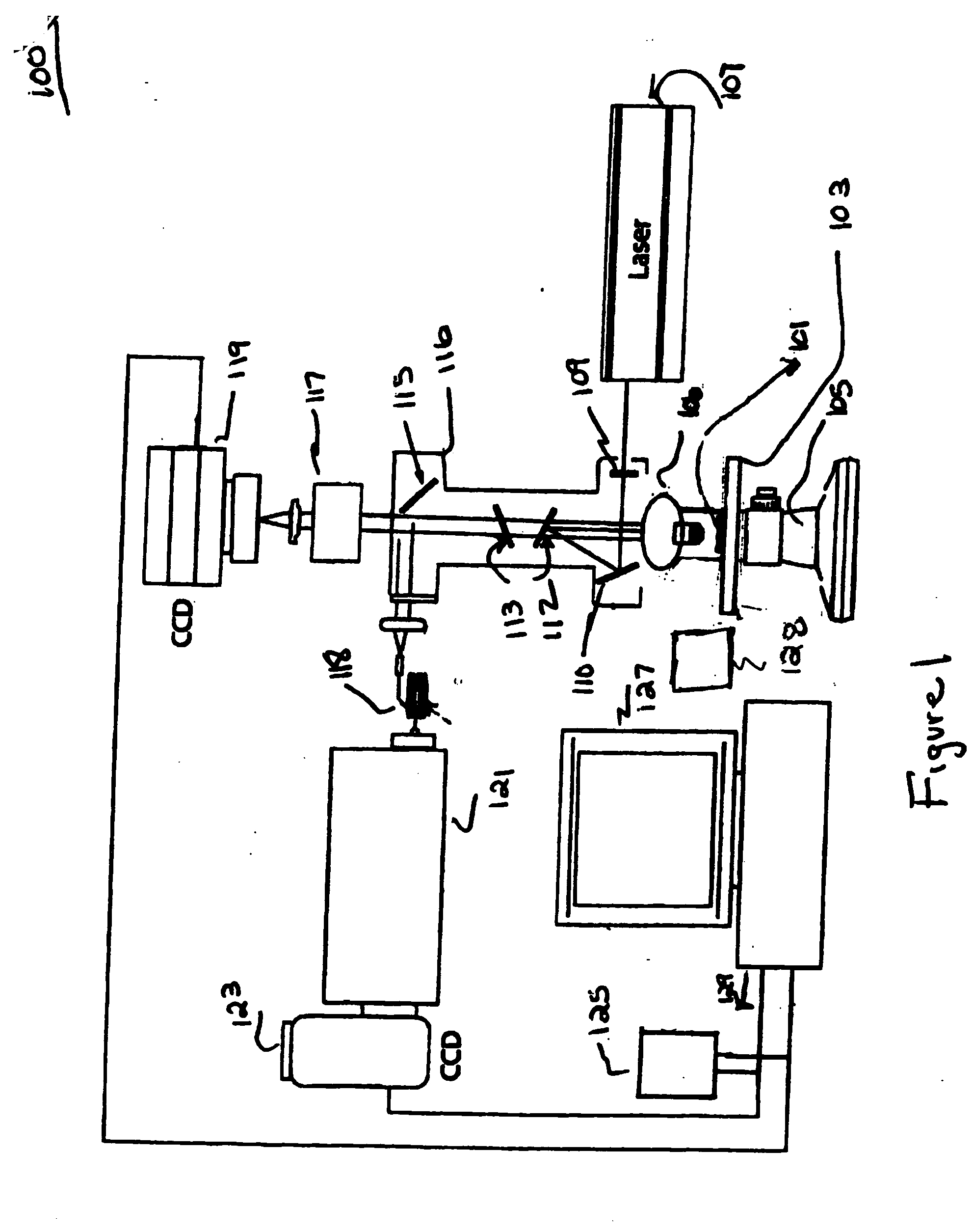
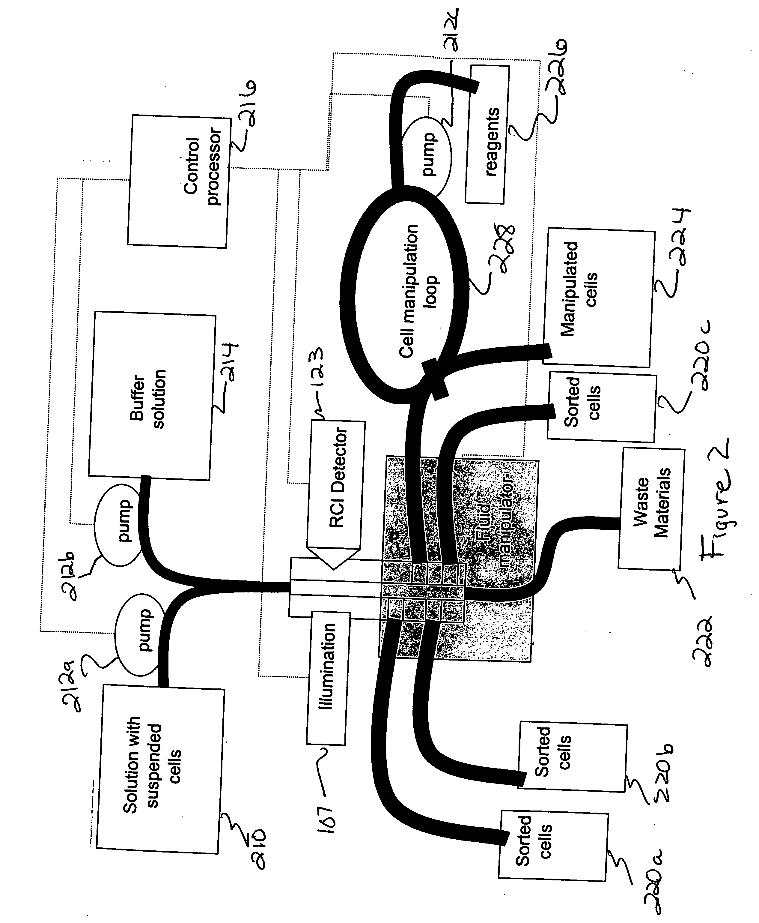
In vivo cytological methods are often impractical owing, for example, to relative inaccessibility of the cells of interest and unsuitability of staining or labeling reagents for in vivo use.
However, other disease states (including many physiological states which precede or indicate a predisposition to develop a disease state) cannot be readily detected by ordinary cytological methods.
A further shortcoming of many cytological methods is that, even when cytological identification of a disease state is possible, the time, expense, and expertise necessary to perform the cytological analysis can make it impractical or impossible to perform that analysis.
Cancer is significant, not only in terms of mortality and morbidity, but also in terms of the cost of treating advanced cancers and the reduced productivity and quality of life achieved by advanced cancer patients.
Because cancers arise from cells of normal tissues, cancer cells usually initially closely resemble the cells of the original normal tissue, often making detection of cancer cells difficult until the cancer has progressed to a stage at which the differences between cancer cells and the corresponding original normal cells are more pronounced.
Communication of results from the pathologist to the physician and to the patient can further slow the diagnosis of the cancer and the onset of any indicated treatment.
Because of the tissue preparation required, this process is relatively slow.
Moreover, the differentiation made by the pathologist is based on subtle morphological and other differences among normal, malignant, and benign cells, and such subtle differences can be difficult or time-consuming to detect, even for highly experienced pathologists.
Such differences are even more difficult for relatively inexperienced pathologists to detect.
Sickle-shaped RBCs are not able

Method used


Image

  • System and method for cytological analysis by raman spectroscopic imaging
  • System and method for cytological analysis by raman spectroscopic imaging
  • System and method for cytological analysis by raman spectroscopic imaging

Examples


example 1

Raman Scattering Analysis of Bladder Cancer Cells.

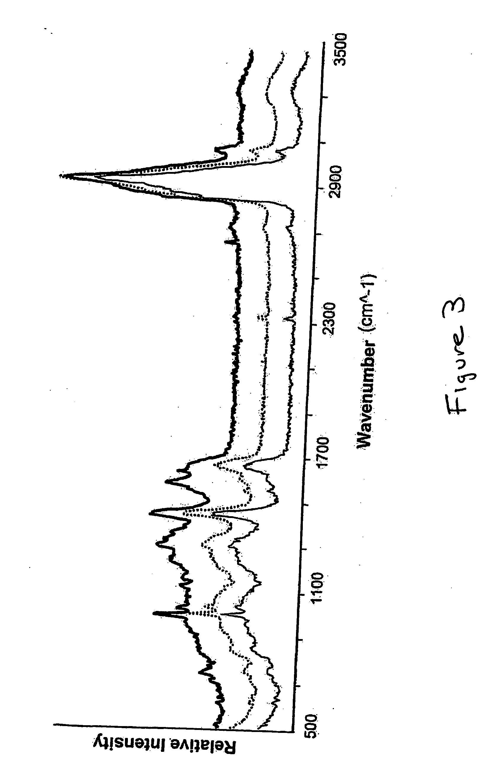
[0157] Raman molecular imaging (RMI) was used to distinguish cancerous and non-cancerous bladder cancer cells to demonstrate that RMI is useful for detection of bladder cancer. RMI is an innovative technology that combines the molecular chemical analysis capacity of Raman spectroscopy with the power of high definition digital image microscopic visualization. This platform enables physicians and their assistants to identify both the physical architecture and molecular environment of cells in a urine sample and can complement or be used in place of current histopathological methods.

[0158] The data presented in this example demonstrate that the Raman scattering signal from bladder cancer tissue and cells voided in the urine can be identified and be distinguished from normal bladder tissue and cells. Detectable differences between high and low grade tumor cells were observed. These data establish that RMI signatures of bladder cancer ...

example 2

Raman Scattering Analysis of Red Blood Cells.

[0193] Raman molecular imaging (RMI) was used to distinguish normal and sickled human red blood cells (RBCs).

[0194] Individual RBCs were obtained from two patients, one of whom was known to be afflicted with sickle cell disease (i.e., homozygous for the sickle cell trait gene) and the other of whom was known not to harbor an allele of the gene for the sickle cell trait. Prior to analysis, RBCs were treated by smearing onto an aluminum-coated glass slide and air dried. For each RBC, a visual microscopic determination was made of whether the cell was normal (i.e., normally-shaped) or sickled (i.e., sickle-shaped) using a FALCON™ Raman imaging microscope obtained from ChemImage Corp. (Pittsburgh, Pa.). A single Raman spectrum was obtained from a field of view that included 3-5 RBCs using the Raman scattering channel of the FALCON instrument. For samples of sickled RBCs, each field included at least one RBC that exhibited the crescent shap...

example 3

Raman Scattering Analysis of Cardiac Tissue.

[0197] Raman molecular imaging (RMI) was used to assess cardiac muscle tissue and connective tissue in cardiac tissue samples obtained from patients afflicted with either idiopathic heart failure or ischemic heart failure.

[0198] Human cardiac tissue samples were obtained from five patients afflicted with ischemic heart failure and from five other patients afflicted with idiopathic heart failure. The tissue samples were obtained in the form of small tissue fragments fractured from explanted hearts which were frozen immediately after removal. Approximately 5 millimeter square tissue fragments were embedded in OCT and sliced into 5-10 micron sections: Tissue slices were placed on an aluminum coated slide. Excess OCT was removed with distilled water. Samples were air-dried and evaluated using a FALCON (TM, ChemImage Inc., Pittsburgh, Pa.) Raman microscope.

[0199] Each tissue sample was sighted by visible light microscopy a Raman spectrum wa...


PUM

PropertyMeasurementUnit
Stress optical coefficientaaaaaaaaaa
Cytotoxicityaaaaaaaaaa

Abstract

A method and system of differentially manipulating cells where the cells, suspended in a fluid, are irradiated with substantially monochromatic light. A Raman data set is obtained from the irradiated cells and where the data set is characteristic of a disease status. The data set is assessed to identify diseased cells. A Raman chemical image of the irradiated cells is also obtained. Based on the assessment and the Raman chemical image, the fluid in which the cells are suspended is differentially manipulated. The diseased cells are directed to a first location and other non-diseased cells are directed to a second location as part of the differential manipulation. The diseased cells may be treated with a physical stress, a chemical stress, and a biological stress and then returned to a patient from whom the diseased cells were obtained prior to the irradiation.

Description

RELATED APPLICATIONS [0001] This application claims the benefit of U.S. Provisional Appl. No. 60 / 735,062, filed Nov. 9, 2005, entitled “Cytological Analysis by Raman Spectroscopic Imaging” which is incorporated herein by reference in its entirety. This application is also a continuation-in-part of U.S. application Ser. No. 11 / 269,596, filed Nov. 9, 2005, entitled “Cytological Methods for Detecting a Disease Condition Such as Malignancy By Raman Spectroscopic Imaging” which is incorporated herein by reference in its entirety.FIELD OF THE DISCLOSURE [0002] This application generally relates to cytological analysis, immunization, modification and treatment of diseased cells enabled by the use of Raman spectroscopic techniques. BACKGROUND OF THE DISCLOSURE [0003] The disclosure relates generally to the field of mammalian cellular evaluation and to correlation of cellular physiological status and diagnosis of disease based on such evaluation. In one embodiment the disclosure relates to m...

Claims


Application Information

Patent Timeline
IPC IPC(8): A61K48/00C12Q1/00G06F19/00
CPCG01N15/1459G01N21/65G01N2021/656G01N33/574G01N33/57407G01N33/5005
Inventor MAIER, JOHNDEMUTH, JOSEPHCOHEN, JEFFREYMCCLELLAND, LINDYSTEWART, SHONA
Owner CHEMIMAGE
Why Eureka
Try Eureka