Method of producing three-dimensional tissue and method of producing extracellular matrix used in the same

a three-dimensional tissue and extracellular matrix technology, applied in general culture methods, non-embryonic pluripotent stem cells, biochemistry apparatus and processes, etc., can solve the problems of difficult to realize three-dimensional tissue construction with cells merely by cell culture, difficult to construct the tissues of such organs by cell culture, and the layer itself has a very low mechanical strength, etc., to achieve excellent reproducibility and efficiency, simple

Active Publication Date: 2007-09-06
OSAKA UNIV
View PDF1 Cites 27 Cited by
  • Summary
  • Abstract
  • Description
  • Claims
  • Application Information

AI Technical Summary

Benefits of technology

[0010]Therefore, with the foregoing in mind, it is an object of the present invention to provide a novel method of producing a three-dimensional tissue by which cell lamination can be carried out easily and to provide a method of producing an extracellular matrix used in the method.
[0013]According to the ECM production method of the present invention, a nanoscale-thick ECM for adhering cell layers can be produced merely by bringing a cell layer into contact with a first substance-containing solution and a second substance-containing solution alternately. Thus, according to the three-dimensional tissue production method of the present invention that uses this ECM production method, merely by repeating the step of forming a cell layer and the step of bringing the cell layer into contact with a first substance-containing solution and a second substance-containing solution alternately, it is possible to laminate a plurality of cell layers successively with a nanoscale-thick ECM intervening between each pair of the adjacent cell layers. Thus, unlike the conventional methods, the method according to the present invention does not require separating a single cell sheet, laminating a plurality of separated cell sheets, etc., so that the three-dimensional tissue can be produced with excellent reproducibility and efficiency in a very simple manner. Therefore, the three-dimensional tissue production method according to the present invention is particularly suitable for reconstruction of tissues having a complex three-dimensional structure and thus is a very useful technique in the field of regenerative medicine.

Problems solved by technology

However, organs such as kidney and liver have a complex three-dimensional tissue structure composed of many types of cells, and hence, unlike the case of skin or the like, it is difficult to construct the tissues of such organs merely by cell culture.
In particular, when cells are cultured artificially, they proliferate two-dimensionally (i.e., in the plane direction) but hardly proliferate three-dimensionally (i.e., in the height direction), which makes it difficult to realize three-dimensional tissue construction with cells merely by cell culture.
However, since the cell layer itself has a very low mechanical strength, it is difficult to separate the cell layer from the support together with the carrier or the fibrin gel layer without losing the shape of the cell layer resulting from the culture on the support.
However, in the case where the mechanical strength of the fibrin gel layer is improved by increasing its thickness, there arises a problem in that the thick fibrin gel layer intervenes between the cell layers.
Moreover, the intervention of the thick fibrin gel layer may bring about the risk that, for example, after the laminate of the cell sheets has been grafted in a living body, the vascular invasion, which is very important in supplying nutrients and enzymes to the cells inside the laminate, is inhibited by the fibrin gel layer, which may lead to necrosis of the cell layers included in the laminate.
Furthermore, the methods that require separating the cell sheet or laminating the plurality of cell sheets as described above also have a problem in that construction of tissues having a complex shape is difficult.
That is, when a tissue to be constructed has a complex shape, the planar shape of a cell sheet to be used also becomes complex.
However, separating the cell sheet with high reproducibility while maintaining such a complex planar shape itself is very difficult.
This, however, still poses a problem because there is a limit on the shape that can be achieve by cutting.
However, such a technique is very complicated and thus is not practical.
However, there have been problems in that, for example, tissue construction using different types of cells, tissue construction into a desired size, etc. are difficult (see JP 2005-278608 A; Yamada N, Okano Y. Sakai H, Karikusa F. Sawasaki Y, Sakurai Y, Macromol Chem Rapid Commun.

Method used


Image

  • Method of producing three-dimensional tissue and method of producing extracellular matrix used in the same
  • Method of producing three-dimensional tissue and method of producing extracellular matrix used in the same
  • Method of producing three-dimensional tissue and method of producing extracellular matrix used in the same

Examples


example 1

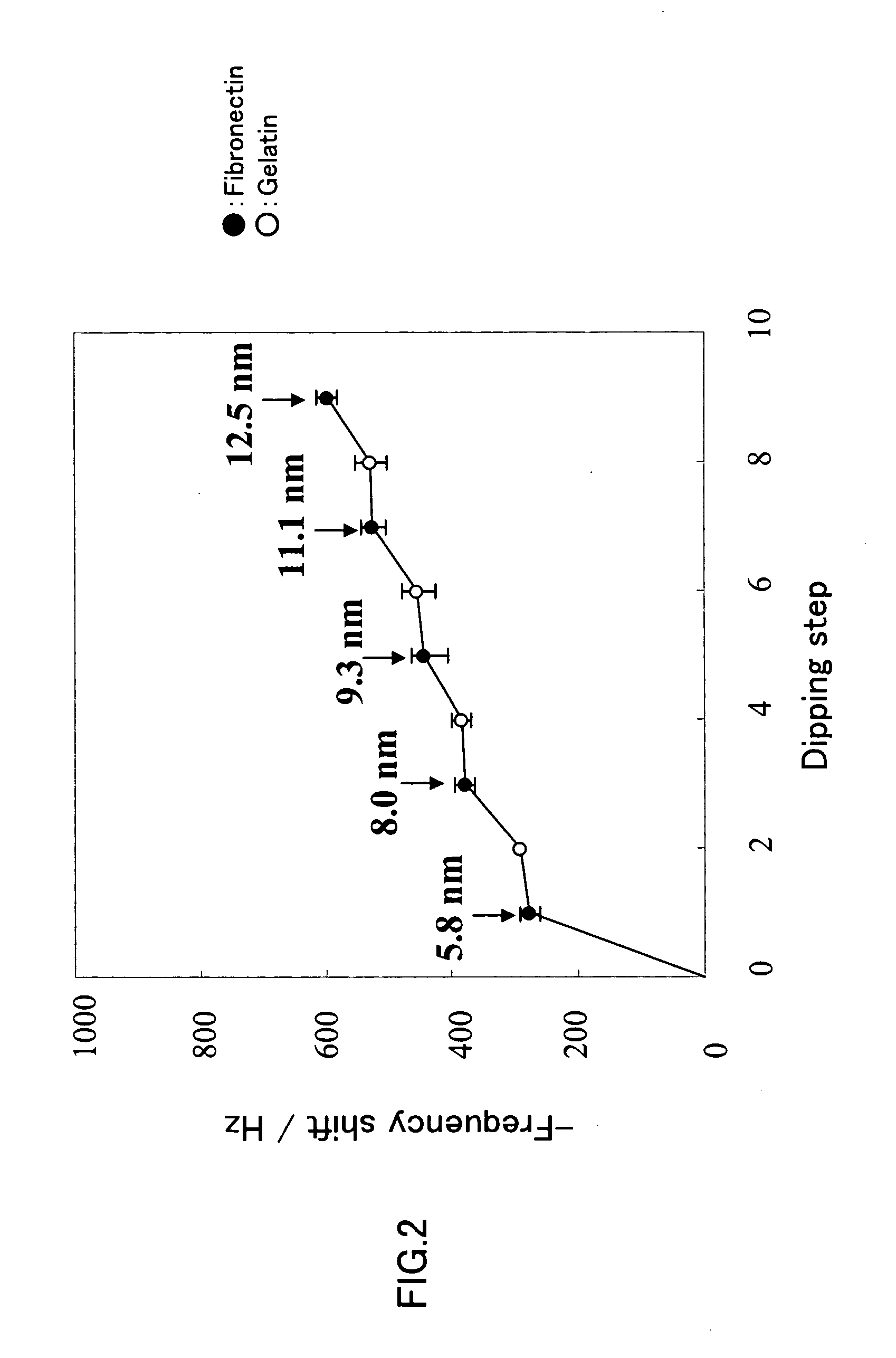
[0069]A nanoscale-thick nano-ECM in which fibronectin and gelatin were laminated alternately was produced. The substrate used for the nano-ECM formation was a quartz crystal microbalance (QCM) substrate, and the thickness of the matrix being formed was determined by measuring a frequency shift.

[0070]First, the QCM substrate was washed with a piranha solution for 1 minute and then dipped in a 0.5 M Tris buffer containing 0.2 mg / ml (0.02 wt %) fibronectin (pH 7.4, hereinafter the same) at 37° C. for 15 minutes. Subsequently, the substrate was washed with a 1 mM Tris buffer (pH 7.4) and then air-dried, after which the frequency shift was measured. Next, the substrate was dipped in a 0.05M Tris buffer containing 1 mg / ml (0.1 wt %) gelatin (pH 7.4, hereinafter the same) at 37° C. for 15 minutes. Again, the substrate was washed with a 1.0 mM Tris buffer (pH 7.4) and then air-dried, after which the frequency shift was measured. By repeating these steps alternately, nano-ECMs (n=3) were for...

example 2

[0072]Nano-ECMs each composed of gelatin and fibronectin were produced to construct a three-dimensional tissue with mouse fibroblasts (L929).

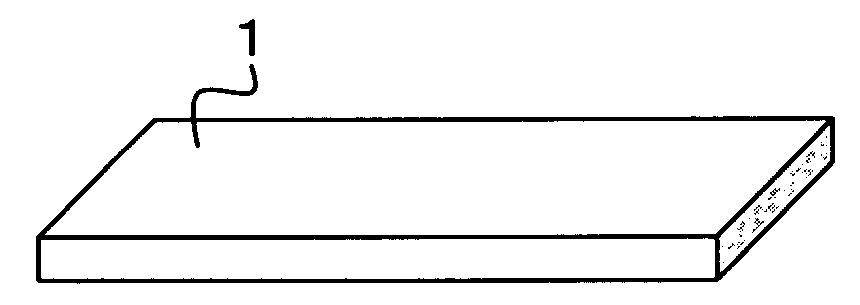
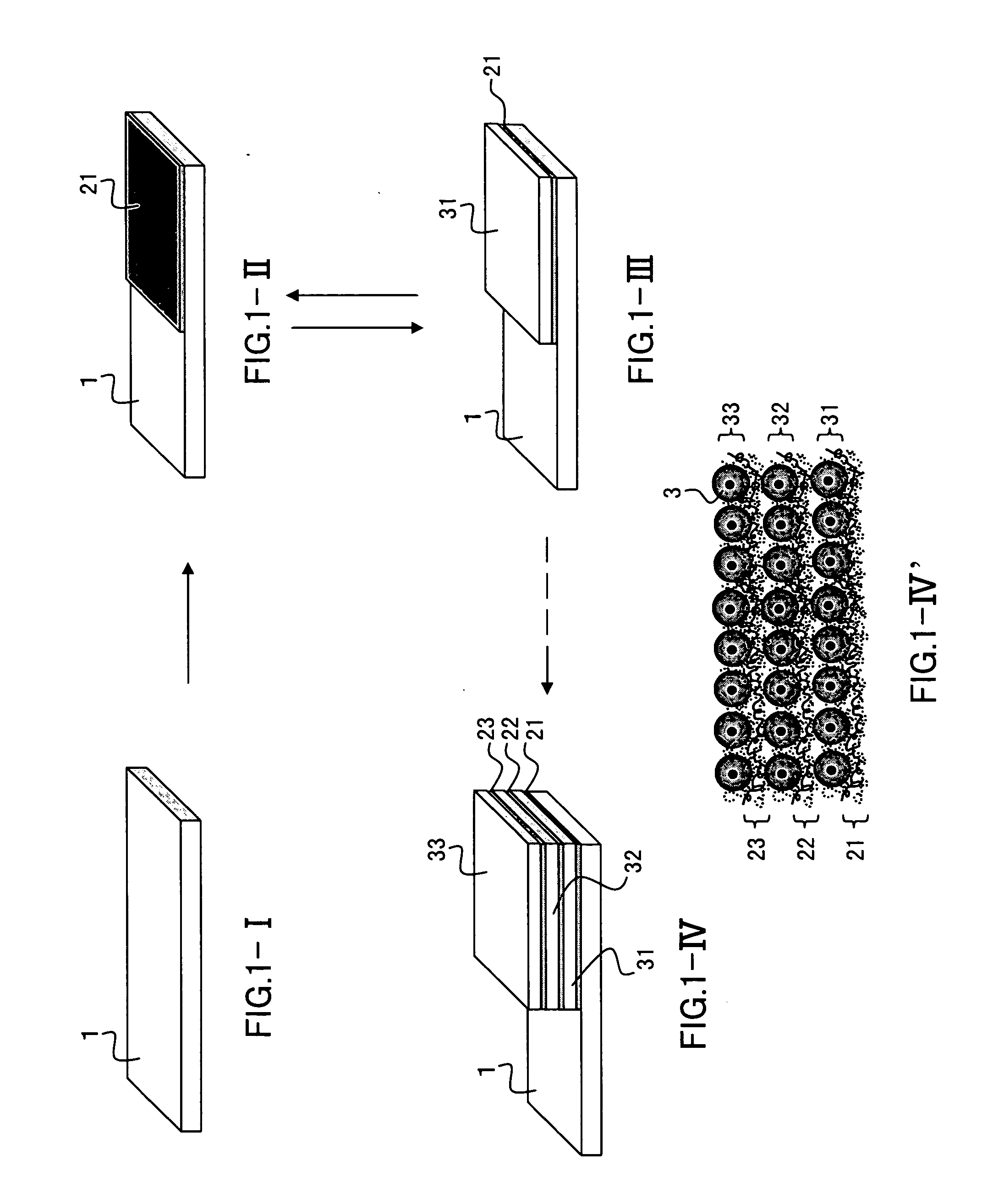
[0073]A first nano-ECM was formed on a slide glass (1.5 cm in width×2 cm in length) in the following manner.

[0074]The first nano-ECM was formed by dipping the slide glass in the fibronectin-containing buffer and the gelatin-containing buffer. Note here that the seven dipping steps were performed in total with the buffer in which the slide glass was dipped first being the fibronectin-containing buffer. After the slide glass had been dipped in the respective buffers, the slide glass was washed by being dipped in a 50 mM Tris buffer (pH 7.4) for 1 minute. As a result, about 10 nm thick nano-ECM whose lowermost layer and uppermost layer were both fibronectin layers was formed on the slide glass. Note here that dipping of the slide glass in either of the buffers at 37° C. for 15 minutes was regarded as one dipping step. Also note that only an end po...

example 3

[0081]Nano-ECMs each composed of fibronectin and gelatin were produced to laminate mouse fibroblasts (L929) three-dimensionally. The resultant three-dimensional tissue was evaluated using a confocal laser microscope.

[0082]Nano-ECMs and cell layers were laminated on a slide glass in the same manner as in Example 2, thus forming a three-dimensional tissue having a layered structure composed of first nano-ECM / first cell layer / second nano-ECM / second cell layer. The thickness of the first nano-ECM was about 13 nm and the thickness of the second nano-ECM was about 13 nm as in Example 2.

[0083]The three-dimensional structure of the thus-obtained three-dimensional tissue was observed with a confocal laser microscope. The results are shown in FIGS. 5 and 6. In FIG. 5, the micrograph (A) shows the X-Y plane of the three-dimensional tissue, the micrograph (B) shows the X-Z plane of the three-dimensional tissue, and the micrograph (C) shows the Y-Z plane of the three-dimensional tissue. The micr...


PUM


Abstract

The present invention provides a novel method of producing a three-dimensional tissue by which cell lamination can be carried out easily. According to the method, a three-dimensional tissue in which cell layers are laminated with an extracellular matrix intervening between each pair of the adjacent cell layers is produced by: (A) forming a cell layer on a substrate; (B) bringing the cell layer formed on the substrate into contact with a solution containing a first substance and a solution containing a second substance alternately, thus forming, on the cell layer, an extracellular matrix in which the first substance and the second substance are laminated alternately; and (C) culturing a cell on the extracellular matrix to form a further cell layer. In the present invention, a combination of the first substance and the second substance is (a) a combination of a protein or polymer having an RGD sequence and a protein or polymer that interacts with the protein or polymer having the RGD sequence or (b) a combination of a protein or polymer that is positively charged and a protein or polymer that is negatively charged.

Description

[0001]The patent or application file contains at least one drawing executed in color. Copies of this patent or patent application publication with color drawing(s) will be provided by the Office upon request and payment of the necessary fee.BACKGROUND OF THE INVENTION[0002]1. Field of the Invention[0003]The present invention relates to a method of producing a three-dimensional tissue in which cell layers are laminated and also to a method of producing an extracellular matrix.[0004]The present disclosure relates to subject matter contained in priority Japanese Application No. 2006-56836, filed on Mar. 2, 2006, which is herein expressly incorporated by reference in its entirety.[0005]2. Description of Related Art[0006]In recent years, regenerative medicine has been attracting great attention. Regenerative medicine has become feasible and actually is in the stage of clinical application with regard to regeneration of tissues with a relatively simple structure, such as bone, cartilage, ...

Claims


Application Information

Patent Timeline
Patent Type & Authority Applications(United States)
IPC IPC(8): C12N5/06C12N5/07C12N5/074C12N5/0789
CPCC12N5/0062C12N2533/54C12N2533/52C12N5/0068Y10S977/902
Inventor AKASHISAWA, YOSHIKIMATSUSAKI, MICHIYANAKAHARA, YOSHIO
Owner OSAKA UNIV
Why Eureka
Try Eureka