
Protein isoform discrimination and quantitative measurements thereof

a technology of applied in the field of protein isoform discrimination and quantitative measurement, can solve the problems of limited unique regions, difficult discrimination of these proteins, and insufficient present methods for analyzing distinct proteins with similar amino acid sequences in a sampl

Inactive Publication Date: 2007-09-27
MILLIPORE CORP
View PDF53 Cites 38 Cited by
  • Summary
  • Abstract
  • Description
  • Claims
  • Application Information

AI Technical Summary

Benefits of technology

[0124] An important advantage of the invention is that useful capture agents can be identified and / or synthesized even in the absence of a sample of the protein to be detected. With the completion of the whole genome in a number of organisms, such as human, fly (e.g., Drosophila melanogaster) and nematode (e.g., C. elegans), PETs of a given length or combinations thereof can be identified for any single given protein in a certain organism, and capture agents for any of these proteins of interest can then be made without ever cloning and expressing the full length protein.
[0125] In addition, the suitability of any PET to serve as an antigen or target of a capture agent can be further checked against other available information. For example, since amino acid sequence of many proteins can now be inferred from available genomic data, sequence from the structure of the proteins unique to the sample can be determined by computer aided searching, and the location of the peptide in the protein, and whether it will be accessible in the intact protein, can be determined. Once a suitable PET peptide is found, it can be synthesized using known techniques. With a sample of the PET in hand, an agent that interacts with the peptide such as an antibody or peptidic binder, can be raised against it or panned from a library. In this situation, care must be taken to assure that any chosen fragmentation protocol for the sample does not restrict the protein in a way that destroys or masks the PET. This can be determined theoretically and / or experimentally, and the process can be repeated until the selected PET is reliably retrieved by a capture agent(s).
[0126] The PET set selected according to the teachings of the present invention can be used to generate peptides either through enzymatic cleavage of the protein from which they were generated and selection of peptides, or preferably through peptide synthesis methods.
[0127] Proteolytically cleaved peptides can be separated by chromatographic or electrophoretic procedures and purified and renatured via well known prior art methods.
[0128] Synthetic peptides can be prepared by classical methods known in the art, for example, by using standard solid phase techniques. The standard methods include exclusive solid phase synthesis, partial solid phase synthesis methods, fragment condensation, classical solution synthesis, and even by recombinant DNA technology. See, e.g., Merrifield, J. Am. Chem. Soc., 85:2149 (1963). Solid phase peptide synthesis procedures are well known in the art and further described by John Morrow Stewart and Janis Dillaha Young, Solid Phase Peptide Syntheses (2nd Ed., Pierce Chemical Company, 1984).
[0129] Synthetic peptides can be purified by preparative high performance liquid chromatography [Creighton T. (1983) Proteins, structures and molecular principles. WH Freeman and Co. N.Y.] and the composition of which can be confirmed via amino acid sequencing.

Problems solved by technology

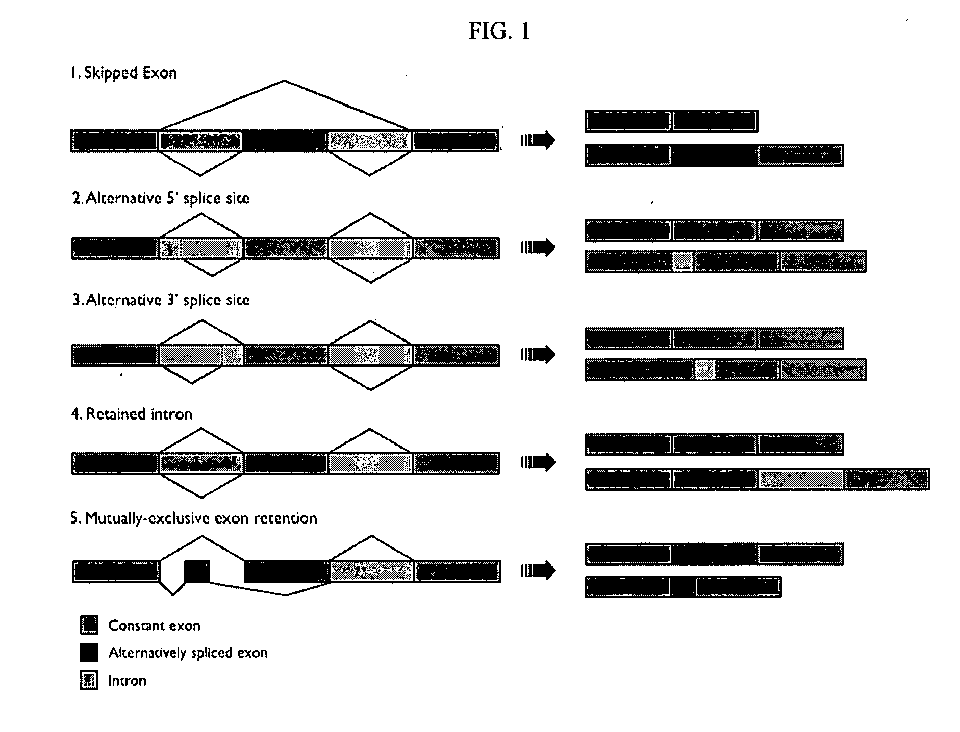
However, mammalian genes can undergo modifications that yield modified protein forms with similar amino acid sequences, and only limited unique regions.
Since one or more distinct proteins with similar amino acid sequences may be present in the same protein sample to be analyzed, discrimination of these proteins is a challenging application.
Unfortunately, the present methods for analyzing distinct proteins with similar amino acid sequences in a sample are inadequate.
However, the choice of sequences for raising antibodies that recognize the uniqueness at the junction region is limited based on the amino acids comprising the junction region, and such limited choices may not even be desirable.
For example, the junction region may be too hydrophobic, too short, etc., making such regions poor candidates for raising effective capture agents (e.g., antibodies or functional fragments thereof).
Consequently, it is not always possible to develop antibodies to the junction region.
Further, while sequences at the junction region are generally unique relative to the isoform family, they may not be unique across the entire proteome.
In fact, they may not even be unique for all the other proteins in a given sample to be analyzed.

Method used


Image

  • Protein isoform discrimination and quantitative measurements thereof
  • Protein isoform discrimination and quantitative measurements thereof
  • Protein isoform discrimination and quantitative measurements thereof

Examples


example 1

Bcl-x Isoform Detection

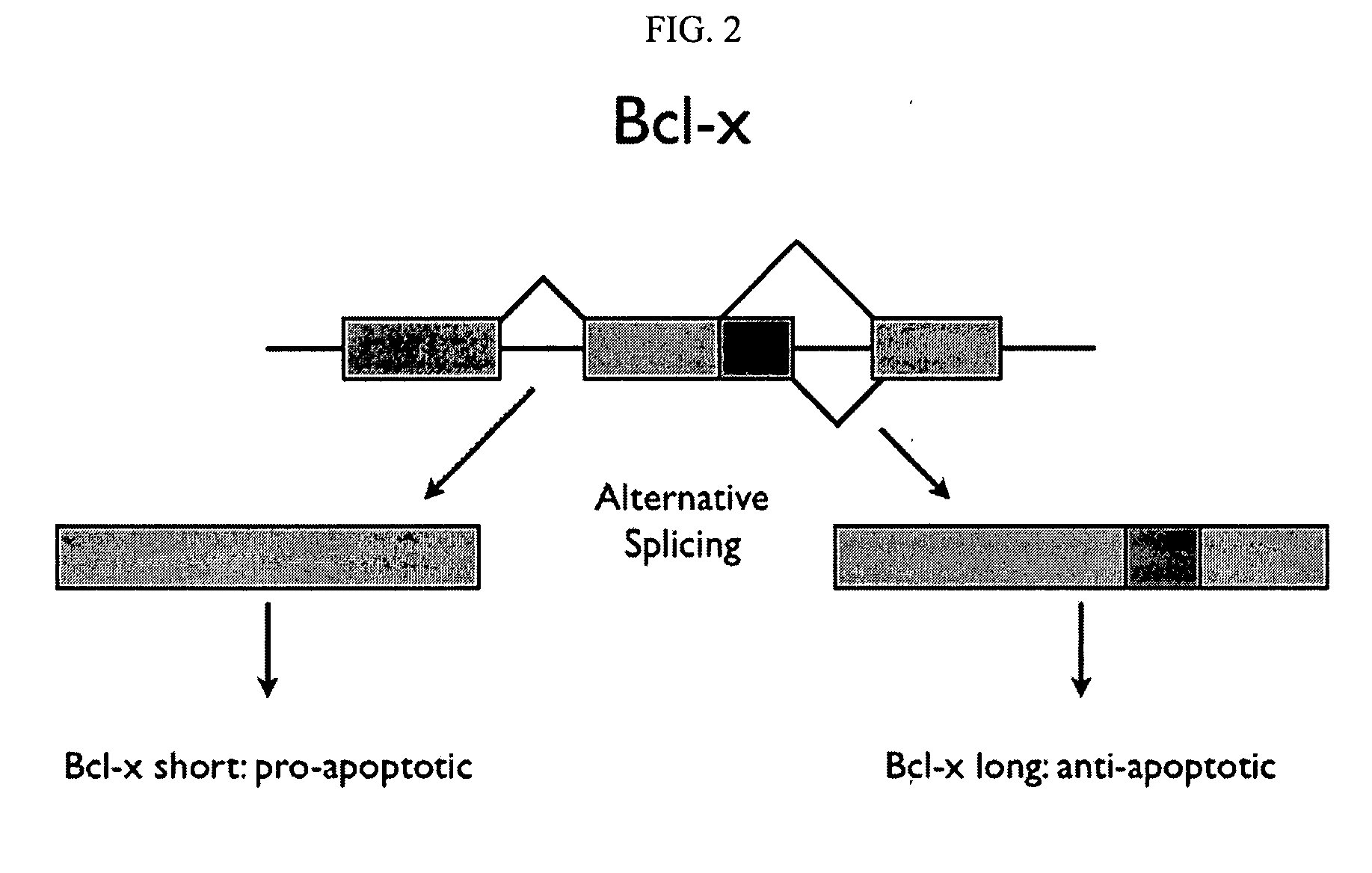
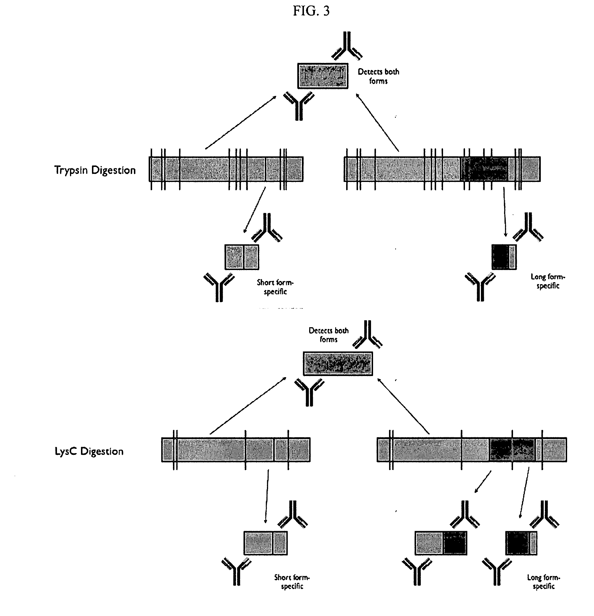
[0285] Two forms of the protein Bcl-x (isoform 1: NP—612815; isoform 2: NP—001182) have been identified and are shown below in schematic representation (see FIGS. 2 and 3) and in sequence alignment (FIG. 4). Bcl-x is a member of the Bcl-2 family of apoptotic factors. Alternative splicing of Bcl-x results in two isoforms that have opposing apoptotic activities. The Bcl-x long isoform is anti-apoptotic, while the Bcl-x short isoform is pro-apoptotic. Also shown in the schematic are protease cleavage sites and the resulting protein fragments for both trypsin and Lys C digestion, respectively (FIG. 3). A protein fragment is selected for each of the two protein isoforms that is unique to that form. As can be seen in the figures, both trypsin and lysC digestion results in the creation of at least one proteolytic fragment unique to each isoform and one that is common to both. Using established PET (peptide epitope tag) picking algorithms, 8-mer PET sequences were id...

example 2

CD44 Isoform Detection

[0287] For some isoform families (especially ones that incorporate many variable exons), it may not be as straightforward as the case described above to uniquely discriminate among all isoforms (should all or several isoforms be present in a given sample). In more complicated cases, isoforms will fall into groups that share common peptide fragments. A good example of this application is the CD44 isoform family.

[0288] CD44 is a cell surface receptor with a variety of roles in cell adhesion, lymphocyte activation, cell-cell and cell-extracellular matrix interactions, and tumor growth and progression. The CD44 gene consists of 20 exons, the central ten of which are subject to alternative splicing designated by a number followed by “v” for “variable”—(See FIG. 7.) Exons 1-5 and 16-20 are invariant and occur in all known isoforms. While only a dozen or so CD44 isoforms have been identified, alternative splicing of CD44 has the potential to produce hundreds of isof...

example 4

Exemplary Sample Preparation

[0309] Samples for the methods of the invention may be prepared according to any of the methods described herein. This example provides a specific preparation method that is preferred for certain embodiments, such as the sandwich immunoassay. However, it should be understood that it is by no means limiting.

[0310] A typical sample was prepared in 5 mM TCEP (Tris(2-Carboxyethyl)Phosphine), 0.05% (w / v) SDS, and approximately 20 mM triethanolamine, pH 8.5. The mixture was heated at about 100° C. for about 5 minutes, and then allowed to cool back to room temperature (about 25° C.). Iodoacetamide was then added to a concentration of about 10 mM, and the sample was alkylated for about 30 minutes at room temperature (usually in the dark). Then about 1 / 20 (w / w) trypsin relative to the amount of protein in the sample was added (e.g., if the total protein concentration was about 1 mg / ml, add 0.05 mg / ml trypsin). Digestion was allowed to proceed for between 2 hours...


PUM

PropertyMeasurementUnit
pHaaaaaaaaaa
temperaturesaaaaaaaaaa
concentrationaaaaaaaaaa

Abstract

The invention relates to methods, reagents and apparatus for detecting protein isoforms (e.g., those due to alternative splicing, or different disease protein isoforms or degradation products) in a sample, including using combinations of capture agents to identify the isoforms to be detected / measured.

Description

CROSS-REFERENCE TO RELATED APPLICATIONS [0001] The present application is a continuation-in-part of U.S. patent application Ser. No. 11 / 387,389, filed Mar. 23, 2006, the entire disclosure of which is incorporated by reference herein for all purposes.BACKGROUND OF THE INVENTION [0002] Antibody arrays are useful for detecting multiple proteins simultaneously in a biological sample. An important and growing application area for the study of multiple proteins is the identification and quantitative measurement of proteins having similar amino acid sequences, for example, protein isoforms resulting from gene-level events, for example, gene splicing and genetic translocations. [0003] U.S. patent application publication number US 2004-0029292 A1 describes technology that can be used to identify multiple proteins using an array. Briefly, starting from the primary amino acid sequence of any protein, one can identify a series of linear epitopes (PETs) that uniquely represent the protein. By de...

Claims


Application Information

Patent Timeline
Patent Type & Authority Applications(United States)
IPC IPC(8): C12Q1/68G01N33/53C12Q1/37G01N33/543
CPCG01N33/6878G01N33/6803
Inventor GORDON, NEAL F.GRAHAM, JAMES R.
Owner MILLIPORE CORP
Learn More