
Methotrexate-modified nanoparticles and related methods

a technology of metastatic nanoparticles and nanoparticles, which is applied in the field of metastatic nanoparticles, can solve the problems of insufficient delivery of chemotherapeutic agents to target cells, limited resolution of instruments, and ineffective gd) complex contrast agents, and achieve the effect of inhibiting the invasive activity of neoplastic cells

Inactive Publication Date: 2007-11-22
IND TECH RES INST +1
View PDF3 Cites 29 Cited by
  • Summary
  • Abstract
  • Description
  • Claims
  • Application Information

AI Technical Summary

Benefits of technology

[0034] In one embodiment, the invention provides a method for treating cancer, comprising administering to a patient in need thereof an effective amount of a pharmaceutical composition comprising a methotrexate-modified nanoparticle and a pharmaceutically acceptable carrier.
[0035] In one embodiment, the invention provides a method for inhibiting invasive activity of neoplastic cells, comprising administering to neoplastic cells an effect

Problems solved by technology

While the imaging capabilities of these instruments have revolutionized imaging technology, the resolution of the instrument is limited to the elucidations of lesions within the body on the order of 1 mm with a clinical scanner.
This limitation of the instrument has led to the development of several types of contrast enhancement agents including magnetite / dextran-based nanoparticles and chelated gadolinium contrast agents, which are currently available on the market and used widely in clinical applications.
However, gadolinium (Gd) complex contrast agents are effective only when present in millimolar concentrations.
However, identification of specific target agents and drugs which are capable of being released inside target cells remain as a challenge and is the central focus of the current studies in the field.
Delivery of chemotherapeutic agents to target cells is not sufficient to induce cell death.
However, the clinical application of this drug is limited by its toxic dose-related side effects and drug resistance by target cells.
These conjugates have been shown to increase cellular cytotoxicity and increase cellular mortality.
Despite the success of these conjugates in vitro, there is still no clinical method of detecting the levels of methotrexate taken up by the target cells, which may reduce the efficacy of the treatment.
However, the large size of the drug conjugate does not facilitate intravenous drug delivery.
Thus, the large size of these conjugates limits the administration of the drug carriers to direct injection into the tumor site.

Method used


Image

  • Methotrexate-modified nanoparticles and related methods
  • Methotrexate-modified nanoparticles and related methods
  • Methotrexate-modified nanoparticles and related methods

Examples


example 1

Synthesis of Methotrexate-Modified Nanoparticles with Alkyl Linker

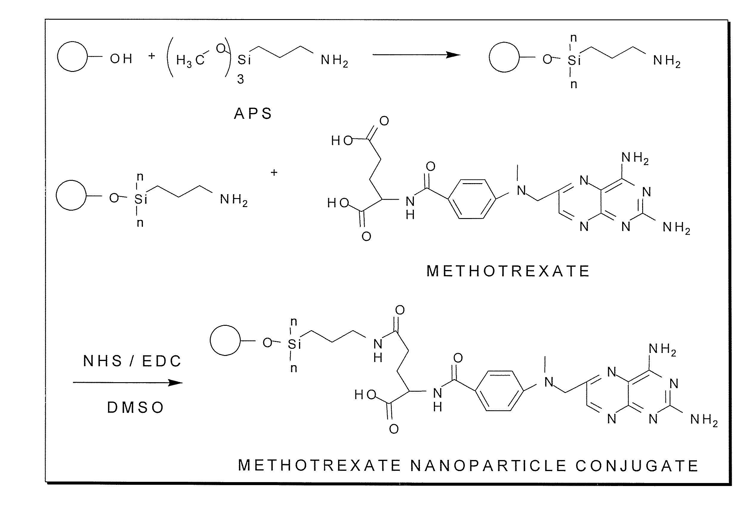
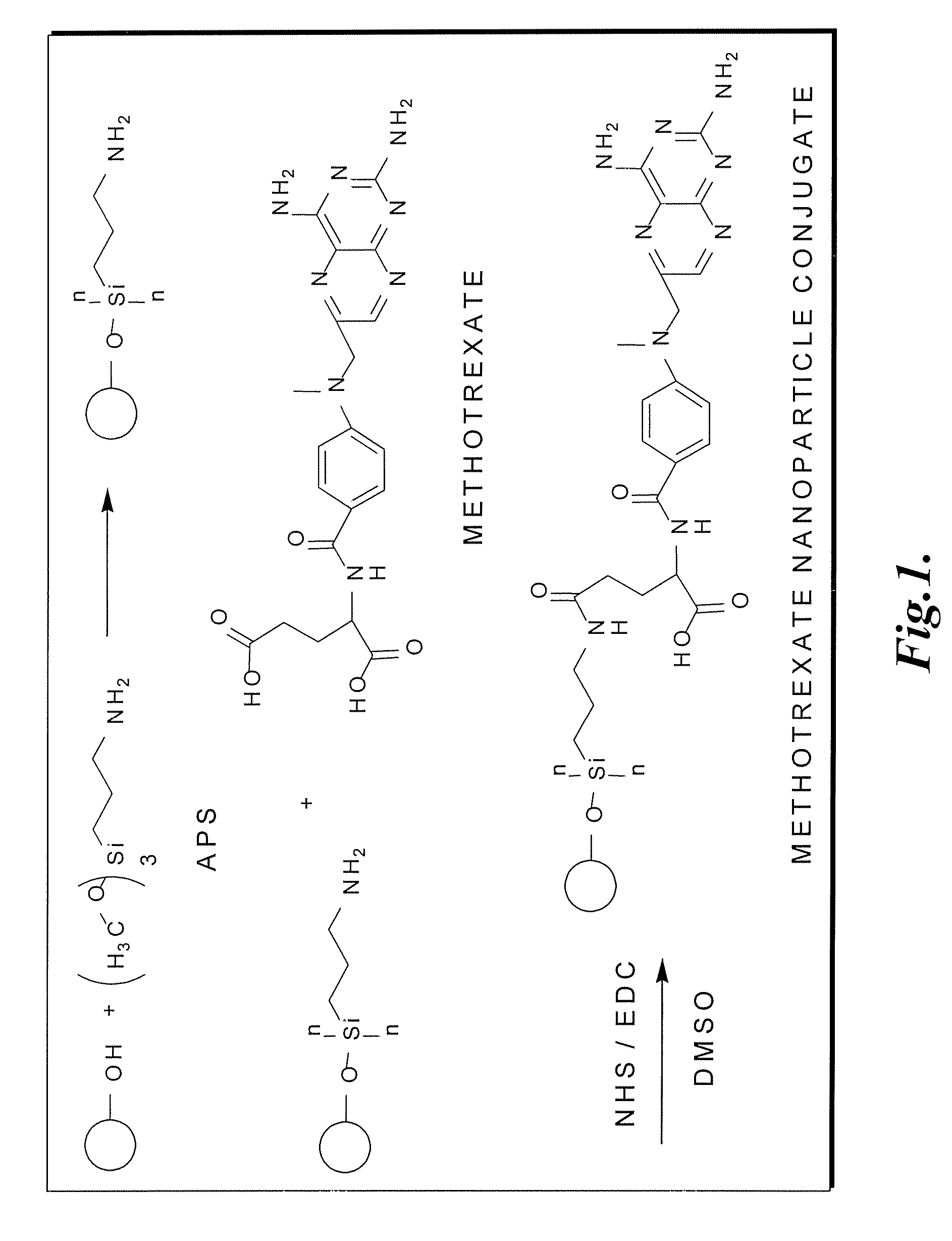
[0133] Surface modification of nanoparticles with 3-aminopropyl trimethoxysilane and methotrexate. Magnetite nanoparticles were synthesized by a co-precipitation method with minor modifications outlined previously (Chang, Y.; Kohler, N.; Zhang, M., Surface Modification of Superparamagnetic Magnetite Nanoparticles and Their Intracellular Uptake, Biomaterials 2002, 23, 1553-1561). The magnetite nanoparticles were surface-modified with methotrexate via a chemical scheme outlined in FIG. 1. The nanoparticles were first surface-modified with 3-aminopropyltrimethoxysilane (APS) to form a self-assembled monolayer (SAM) and subsequently conjugated with methotrexate through amidation between the carboxylic acid end group on methotrexate and the amine groups on the particle surface. The methotrexate conjugation reaction may occur through either the a or B carboxylic acid groups on the glutamic acid residue. One milliliter of A...

example 2

Synthesis of Methotrexate-Modified Nanoparticles with PEG Linker

[0135] Dulbecco's phosphate buffered saline (PBS), N-Hydroxysuccinimide 97% (NHS), 1-Ethyl-3-(3-dimethylamino-propyl) carbodiimide (EDAC), iron (II) and iron (III) chloride were purchased from Sigma (St. Louis, Mo.). All other solvents were purchased from Fisher Scientific (Hampton, N.H.) or Aldrich (Milwaukee, Wis.).

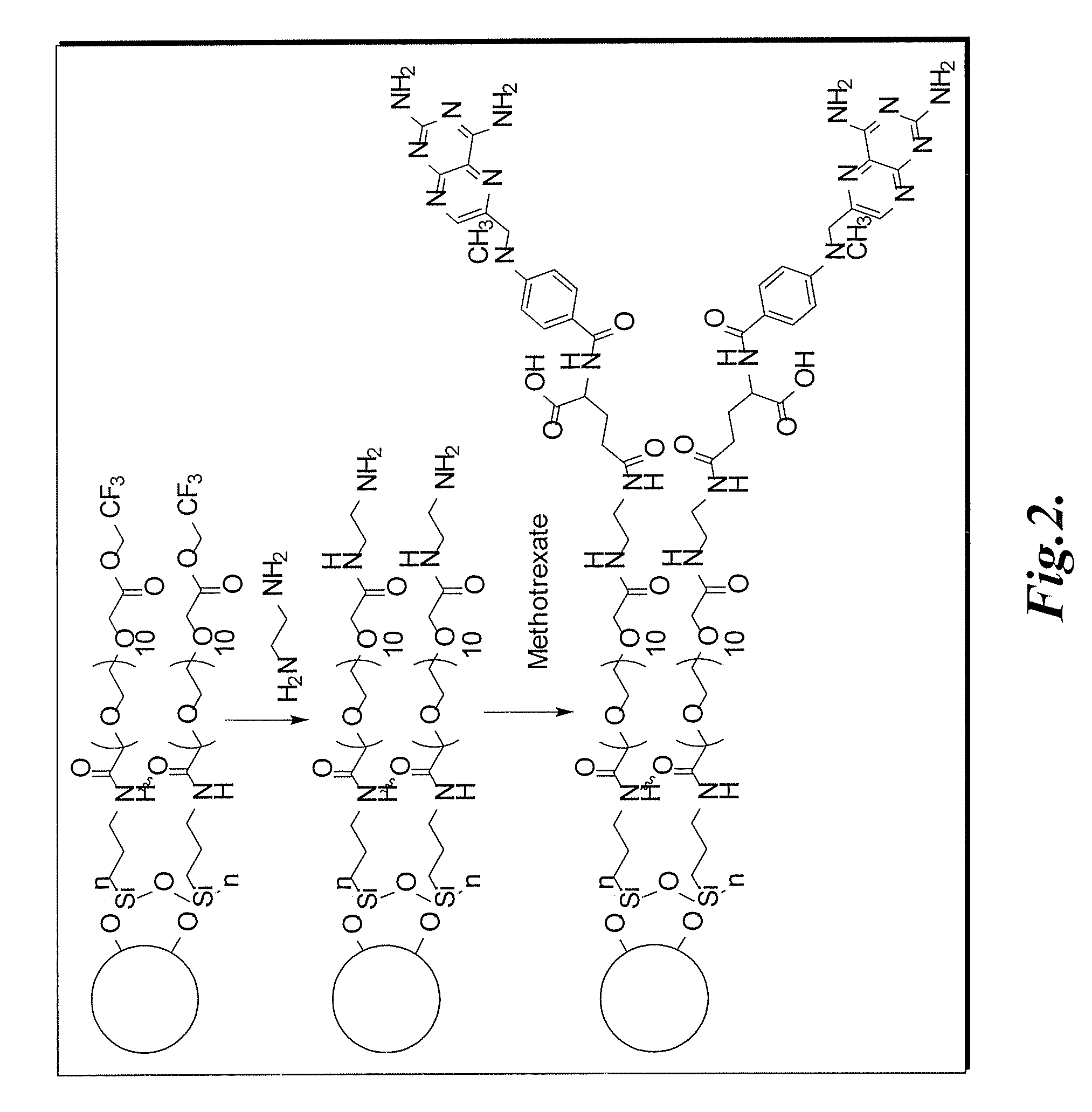
[0136] Magnetite nanoparticles were synthesized by a co-precipitation method. The acidic iron chloride solution was prepared by dissolution of 3.09 g (24.37 mM) of FeCl3 and 5.2 g (32.06 mM) of FeCl2 in 100 mL of 0.96 M hydrochloric acid solution. The resultant solution was placed in a sonicating bath and stirred for five hours during which time 500 mL of 1.5M NaOH solution was introduced drop-wise via a peristaltic pump. Particle synthesis temperature was controlled by an external water-circulator connected to the sonicating bath containing a solution of 50% ethylene glycol and 50% DI water. To prevent n...

example 3

Analysis of Methotrexate Release from Magnetite Nanoparticles

[0140] To simulate intracellular lysosomal conditions, methotrexate modified nanoparticles, NP-propyl-methotrexate, at a concentration of 0.1 mg / mL were suspended in a solution of 0.1 mg / mL crude protease from bovine pancreas (Sigma) in 5 mL of phosphate-buffered saline (PBS) solution at 37° C. under constant stirring. The solution pH was adjusted by the titration of 1.0M HCl and 1.0 M NaOH to achieve pH's of 2, 3, 4, 5.6, and 7.44 respectively. Following incubation for 8, 24, 48, and 72 hours, the nanoparticle suspensions were centrifuged at 2000 rpm to isolate the particles from methotrexate, PBS, and protease solutions. Methotrexate cleavage from nanoparticles was then quantified with UV spectroscopy at wavelength of 304 nm.


PUM

PropertyMeasurementUnit
Diameteraaaaaaaaaa
Diameteraaaaaaaaaa
Diameteraaaaaaaaaa

Abstract

Methotrexate-modified nanoparticles that target tumors, compositions that include the nanoparticles, methods of imaging tissues using the nanoparticles, and methods for treating tissues using the nanoparticles.

Description

CROSS-REFERENCE TO RELATED APPLICATION [0001] This application claims the benefit of U.S. Provisional Application No. 60 / 725,913, filed Oct. 11, 2005, incorporated herein by reference in its entirety.STATEMENT OF GOVERNMENT LICENSE RIGHTS [0002] This invention was made with government support under Contract No. NO1-C037122 awarded by National Institute of Health. The Government has certain rights in the invention.BACKGROUND OF THE INVENTION [0003] Over the past several years, there has been a growing interest in developing nanoparticle-based targeting agents for tumor diagnostics and therapeutics. It is recognized that with these targeting agents, tumors or other lesions can be detected at the cellular or molecular level. Two major applications associated with these systems are magnetic resonance imaging (MRI) and controlled drug release (CDR). [0004] Magnetic resonance imaging (MRI) is an appealing non-invasive approach for early cancer diagnostics and therapeutics. While the imagi...

Claims


Application Information

Patent Timeline
IPC IPC(8): A61K31/4985A61K9/14A61P35/00C07D475/08G01N33/574
CPCA61K31/4985A61K47/48861A61K47/48884G01N33/57492A61K49/1848A61K49/186A61K49/1833A61K47/6923A61K47/6929
Inventor ZHANG MIQINWANG SHIAN-JYKOHLER NATHANLIN YUH-JIUANSUN CONROY
Owner IND TECH RES INST
Learn More