
Monitoring gene silencing and annotating gene function in living cells

a gene function and gene silencing technology, applied in the field of biological, molecular biology, chemistry and biochemistry, can solve the problems of not being readily adapted to large-scale studies or automated analyses of hundreds or thousands of potential drug targets, and none of the above methods can be applied to intact cells

Inactive Publication Date: 2008-03-13
ODYSSEY THERA INC
View PDF11 Cites 1 Cited by
  • Summary
  • Abstract
  • Description
  • Claims
  • Application Information

AI Technical Summary

Benefits of technology

[0022] It is an object of the present invention to provide methods for mapping genes and proteins into biochemical pathways, and for validating novel pharmaceutical targets.
[0023] It is an additional object of the present invention to provide methods for directly measuring the effects of an annotation reagent on any protein or pathway in an intact cell.
[0024] It is an additional object of the present invention to show that the methods provided herein can be applied to any target class, cell type, species, disease mechanism, or organism.
[002...

Problems solved by technology

Typically these assays require the preparation and analysis of cell lysates following the cell treatment; none of the above methods can be applied to intact cells.
Such methods are difficult to scale up or automate and are semi-quantitative at best, and are not readily adapted to large-scale studies or to automated analyses of hundreds or thousands of potential drug targets.
However, mapping pathways is not possible with transcription reporter assays except through inference.
It is often not possible to determine whether the effect on transcription is a direct vs. an indirect effect of the gene on the pathway of interest or to determine the mechanism by which a particular gene affects transcription.

Method used


Image

  • Monitoring gene silencing and annotating gene function in living cells
  • Monitoring gene silencing and annotating gene function in living cells
  • Monitoring gene silencing and annotating gene function in living cells

Examples


example 1

DNA Constructs

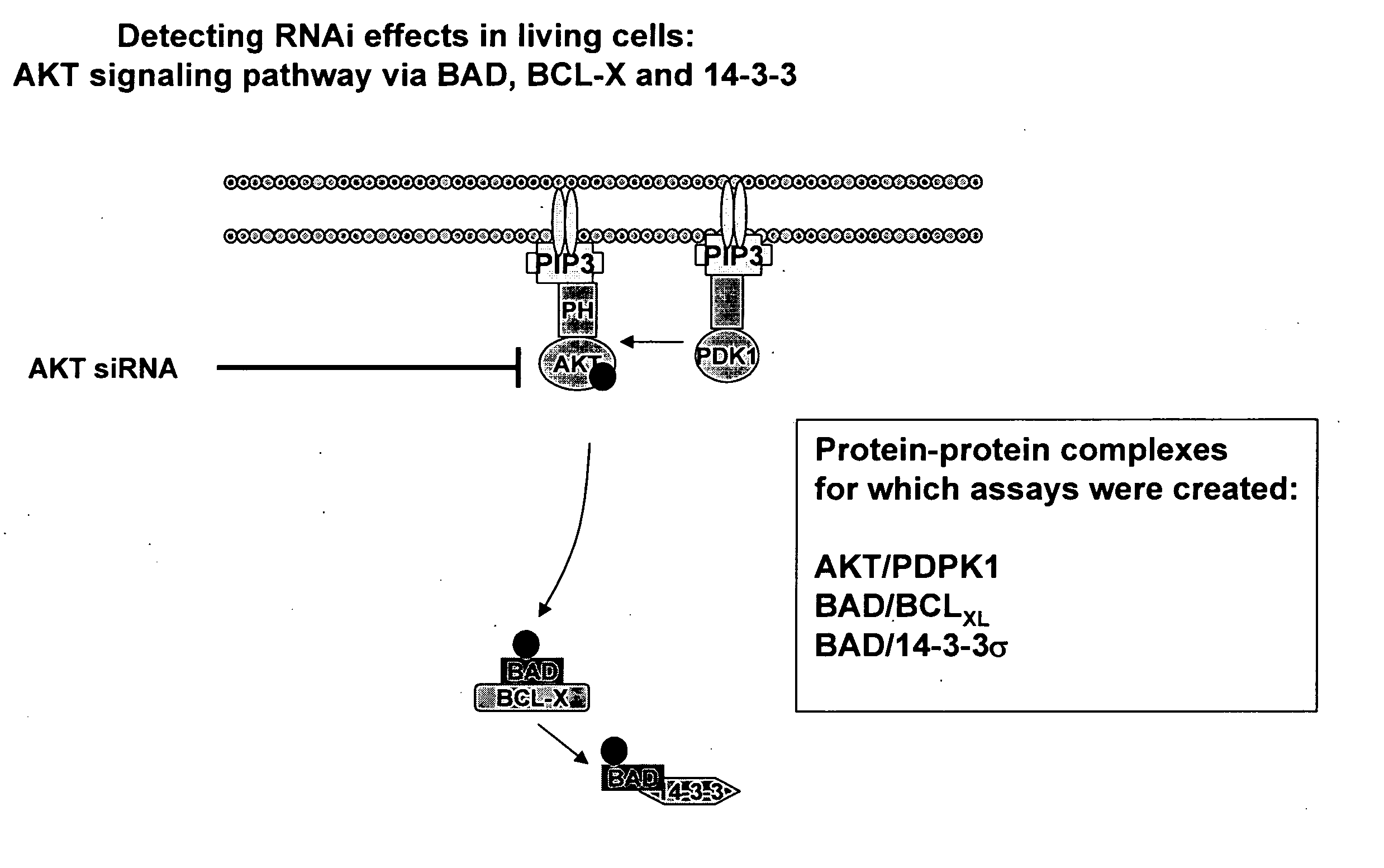
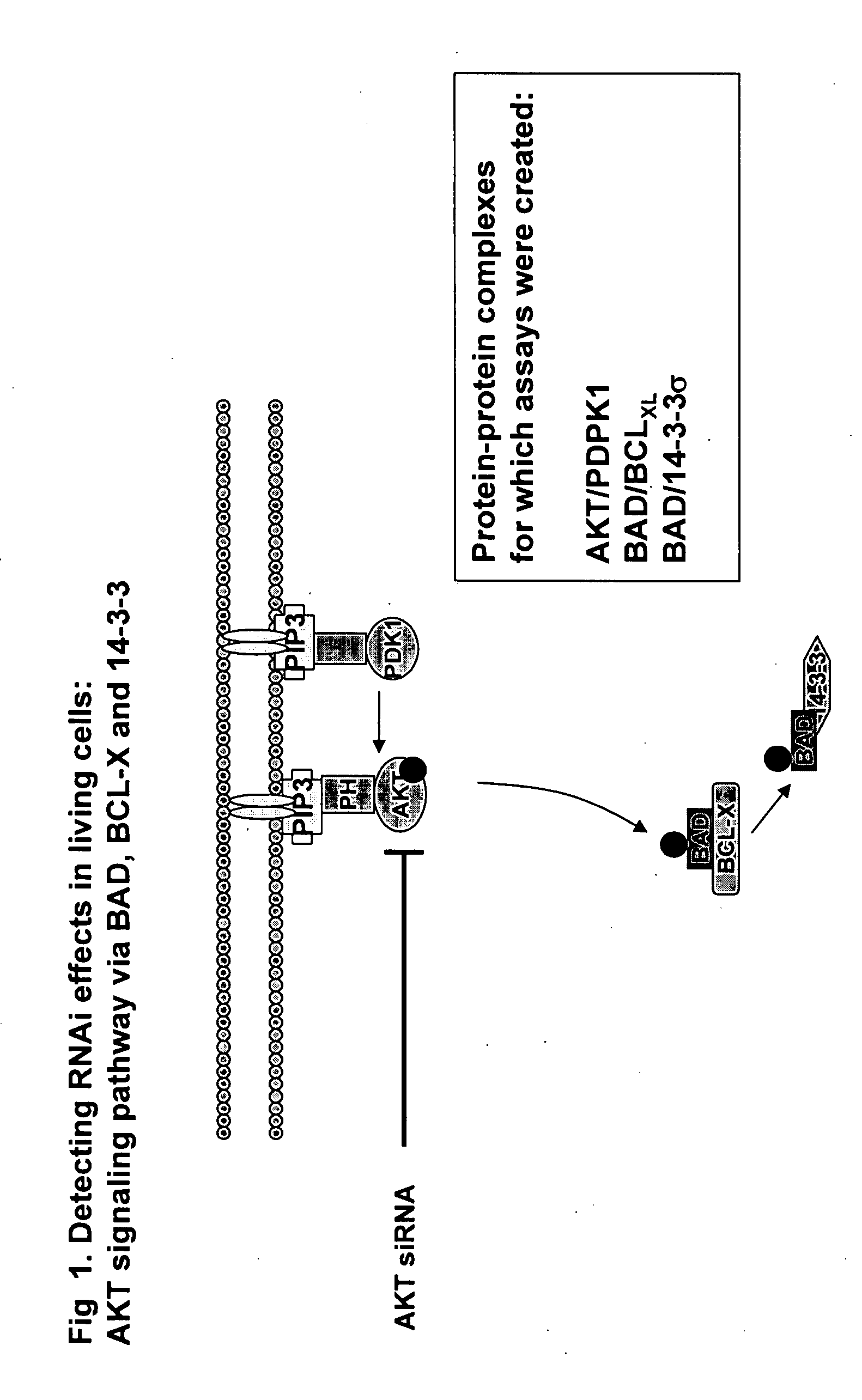
[0070] The full-length cDNAs encoding PDPK1, AKT1, BAD, BCL-xL and 14-3-3 sigma (all of which are known proteins and the corresponding full-length sequence of each is known in the art) were amplified by PCR and subcloned into a mammalian expression vector (pcDNA3.1Z, Invitrogen) which had been previously modified to contain either fragment 1 (Y1) of a yellow variant of E. victoria GFP (EYFP-F1, corresponding to GFP amino acids 1 to 158; the protein and fragment sequences being previously disclosed in commonly assigned pending U.S. application Ser. No. 10 / 772,021 filed Feb. 5, 2004) or fragment 2 (Y2) of EYFP (EYFP-F2, corresponding to GFP amino acids 159 to 239; the protein and fragment sequences being previously disclosed in commonly assigned pending U.S. application Ser. No. 10 / 724,178 filed Dec. 1, 2003). In all constructs, a 10 amino acid flexible linker consisting of glycine and serine residues [(Gly. Gly. Gly. Gly. Ser)2] such as that previously disclosed and em...

example 2

Large-Scale Mapping of Biochemical Pathways in High Throughput Formats

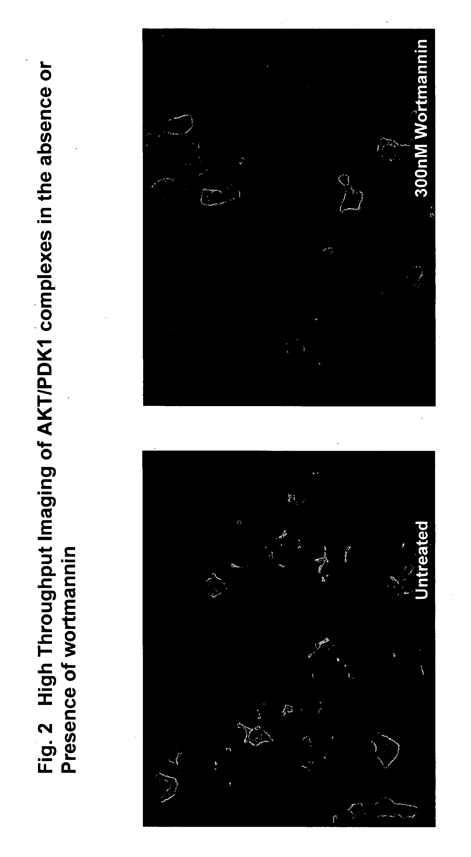
[0078] In mammalian cells, AKT is protected from degradation and dephosphorylation by binding to a heat shock protein (HSP90) which acts as a chaperone. HSP binding to AKT is thought to occur via the Cdc37 co-chaperone (Brazil et al., 2002). AKT present in protein complexes containing HSP90 and CDC37 was catalytically active and phosphorylated GSK3beta in vitro. HSP90 complex formation with AKT may therefore facilitate kinase activation by preventing both dephosphorylation and AKT degradation. Perturbation of this interaction, for example by ansamycin antibiotics that act as inhibitors of HSP90, or by a expressing a dominant negative form of PKB, leads to inactivation of AKT by dephosphorylation or proteasome-mediated degradation. HSP90 also stabilizes PDK1 in cells, enhancing the phorphorylation of PKB. HSP90 may also act as a scaffold protein, presenting substrates such as eNOS to PKB for phosphorylation. We us...

example 3

[0088] In a further demonstration of large-scale pathway mapping, we demonstrated the effects in human cells of an siRNA pool that silences the gene encoding the known co-chaperone protein, Cdc37.

[0089]FIG. 8 shows the results of a single annotation reagent, an optimized siRNA pool designed to silence Cdc37, against a panel of 32 assays. These assays report on diverse cellular activities. The effect of the Cdc37 siRNA (40 nM) on the fluorescence intensity of the different protein complexes (assessed by PCA) is plotted relative to the effect of a negative control siRNA. A value of 1 represents no change relative to the control. The 32 assays are represented on the x-axis, and are also listed in Table 2. Silencing of the Cdc37 gene by siRNA significantly decreased the fluorescence intensity of 8 different protein complexes in human cells, many containing proteins that are known to bind to Cdc37 (e.g. Akt1, Raf1, Chk1, etc). Representative images of the effect of siCdc37 on the Chk1:Cd...


PUM

PropertyMeasurementUnit
Fractionaaaaaaaaaa
Timeaaaaaaaaaa
Sensitivityaaaaaaaaaa

Abstract

The cell-based assays described in the present invention can be used to directly assess the sensitivity and specificity of the gene annotation reagent against its target, and to determine if a non-targeted gene participates in a pathway of interest or is functionally linked to another gene or protein. The combination of annotation reagents with such cell-based assays is useful for mapping genes (proteins) into cellular pathways on a genome-wide scale. Preferred assay embodiments include fluorescence or luminescence assays in intact (live or fixed) cells. Such fluorescence or luminescence assays include high-throughput or high-content assays for protein activity, subcellular localization, post-translational modifications, or interactions of proteins. Suitable assays may include protein-protein interaction assays; protein translocation assays; and post-translational modification assays. The invention can be used to assess the efficacy of any gene silencing experiment, to determine the level of gene silencing that is achieved, and to map novel genes into biochemical pathways, and to identify novel pharmaceutical targets. The results also demonstrate the feasibility of employing this strategy in genome-wide functional annotation efforts.

Description

[0001] This application claims the priority benefit under 35 U.S.C. section 119 of U.S. Provisional Patent Application No. 60 / 474,283 entitled “Monitoring Gene Silencing And Annotating Gene Function In Living Cells”, filed May 30, 2003, which is in its entirety herein incorporated by reference.BACKGROUND OF THE INVENTION [0002] This invention relates generally to the fields of biology, molecular biology, chemistry and biochemistry. Specifically, the invention is directed to methods for annotating gene function, mapping disease pathways, and validating pharmaceutical targets. In particular, the invention provides for the use of annotation technologies in combination with cell-based high-content or high-throughput assays as a method to map genes and proteins into biochemical pathways; to identify novel disease pathways; and to identify and validate novel pharmaceutical targets. [0003] A wide range of biochemical tools, reagents, and technologies are now available for use in target val...

Claims


Application Information

Patent Timeline
IPC IPC(8): C12Q1/68G01N33/50
CPCC12Q1/6897G01N33/5041G01N33/502G01N33/5008
Inventor WATSON MICHNICK, STEPHEN WILLIAMBELISLE, BARBARAMACDONALD, MARNIE L.WESTWICK, JOHN K.LAMERDIN, JANE ELIZABETH
Owner ODYSSEY THERA INC
Learn More