Uveoscleral shunt and methods for implanting same

a uveoscleral shunt and uveoscleral shunt technology, applied in the field of reducing intraocular pressure within, can solve the problems of large blindness if untreated, patients may suffer substantial, and sometimes associated with significant side effects of drug therapies for glaucoma, and achieve the effect of small cross section

Active Publication Date: 2008-09-18
GLAUKOS CORP
View PDF41 Cites 380 Cited by
  • Summary
  • Abstract
  • Description
  • Claims
  • Application Information

AI Technical Summary

Benefits of technology

[0030]A system for treating an ocular disorder in a patient is disclosed in accordance with some embodiments of the present invention. In some embodiments, the system comprises a drainage implant which, following implantation at an implantation site, conducts fluid from the anterior chamber to a uveoscleral outflow pathway, such as the supraciliary space and a delivery instrument for implanting the drainage implant. The instrument has a distal end sufficiently sharp to penetrate eye tissue at an insertion site near the limbus of the patient's eye, and is sufficiently long to advance the implant transocularly from the insertion site across the anterior chamber to the implantation site. The instrument also has a sufficiently small cross section such that the insertion site self seals without suturing upon withdrawal of the instrument from the eye. The instrument comprises a plurality of members longitudinally moveable relative to each other and a seal between the members to prevent aqueous humor from passing between the members proximal the seal when the instrument is in the eye.

Problems solved by technology

Glaucoma causes pathological changes in the optic nerve, visible on the optic disk, and it causes corresponding visual field loss, resulting in blindness if untreated.
Patients may suffer substantial, irreversible vision loss prior to diagnosis and treatment.
However, there are secondary open-angle glaucomas that may include edema or swelling of the trabecular spaces (e.g., from corticosteroid use), abnormal pigment dispersion, or diseases such as hyperthyroidism that produce vascular congestion.
However, drug therapies for glaucoma are sometimes associated with significant side effects.
The most frequent and perhaps most serious drawback to drug therapy, especially the elderly, is patient compliance.
Patients often forget to take their medication at the appropriate times or else administer eye drops improperly, resulting in under- or overdosing.
Patient compliance is particularly problematic with therapeutic agents requiring dosing frequencies of three times a day or more, such as pilocarpine.
Because the effects of glaucoma are irreversible, when patients dose improperly, allowing ocular concentrations to drop below appropriate therapeutic levels, further permanent damage to vision occurs.
And current therapies do not provide for a continuous slow-release of the drug.
However, long-term review of surgical results showed only limited success in adults.
In retrospect, these procedures probably failed due to cellular repair and fibrosis mechanisms and a process of “filling in.” Filling in is a detrimental effect of collapsing and closing in of the created opening in the trabecular meshwork.
Once the created openings close, the pressure builds back up and the surgery fails.
However, the relatively small hole created by this trabeculopuncture technique exhibits a filling-in effect and fails.
This method did not succeed in a clinical trial.
Although ocular morbidity was zero in both trials, success rates did not warrant further human trials.
Failure was again from filling in of surgically created defects in the trabecular meshwork by repair mechanisms.
Neither of these is a viable surgical technique for the treatment of glaucoma.
The risk of placing a glaucoma drainage device also includes hemorrhage, infection, and diplopia (double vision).
All of the above embodiments and variations thereof have numerous disadvantages and moderate success rates.
They involve substantial trauma to the eye and require great surgical skill in creating a hole through the full thickness of the sclera into the subconjunctival space.
However, in severe cases, it has been found that these procedures do not sufficiently reduce intraocular pressure.
However, modifying existing filtering surgery techniques in any profound way to increase their effectiveness appears to have reached a dead end.

Method used


Image

  • Uveoscleral shunt and methods for implanting same
  • Uveoscleral shunt and methods for implanting same
  • Uveoscleral shunt and methods for implanting same

Examples


Embodiment Construction

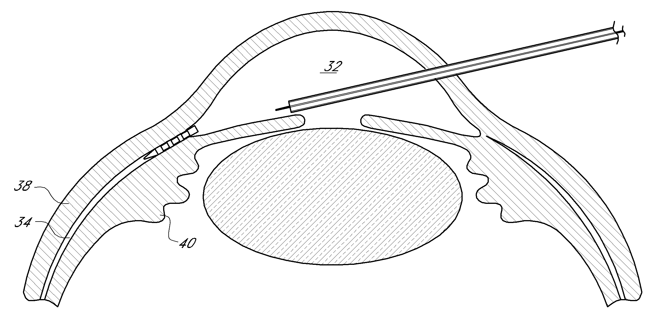
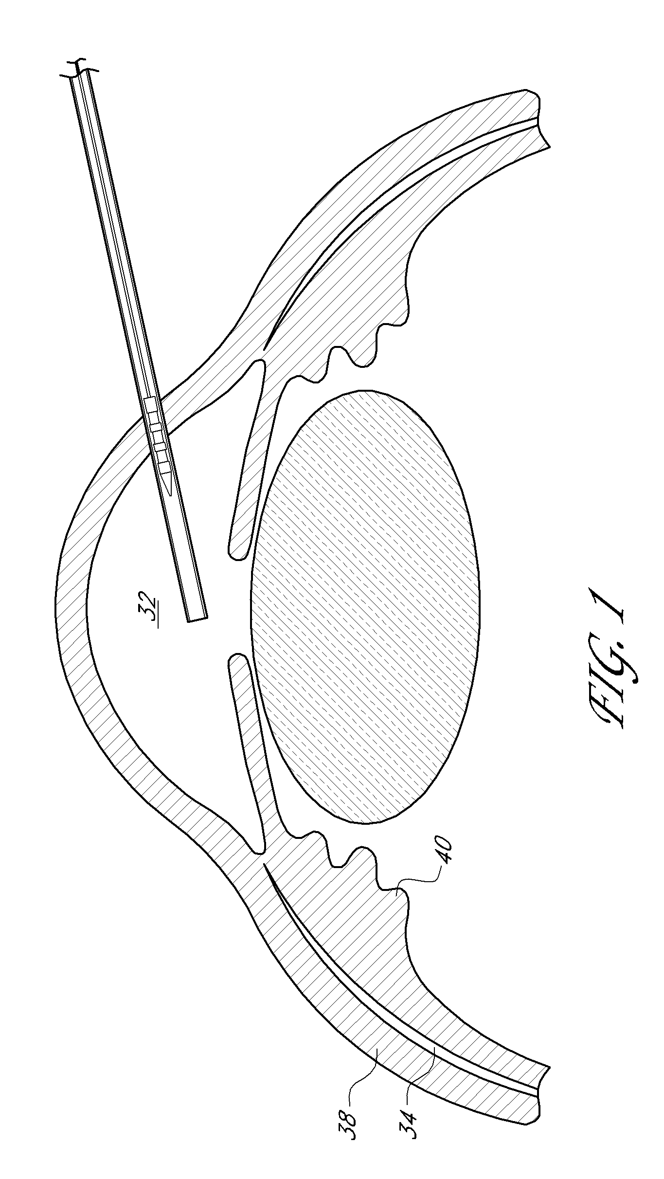
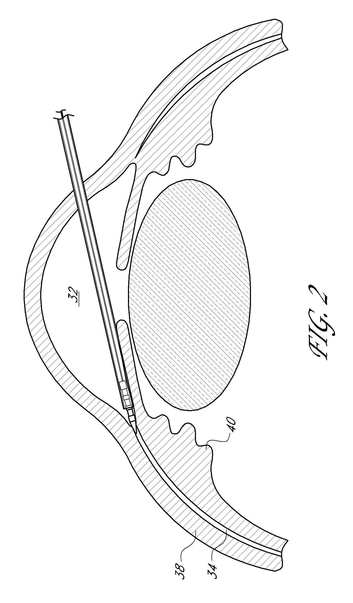
[0079]An ophthalmic implant system is provided that comprises a shunt and a delivery instrument for implanting the shunt. While this and other systems and associated methods are described herein in connection with glaucoma treatment, the disclosed systems and methods can be used to treat other types of ocular disorders in addition to glaucoma.

[0080]The shunt, following implantation at an implantation site, drains fluid from the anterior chamber into a physiologic outflow space. In some embodiments, the shunt is configured to provide a fluid flow path for draining aqueous humor from the anterior chamber of an eye to the uveoscleral outflow pathway to reduce intraocular pressure. In some embodiments, an instrument is provided for delivering and / or implanting the drainage shunt ab interno in an eye to divert aqueous humor from the anterior chamber to the uveoscleral outflow pathway. In some embodiments, a method is provided for implanting a drainage shunt ab interno in an eye to divert...


PUM


Abstract

Devices and methods for treating intraocular pressure are disclosed. The devices include shunts for draining aqueous humor from the anterior chamber to the uveoscleral outflow pathway, including the supraciliary space and the suprachoroidal space. The shunts are preferably implanted by ab interno procedures.

Description

RELATED CASES[0001]This application claims priority and the benefit under 35 U.S.C. § 119(e) to Provisional Patent Application Ser. No. 60 / 857,872, filed Nov. 10, 2006, Ser. No. 60 / 880,091, filed Jan. 11, 2007, Ser. No. 60 / 890,610, filed Feb. 19, 2007, and Ser. No. 60 / 947,942, filed Jul. 3, 2007. Each of these disclosures is hereby incorporated by reference herein in its entirety.BACKGROUND OF THE INVENTION[0002]1. Field of the Invention[0003]This disclosure relates to reducing intraocular pressure within the eye. This disclosure also relates to a treatment of glaucoma and / or other ocular disorders wherein aqueous humor is permitted to flow out of an anterior chamber of the eye through a surgically implanted pathway.[0004]2. Description of the Related Art[0005]A human eye is a specialized sensory organ capable of light reception and is able to receive visual images. Aqueous humor is a transparent liquid that fills at least the region between the cornea, at the front of the eye, and ...

Claims


Application Information

Patent Timeline
Patent Type & Authority Applications(United States)
IPC IPC(8): A61M27/00A61B17/00
CPCA61F9/00781A61P27/06
Inventor BURNS, THOMAS W.HAFFNER, DAVIDHEITZMANN, HAROLD A.FJIELD, TODD N.HILL, RICHARD A.
Owner GLAUKOS CORP
Why Eureka
Try Eureka