
Methods and compositions involving bacteriophage isolates

a technology of bacteriophage isolates and compositions, applied in the field of infectious diseases, can solve the problems of increasing the number of bacterial strains, narrow host range of any one bacteriophage, and raising widespread fears of a pre-antibiotic era. , to achieve the effect of improving the detection of lytic phages and increasing the lytic potential of phages

Inactive Publication Date: 2009-03-26
BOARD OF RGT THE UNIV OF TEXAS SYST
View PDF10 Cites 22 Cited by
  • Summary
  • Abstract
  • Description
  • Claims
  • Application Information

AI Technical Summary

Benefits of technology

[0014]The present invention provides for methods of (1) improving the detection of lytic phages, (2) methods of propagating and isolating therapeutic bacteriophage and (3) increasing phage lytic potential.

Problems solved by technology

A major limitation was narrow host range for any one bacteriophage.
Subsequently, extensive use of antibiotics led to an increase in the number of bacterial strains resistant to most or all available antibiotics, causing increasingly serious problems and raising widespread fears of return to a pre-antibiotic era of untreatable infections and epidemics.
Methods of isolation and characterization of bacteriophages for this purpose have not been adequate.
In addition, particles are tethered to a substrate, i.e., they are not free in solution for further analysis.
Thus, aggregating viruses are apparently often overlooked.
Though approximations of in situ electron microscopy by negative staining have been developed (reviewed in Ackermann and DuBow, 1987), true in situ electron microscopy has not been feasible because (1) the specimen must be dried in the case of procedures other than cryo-electro microscopy, (2) cryo-electron microscopy requires thin specimens (less than about 1 μm) possibly incompatible with large aggregates (reviewed in Jiang and Ludtke, 2005; Grunewald and Cyrklaff, 2006) and (3) all forms of high resolution electron microscopy require immobilization of particles in the specimen.
Some of the more effective phages may aggregate in the wild and, therefore, may not be observable with the traditional procedures.

Method used


Image

  • Methods and compositions involving bacteriophage isolates
  • Methods and compositions involving bacteriophage isolates
  • Methods and compositions involving bacteriophage isolates

Examples


example 1

In Situ Fluorescence Microscopy of Bacteriophage Aggregates

Materials and Methods

[0094]Media and Buffers

[0095]The medium used for growing both environmental bacteriophages and their hosts was the following: 10 g Bacto tryptone, 5 g KCl in 1000 ml water with 0.002 M CaCl2 added post-autoclaving (growth medium). For supporting bacteriophage plaques, the upper gel used in Petri plates was made of agarose (Seakem Gold; Cambrex Corp., Walkersville, Md.; concentration in the text) dissolved in growth medium. Beneath the upper gel, the lower gel was made of 1% Sigma agar in 10 g Bacto tryptone, 5 g NaCl in 1000 ml water.

[0096]Hosts and Bacteriophages

[0097]Bacteriophages 0305φ8-36 (host=Bacillus thuringiensis; Serwer et al., 2007a, b) and G (host=Bacillus megaterium; Fangman, 1978) and their hosts are the strains previously described. Bacteriophage 0905φ8-18 was isolated de novo via overlay of a soil sample with a 0.15% agarose gel that contained the host, B. thuringiensis. Bacteriophage 090...

example 2

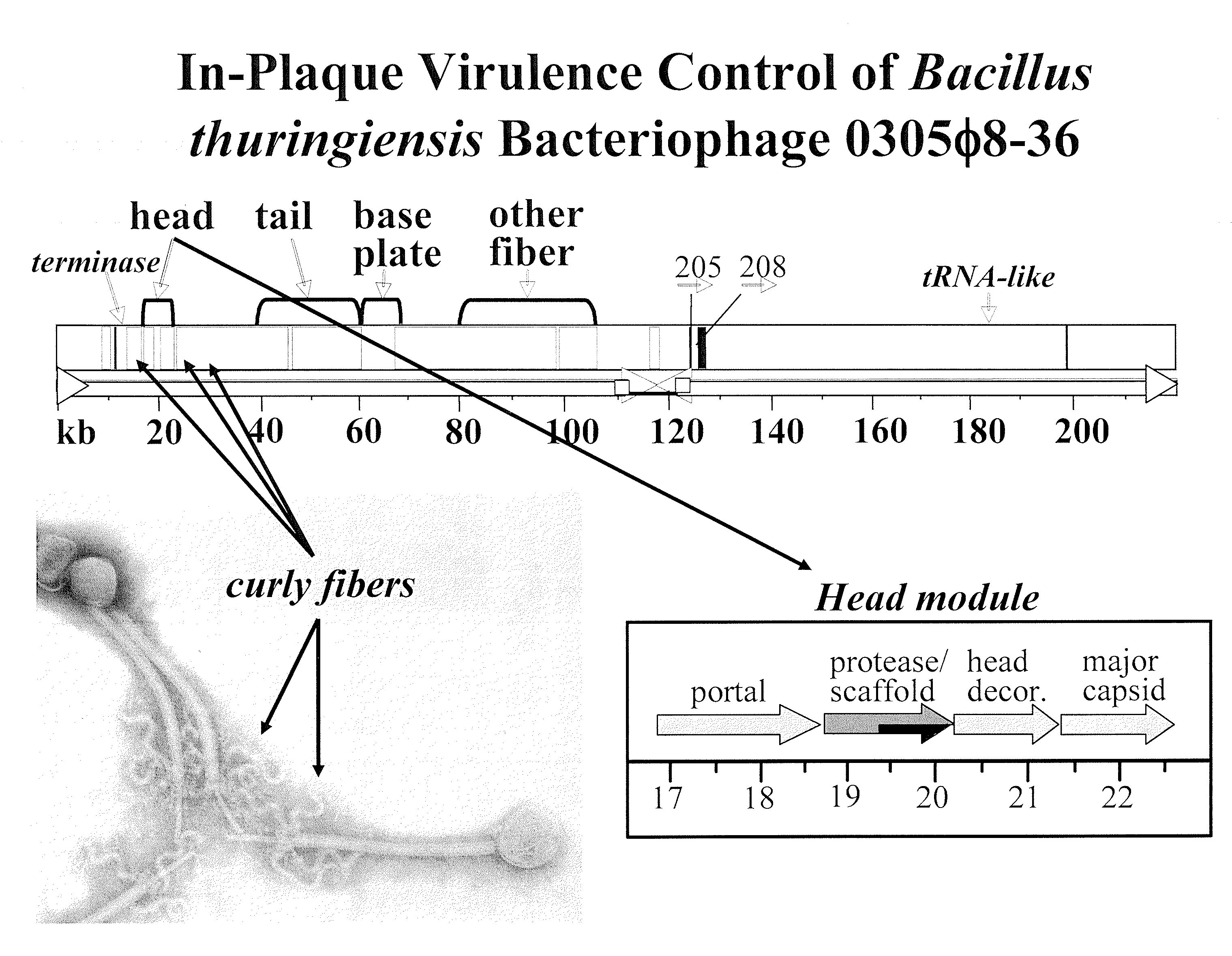
In-Plaque Virulence Control of Bacillus Thuringiensis Bacteriphage 0305φ8-36

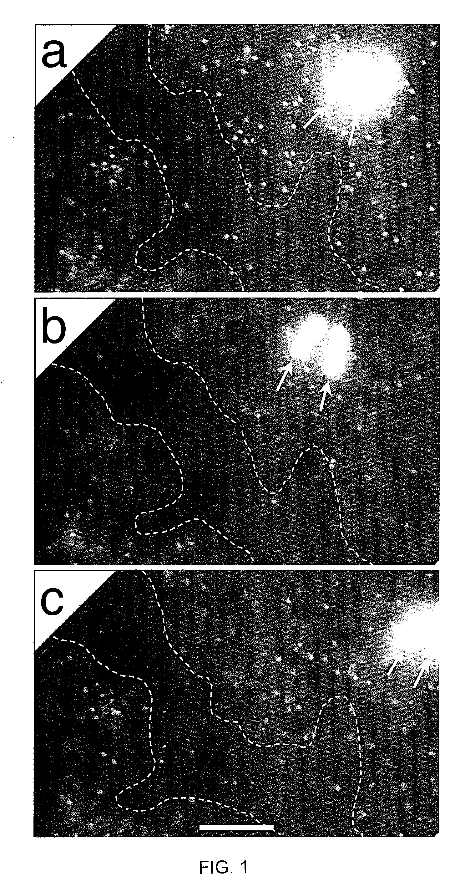
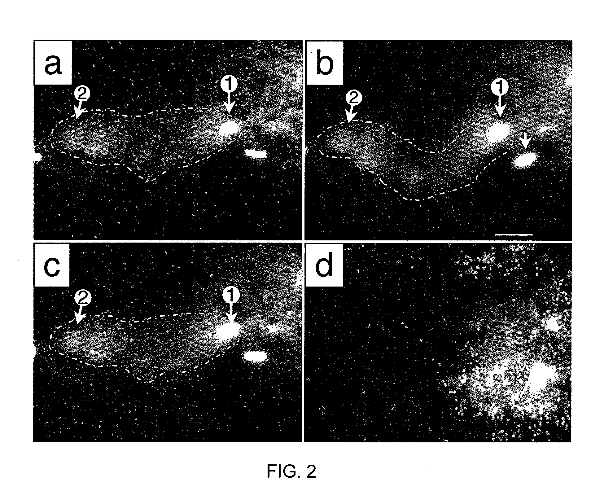
[0149]Bacillus thuringiensis bacteriophage 0305φ8-36 is lytic, based on both genomic sequence and formation of clear plaques in dilute (0.075-0.10%) agarose gels. However, 0305φ8-36 does not clear liquid cultures (Serwer, P. et al. Virol. J. 4, 21). In the present study, in situ fluorescence microscopy reveals that the dilute gel-supported 0305φ8-36 plaques have two phases. One is weakly fluorescent; the other is comparatively dark. Most bacteriophage particles are in the weakly fluorescent phase. Empirically, formation of two phases requires the presence of bacteriophage particles, bacteria and dilute agarose. The weakly fluorescent phase has both a mass density and a viscosity greater than those of the darker phase. Bacteriophage particles do not cross the phase boundary, even when the specimen is distorted by pressing on the cover glass.

[0150]The above data imply that both bacteriophage particles and agar...

example 3

In Situ Fluorescence Microscopy of Bacteriophage 0305φ8-36

[0153]Bacteriophage 0305φ8-36 is a large myovirus with unusual curly fibers (FIG. 5). The genome is 218,948 base pairs long; the tail is 486 nm long. The classical morphogenesis genes of the 8-36 genome are highly diverged and 8-36 has genes in a new category. The genes are called extra or other fiber. The designation “other fiber” comes from homology of some of these proteins with fibronectin 3 domains and von Willebrand factor domain.

[0154]In addition to an unusual genome, 0305φ8-36 has unusual biology, including plaque radius vs. gel concentration plot that is a steep as the plot for bacteriophage G, the largest bacteriophage known (FIG. 6). 8-36 also does not clear liquid culture, though it does grow in liquid culture. In fact, 8-36 co-grows with the host and presumably co-evolves with the host, very unusual (though not unheard of) behavior. Furthermore, 8-36 undergoes aggregation and also a form of phase separation that ...


PUM


Abstract

Disclosed are methods of increasing the virulence of a bacteriophage, comprising contacting a bacteriophage with a composition comprising a bacterium or bacterial extract; and a polymer. Also disclosed are methods of propagating and isolating therapeutic bacteriophages that involve use of dilute polymer compositions. Also disclosed are pharmaceutical compositions the bacteriophage for which virulence has been increased by the methods set forth herein or which have been isolated by the methods set forth herein.

Description

[0001]This application claims the benefit of priority to U.S. Application Ser. No. 60 / 955,277, filed Aug. 10, 2007, which is hereby incorporated by reference in its entirety.[0002]This invention was made with government support under grant RO1 GM24365-27 awarded by the National Institutes of Health. The government has certain rights in the invention.BACKGROUND OF THE INVENTION[0003]1. Field of the Invention[0004]The present invention relates generally to the fields of virology and treatment of infectious diseases. More particularly, it concerns methods of increasing the virulence of bacteriphages, and methods of isolating therapeutic bacteriphages. Therapeutic bacteriophages cure or ameliorate disease caused by infection with bacteria.[0005]2. Description of Related Art[0006]Bacteriophages are viruses that infect bacteria Some phages, called lytic, kill bacteria. This killing is done with specificity and is the basic phenomenon on which the concept of phage therapy is built. Therefo...

Claims


Application Information

Patent Timeline
IPC IPC(8): A61K35/74A61K35/76C12N7/00C12Q1/70A61K35/742A61K35/744
CPCA61K9/0014A61K9/0019A61K35/742A61K35/744A61K35/76C12N7/00C12N2795/10132C12N2795/10151A61K2300/00
Inventor SERWER, PHILIP
Owner BOARD OF RGT THE UNIV OF TEXAS SYST
Learn More