Spore associated display

a technology of spores and display methods, applied in the field of spore display methods, can solve the problems of large number of proteins, and achieve the effect of stable protein display

Inactive Publication Date: 2009-04-16
I2 PHARMA INC
View PDF0 Cites 16 Cited by
  • Summary
  • Abstract
  • Description
  • Claims
  • Application Information

AI Technical Summary

Benefits of technology

[0054]In a still further embodiment, the displayed conjugates are formed by transforming Bacillus with nucleic acid encoding the conjugates, each under control of a sporulation specific promoter, and culturing and harvesting the transformed Bacillus under conditions to support sporulation and stable protein display.
[0055]In a more specific embodiment, colonies of the transformed spores are grown in a sporulation medium for less than 48 hours, such as, for example, for about 14 to about 20 hours, whereupon the spores are liberated retaining the majority of the displayed peptides or polypeptides in an intact, non-degraded form.
[0056]In other embodiments, the method further comprises a step of testing the stability of the display and / or testing the chemical or biological integrity of one or more peptides or polypeptides displayed and / or selecting the Bacillus spores displaying a coat protein-peptide or coat protein-polypeptide conjugate.

Problems solved by technology

Bt produces a large number of proteins that are toxic to insects.

Method used


Image

  • Spore associated display
  • Spore associated display
  • Spore associated display

Examples


example 1

Recombinant CotB / GFP Protein Fusion Constructs and Expression in Bacillus thuringiensis

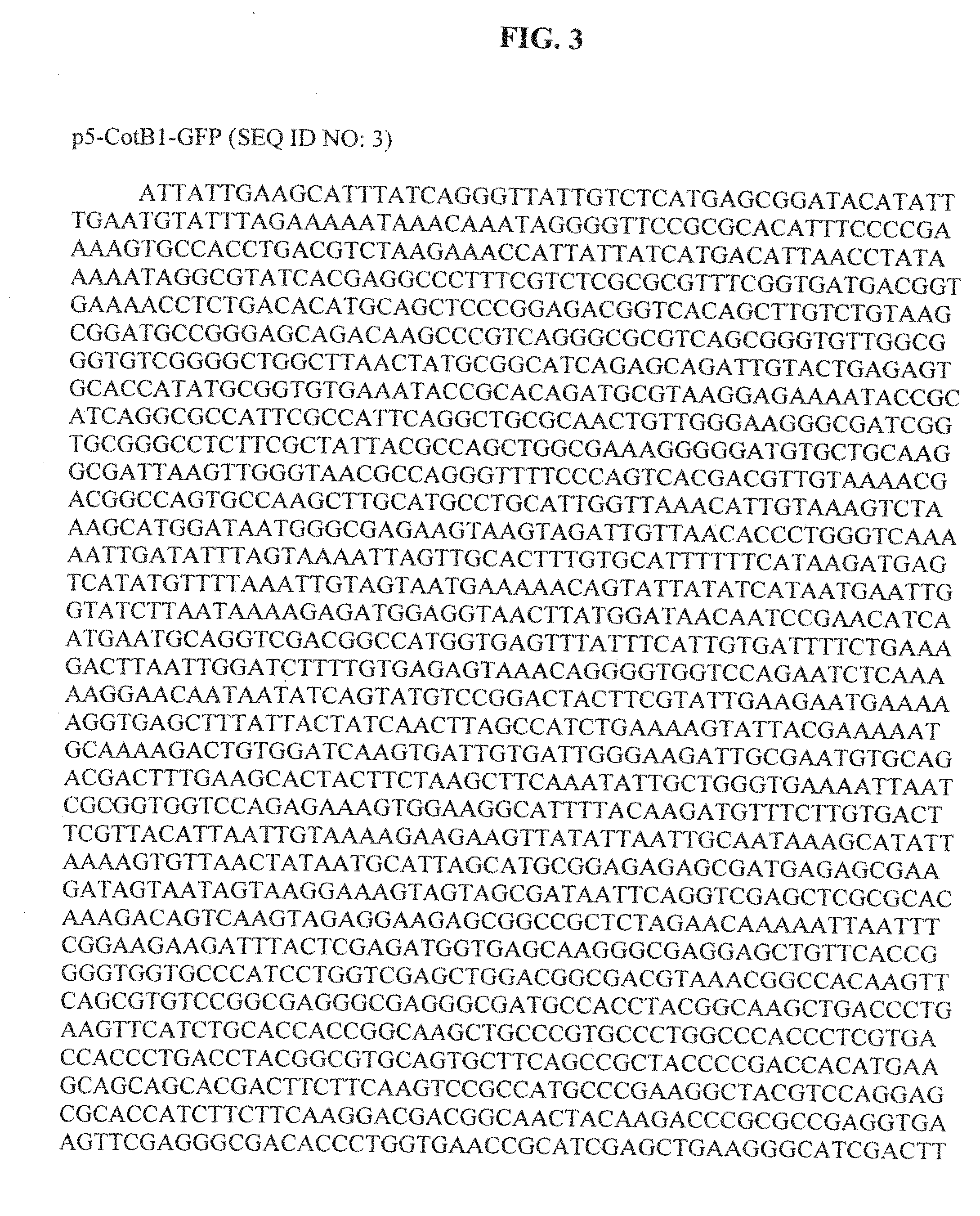
[0178]The main construct, p5-CotB1-GFP (FIG. 3, SEQ ID NO: 3), contains a sporulation specific promoter (BtI-II) from the crystal protein Cry1Ac (coat protein) of Bacillus thuringiensis (Bt) followed by the first eleven amino acids of the Cry1A toxin and the CotB1 gene (FIG. 9, SEQ ID NO: 6). For analytical purposes, a myc tag was inserted between the coat protein and the Green Fluorescent Protein gene (GFP). The p5-CotB1-GFP construct also contains an ampicillin resistant gene for selection in Escherichia coli and an erythromycin gene for selection in Bt, and is graphically illustrated in FIG. 4. CotB1 was obtained by amplification by Polymerase Chain Reaction (PCR) of genomic DNA from the a crystalliferous strain of Bt 4D7. Genomic DNA was obtained by lysis of a colony in water followed by boiling for 5 minutes. For cloning purposes PCR primers

forward:CCATGGTGAGTTTATTTCATTGTG(SEQ ID NO: 11)andr...

example 2

Spore Surface Display of an Anti-TNF-α Antibody

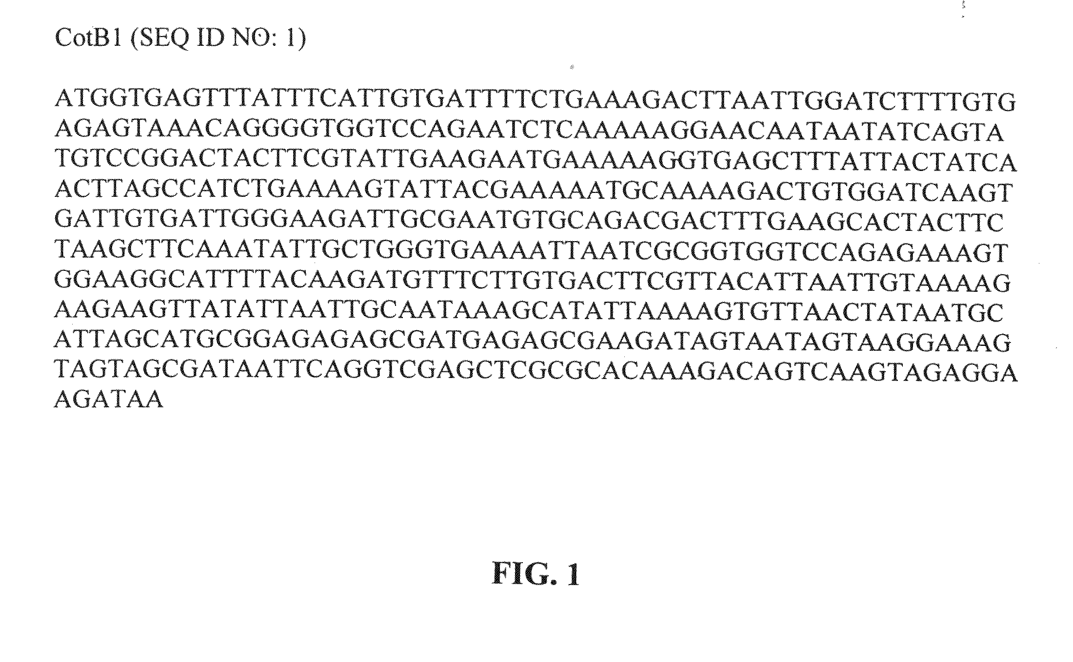
[0181]A scFv construct of the anti-human TNF-α antibody D2E7 (FIG. 6, SEQ ID NO: 5) was cloned as an in-frame fusion to the C-terminus of the coat protein Cot B1 (FIG. 1, SEQ ID NO: 1). Spores were obtained as described in Example 1. Fifty microliters of spores were resuspended in FACS blocking buffer (Phosphate Buffered Saline+0.5% Bovine Serum Albumin) for 10 minutes at 4 C. Spores were washed in FACS buffer and then incubated with Phycoerythrin conjugated Streptavidin for 30 minutes at 4 C, washed and then resuspended in PBS. The resulting spores were next analyzed by western blot (FIG. 7B).

example 3

Magnetic Bead Selection of scFv SPORE

[0182]1×10⁹ washed and blocked spores prepared as described in Example 1 were incubated at 4 C with biotinylated human TNF-α (final concentrations of 50 nM) in a final volume of 1 ml. After two hours, the spores were pelleted, washed 3× with 3 ml PBS and then incubated for 1 hour in 1 ml in the presence of paramagnetic anti-biotin beads (Miltenyi). The bead spore mixture was later applied to a magnetic column, washed, eluted, according to manufacturers instructions, and the eluted spores repropagated as a vegetative cell culture in Brain-Heart Infusion media containing 25 μg / ml erythromycin overnight at 37° C.


PUM

PropertyMeasurementUnit
volumeaaaaaaaaaa
pHaaaaaaaaaa
full-length sequenceaaaaaaaaaa

Abstract

The present invention concerns spore display methods. More specifically, the invention concerns display of heterologous molecules, such as peptides and polypeptides, on spores of bacilli, such as, for example, Bacillus thuringiensis (Bt) or Bacillus cereus (BC), using externally exposed spore coat proteins or fragments or variants thereof.

Description

CROSS REFERENCE TO RELATED APPLICATIONS[0001]This application is a non-provisional application filed under 37 CFR 1.53(b)(1), claiming priority under 35 USC 119(e) to provisional application No. 60 / 995,967 filed Sep. 28, 2007, and provisional application No. 60 / 955,592 filed Aug. 13, 2007, the contents of which are incorporated herein by reference.FIELD OF THE INVENTION[0002]The present invention concerns spore display methods. More specifically, the invention concerns display of heterologous molecules, such as peptides and polypeptides, on spores of bacilli, such as, for example, Bacillus thuringiensis (Bt) or Bacillus cereus (BC), using externally exposed spore coat proteins or fragments or variants thereof.BACKGROUND OF THE INVENTION[0003]There are systems known in the art for display of heterologous proteins on the surfaces of bacteriophage (Scott et al., Science 249:386-390 (1990)), Escherichia coli (Agterberg et al., Gene 88:37-45 (1990); Charbit et al., Gene 70:181-189 (1988)...

Claims


Application Information

Patent Timeline
Patent Type & Authority Applications(United States)
IPC IPC(8): A61K39/07C07K2/00C07K14/00C07K16/12C12N9/96C07H21/04C12N1/21C40B40/10C40B40/06C12N15/74
CPCA61K47/48776A61K47/4833A61K47/646A61K47/6901A61P39/00
Inventor BHATT, RAMESHHOROWITZ, LAWRENCEESTELLES, ANGELES
Owner I2 PHARMA INC
Why Eureka
Try Eureka