
Biofilm formation inhibitor and treatment device thereof

a biofilm and inhibitor technology, applied in the direction of antibacterial agents, prostheses, blood vessels, etc., can solve the problems of obstructive jaundice, too late treatment, difficult early detection, etc., and achieve the effect of suppressing the growth of these bacteria and effectively and continuously inhibiting the formation of biofilm

Inactive Publication Date: 2009-04-16
KYUSHU UNIV
View PDF5 Cites 10 Cited by
  • Summary
  • Abstract
  • Description
  • Claims
  • Application Information

AI Technical Summary

Benefits of technology

[0013]The present invention was made in view of these problems, and it aims to provide a treatment device such as biliary stent capable of effectively and continuously inhibiting biofilm formation, and a biofilm formation inhibitor suitable for coating on the treatment device by killing all bacteria including E. coli and by suppressing the growth of these bacteria.
[0014]Furthermore, another object of the present invention is to provide a treatment device such as a biliary stent and a biofilm formation inhibitor that is suitable for coating on the treatment device, which are capable of continuously preventing biofilm formation and are safe and effective for the human body.

Problems solved by technology

Therefore, early detection is difficult, and when it is diagnosed as a carcinoma, it is often too late to treat it.
On the other hand, there is obstructive jaundice (a symptom of the body turning yellow), which is a symptom related in some degree to these carcinomas.
However, because the life expectancy, for example, of a pancreatic carcinoma patient, is only about 6 months, repeated operations lessen the quality of life for the patient as well as creating a large economic burden.
Moreover, a biofilm formed on a biological surface may cause infectious diseases such as chronic bronchitis and chronic osteomyelitis.
Also, when a biofilm is formed on a prosthetic substrate surface, such as prosthetic heart valve, prosthetic joint, and urethral catheter, etc., sepsis may accompany it, and in the worse case may lead to death.
However, long-term administration of antibiotics has a high probability of causing antibiotic-resistant bacteria to emerge.
Thus currently there is no effective drug for it.
However, these materials cannot suppress bacterial growth.
Therefore, if a small amount of bacteria with a high proliferating ability (the doubling time of the E. coli is 30 minutes) adheres on their surfaces, they cannot prevent the bacteria's growth, and thus they are unable to inhibit biofilm formation.
However, toxicity and effects on the living body pose problems, Thus, they have not been put to practical use for the human body.
However, neither of them is practical in terms of efficacy and safety.

Method used


Image

  • Biofilm formation inhibitor and treatment device thereof
  • Biofilm formation inhibitor and treatment device thereof
  • Biofilm formation inhibitor and treatment device thereof

Examples


example 1

The Inhibitory Effect of Epigallocatechin Gallate (EGCg) on Biofilm Formation

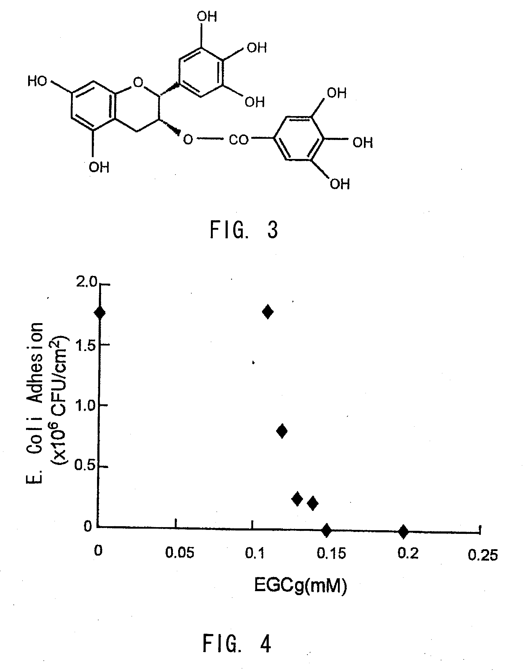
[0047]8 mm diameter disc shaped polyurethanes were cultured with an E. coli suspension (2×10³ CFU / ml) which was mixed with various concentrations of epigallocatechin gallate (EGCg) (0-0.2 mM) under a static condition at 37 degrees Celsius for 24 hours in a culture plate. After incubations, the polyurethane discs were taken out, and having been washed in a phosphate buffer gently, they were moved to a 2 ml phosphate buffer, where they were sonicated to detach the adhered E. coli. Viable E. coli cell counts in the liquid were calculated by the plate count method.

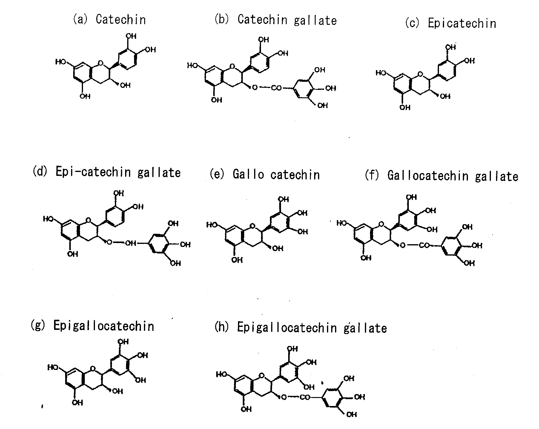
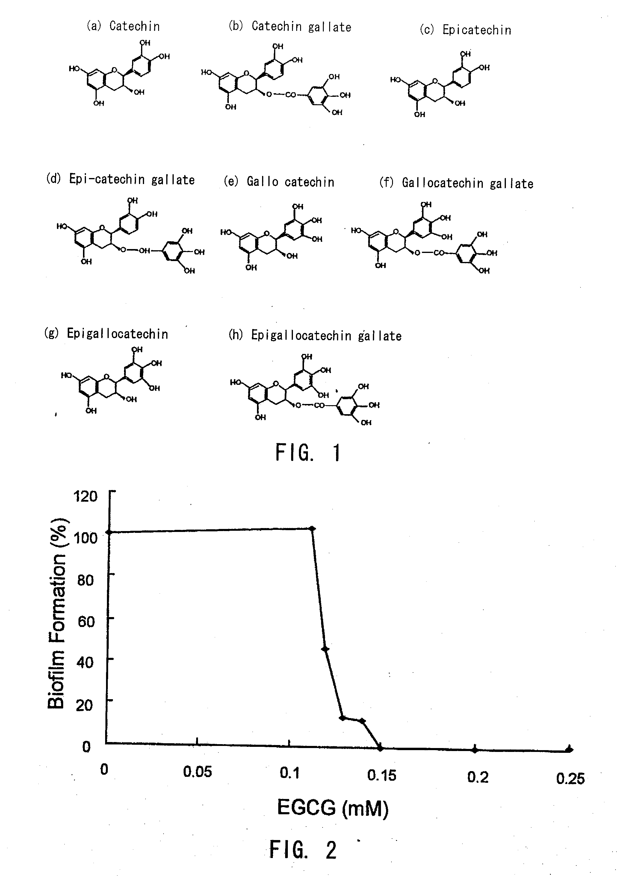
[0048]As shown in FIG. 3 and FIG. 4, epigallocatechin gallate (EGCg) was proved to inhibit biofilm formation significantly at the concentration of 0.12 mM (0.055 mg / ml) and more, and to inhibit completely at the concentration of 0.15 mM (0.069 mg / ml) and more.

example 2

Measurement of Sustained Release Amount of Catechin from Catechin-Loaded Polymer

[0049]Epigallocatechin gallate (EGCg, Sigma—Aldrich, Inc., St. Louis, Mo.: shown in FIG. 3) dissolved in acetone (Sigma—Aldrich) was mixed with three kinds of biodegradable polymers (TMC / LL / PEG1k, TMC / PEG200, TMC / TMP: shown in FIG. 5 (a)-(c)) to make 20 wt % (wt %: weight percent). After acetone was volatilized in a low pressure tank over 24 hours, 250 mg of polymer was coated at the bottom (surface area 1.1 cm2) of a glass bottle, and it was photocured by irradiating with ultraviolet light (SP-V, Ushio, Inc., Yokohama, Japan) for five minutes. 1 ml of phosphate buffer solution (Nissui Pharmaceutical Co., Ltd., Tokyo, Japan) was poured into the glass bottle and stoppered tightly, and kept at 37 degrees Celsius under static conditions for sustained release of EGCg. 100 μl of the solution in the glass bottle was taken out after one, two, and four days, and EGCg levels were calculated by the Folin-Ciocalteu...

example 3

The Inhibitory Effect of Catechin-Loaded Polymer on the Formation of Biofilm by E. coli(Static Conditions)

[0051]EGCg dissolved in acetone was mixed with three kinds of liquid biodegradable polymers (TMC / LL / PEG1k, TMC / PEG200, and TMC / TMP: shown in the FIG. 5 (a)-(c)) to make 0, 5, 10, and 20 wt % (wt %: weight percent). These polymers were spin-coated on polyurethane surfaces, and after having been irradiated with UV for five minutes, and solidified, and 8 mm diameter disc samples were punched out by a belt punch. These samples were incubated at 37 degrees Celsius with an E. coli suspension (2×10³ CFU / ml) in a culture plate for 24 hours. After incubation, the samples were taken out and washed in a phosphate buffer gently, and then they were moved to 2 ml of phosphate buffer solution where they are sonicated to detach the adhered E. coli. Viable E. coli cell count in this liquid was calculated by the plate count method.

[0052]These results were shown in FIG. 7 and FIG. 8. According to ...


PUM

PropertyMeasurementUnit
concentrationaaaaaaaaaa
surface areaaaaaaaaaaa

Abstract

A treatment device such as a biliary stent, capable of effectively and continuously inhibiting biofilm formation, and a biofilm formation inhibitor suitable for coating on the treatment device by killing all bacteria including E. coli and by suppressing the growth of these bacteria, are provided. A biofilm formation inhibitor having an effective amount of catechin for inhibiting biofilm formation and a carrier that is suitable for adhesion of said catechin to a treatment device to be placed in the living body, are provided.

Description

CROSS REFERENCE TO RELATED APPLICATIONS[0001]This application claims priority under 35 U.S.C. 119 based upon Japanese Patent Application Serial No. 2004-337604, filed on Nov. 22, 2004. The entire disclosure of the aforesaid application is incorporated herein by reference.FIELD OF THE INVENTION[0002]The present invention is related to a biofilm formation inhibitor and a treatment device using thereof.BACKGROUND OF THE INVENTION[0003]Progressive pancreatic carcinoma and cholangioma are known for their extreme difficulties in diagnosis and treatment and as so-called intractable cancers. In other words, because the pancreas and bile duct are surrounded by other organs such as the stomach, duodenum, small intestine, large intestine, liver, gall bladder, spleen, etc., it is difficult to detect cancer, and also manifestations that are characteristic to cancer are not found in the early stages. Therefore, early detection is difficult, and when it is diagnosed as a carcinoma, it is often too...

Claims


Application Information

Patent Timeline
Patent Type & Authority Applications(United States)
IPC IPC(8): A61F2/06A61K31/352A61K31/353A61K47/34A61L31/00A61P31/04
CPCA01N65/00A61L31/16A61L2300/404A01N25/24A01N25/10A01N2300/00A01N43/16A01N65/08A61P31/04
Inventor MATSUDA, TAKEHISAMAEYAMA, RYO
Owner KYUSHU UNIV
Learn More