

Methods of reducing a viral infection and kits therefore
a technology of viral infection and reducing kits, which is applied in the field of methods of reducing viral infection and kits therefore, can solve the problems of severe side effects, increased morbidity and mortality of hiv-infected patients, and loss of appetite and mental depression, and achieve the effect of reducing the level of viral particles
- Summary
- Abstract
- Description
- Claims
- Application Information
AI Technical Summary
Benefits of technology
Problems solved by technology
Method used
Image

Examples
example 1
Material and Methods
[0105]Cells and Virus—HuH7 and HEK293T cells were maintained in Dulbecco's modified Eagle medium high glucose (DMEM) (Invitrogen) supplemented with 5 mM sodium pyruvate, 100U of penicillin / ml, 100 μg of streptomycin / ml, and 10% fetal calf serum. All cells were grown at 37° C. in 5% CO2. The HCVcc JFH-1 and J6 / JFH1 were obtained from Dr T. Wakita and R. Bartenschlager, respectively, and were propagated as described (69, 71).
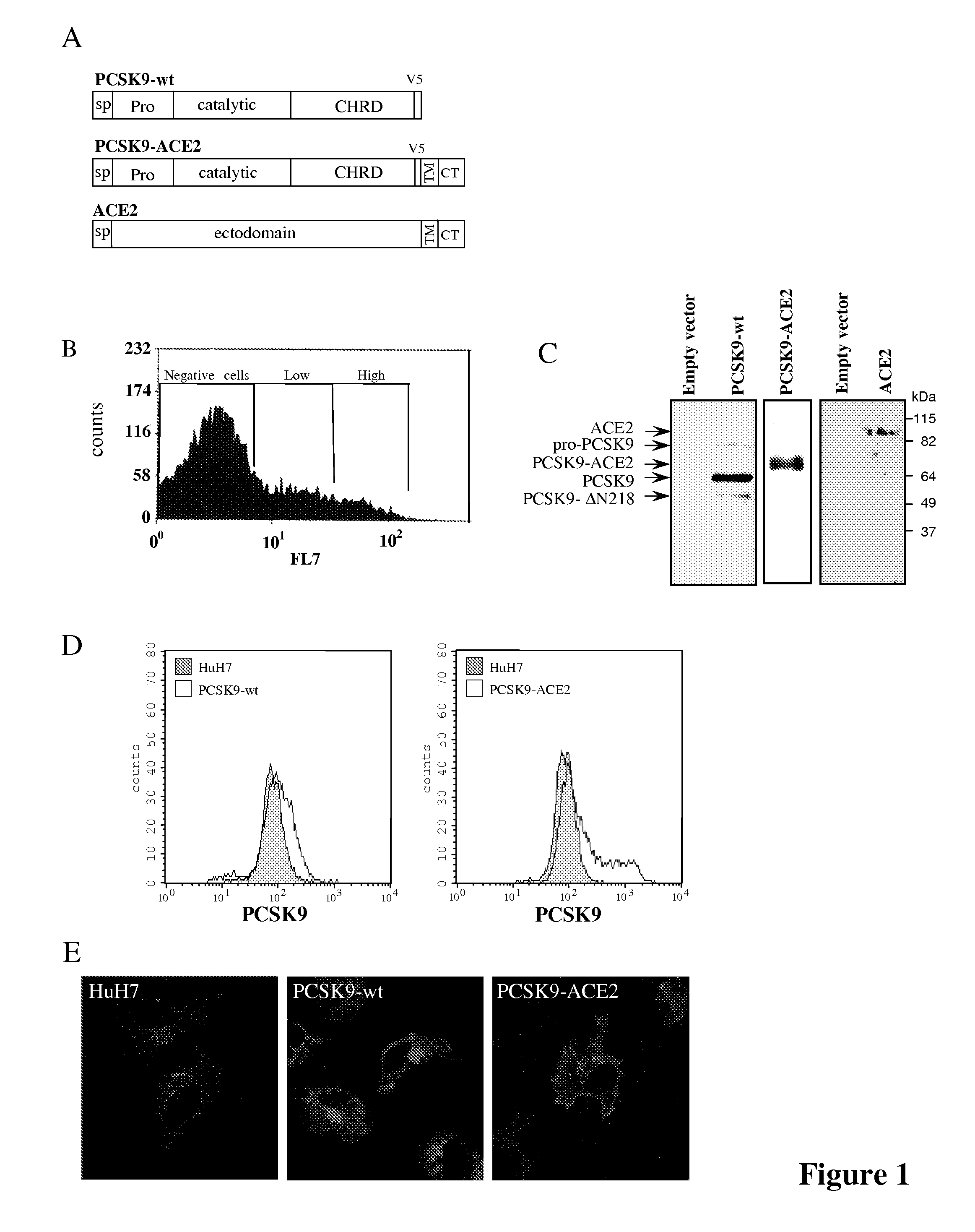
[0106]Plasmids and Stable Cell Lines—HuH7 cell populations expressing PCSK9, PCSK9-TM-CT-ACE2 (PCSK9-ACE2), PCSK9-TM-CT-Lamp1 (PCSK9-Lamp1), ACE2 or the empty phCMV3 vector (Invitrogen) were generated as described (72).
[0107]The PCSK9 sequence used is shown on FIG. 11A and included a V5-tag (italicized in FIG. 11A); the PCSK9-ACE2 sequence used is shown on FIG. 11B and contained the trans-membrane and the cytosolic domains of the protein ACE2 (underlined in FIG. 11B) fused to the carboxy terminal end of PCSK9 wild type sequence including a V5-t...
example 2
PCSK9 Expression in Stable Cell Lines
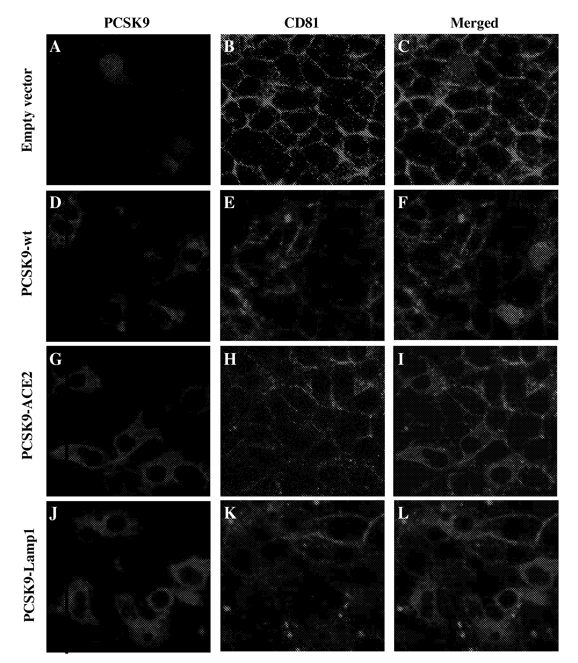
[0119]HuH7 cells stably expressing PCSK9-wt, PCSK9-ACE2, ACE2 or the empty vector phCMV3 were produced as described in Example 1 above in the section Plasmids and Stable Cell lines (FIG. 1A). All cells lines were sorted by FACS for the determination of their respective protein expression as described in Example 1 above in the section Flow cytometry, and the ˜10% highest expressors were collected (FIG. 1B). The expression of the expected protein was confirmed by immunobloting of cell lysates (FIG. 1C). While PCSK9-wt is mostly secreted in the media, PCSK9-ACE2 remained attached to the cell surface through the transmembrane domain of ACE2 (FIG. 1D). Indirect immunofluorescence performed as described in Example 1 above of these stable cells using a PCSK9 polyclonal antibody (1 / 200) and a goat anti-rabbit-Alexa 488 (1 / 1000) using a confocal microscope revealed that just like PCSK9-wt, the chimeric protein PCSK9-ACE2 is detected in perinuclear locatio...
example 3
PCSK9 Expression in HuH7 Cells Inhibits Infection by HCVcc
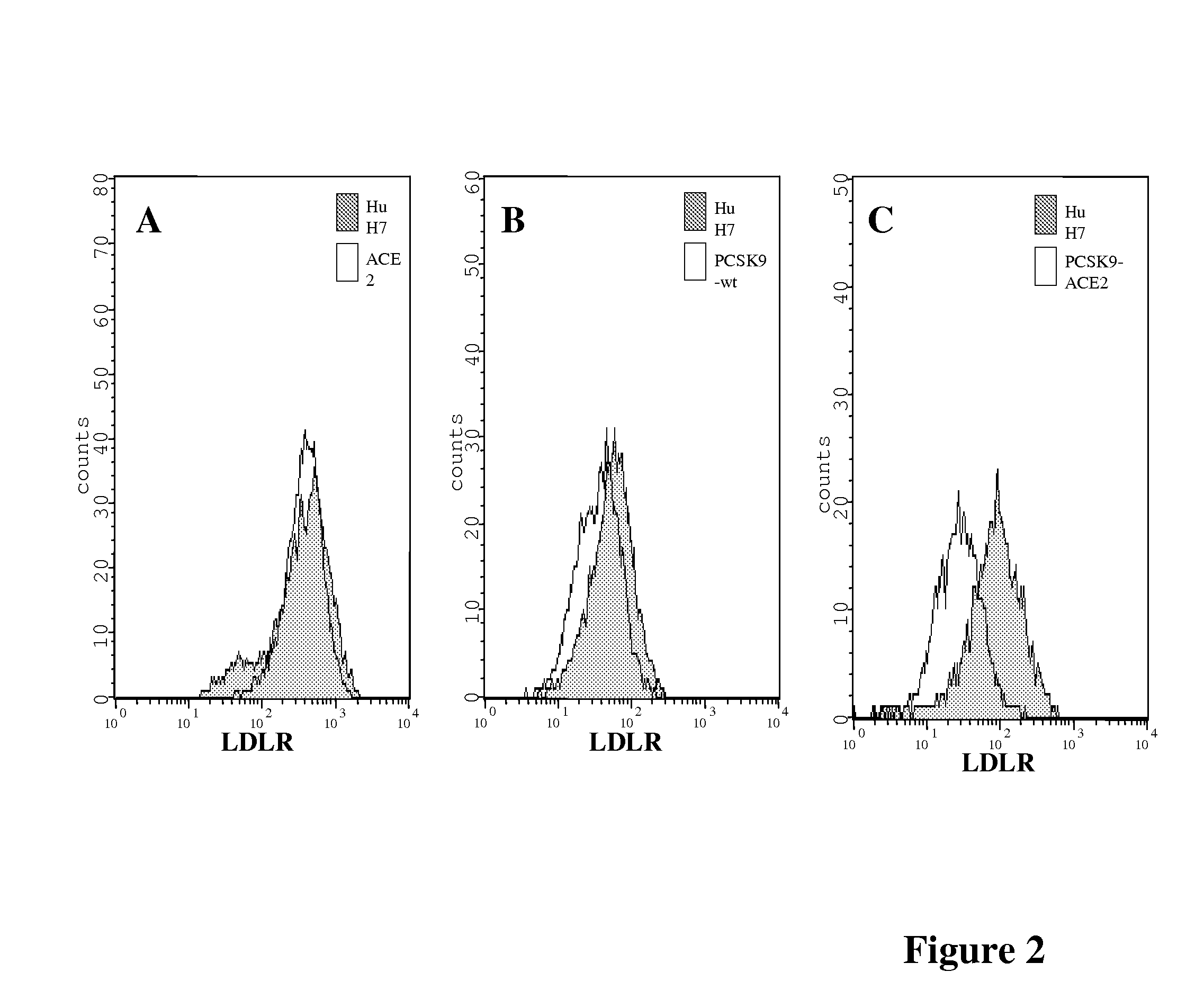
[0121]The effect of PCSK9 on HCV propagation 3 days post-infection was then evaluated by recording the number of foci of infection. The number of foci of infection represents the number of infectious particles that successfully infect cells in a culture. Once within the cell, the virus will replicate and spread to the surrounding cells and form what appear as a cluster of infected cells also called a colony. Immunofluorescence performed as described in Example 1 above revealed that cells expressing PCSK9-ACE2 and, to a lesser extent PCSK9-wt, had a reduced number of foci expressing NS5A, as compared to HuH7 cells expressing ACE2 alone or the empty vector (phcmv3) (FIG. 3). HuH7 cells stably expressing PCSK9-ACE2 are more resistant to HCVcc since only very rare single cells are stained for NS5A and no colony could be detected.
[0122]To ensure that the TM-CT domain of ACE2 in the construct PCSK9-ACE2 was not accountable for the ...
PUM
| Property | Measurement | Unit |
|---|---|---|
| Concentration | aaaaa | aaaaa |
| Biological properties | aaaaa | aaaaa |
| Therapeutic | aaaaa | aaaaa |
Abstract
Description
Claims
Application Information